






Abstract
Objectives
Most airway stenoses are acquired secondary to the use of prolonged endotracheal intubation. Antibiotics have been shown to decrease local inflammation and granulation tissue formation in the trachea. However, antibiotic therapy is not 100% effective in preventing or treating granulation tissue formation. Development of bacterial biofilms may explain this finding. This study evaluates the difference between tracheal stenotic segments and normal trachea in terms of (1) presence of bacterial biofilms, (2) quantitative bacterial counts, and (3) inflammatory markers.
Study Design
Cross-sectional study.
Setting
Tertiary care academic medical center.
Subjects
A total of 12 patients were included in the study. Tissue from stenotic segments from 6 patients with airway stenosis undergoing open airway procedures were compared with tracheal tissue from 6 patients without airway stenosis undergoing tracheostomy.
Methods
Scanning electron microscopy for biofilm detection, quantitative polymerase chain reaction for quantitative analysis of bacterial count, and immunohistochemistry were performed for inflammatory markers transforming growth factor β1 (TGF-β1) and SMAD3.
Results
Compared with the patients without airway stenosis, patients in the airway stenosis group showed presence of bacterial biofilms, a significantly higher expression of 16S rRNA gene copies per microgram of tissue (187.5 vs 7.33, P = .01), and higher expression of TGF-β1 (91% vs 8%, P < .001) and SMAD3 (83.5% vs 17.8%, P < .001).
Conclusion
Bacterial biofilms, increased bacterial counts, and higher expression of TGF-β1 and SMAD3 are associated with airway stenosis.
Most patients with laryngotracheal stenosis have acquired this disease secondary to the use of prolonged endotracheal intubation. 1 The incidence of postintubation stenosis ranges from 0.9% to 3% in United States.2,3 Pressure exerted on the tracheal mucosa by the presence of an endotracheal tube leads to irritation, ulceration, and granulation tissue formation. Granulation tissue itself is an integral part of wound healing and consists of fibroblast, macrophages, loose connective tissue, and newly formed capillaries. Granulation tissue is also commonly encountered in laryngotracheal stents, 4 laryngotracheal reconstructive surgery, 5 and tracheostomy tubes. 6 Studies have demonstrated that tracheal granulation tissue developing after laryngotracheal reconstruction and airway stent placement is associated with increased bacterial colonies.7-9 In these situations, development of granulation tissue appears to be in response to the bacterial infection. 10 Commonly identified aerobic pathogens in tracheal granulation tissues are Pseudomonas aeruginosa, Staphylococcus aureus, Streptococcus viridans, Haemophilus influenzae, and Neisseria spp.8,9 While these organisms are frequently identified among normal airway microflora, these same organisms can create problems if there is damage to mucosal barriers or weakening of the host’s immune system. Granulation tissue formation is also greatly influenced by the host’s own inflammatory response. Antibiotics have been shown to decrease local inflammation and granulation tissue formation in the trachea resulting from the microflora.9,11 However, antibiotic therapy is not 100% effective in preventing or treating tracheal granulation tissue. The development of bacterial biofilms may explain this finding.
Biofilms are composed of a 3-dimensional complex of bacteria in a self-made extracellular matrix of polysaccharides, nucleic acids, and protein. Biofilms help bacteria survive and prolong infection by enabling attachment to mucosal surfaces, conferring stability, promoting community gene-pool sharing, and providing resistance from host immune response, resulting in high degrees of antibiotic resistance.12-15 Prior studies have shown biofilm formation on the internal and external surfaces of the endotracheal tubes of intubated patients.16-18 However, no study has demonstrated the presence of bacterial biofilms on the tracheal tissue itself.
The 16S ribosomal RNA (16S rRNA) is a component of the 30S small subunit of prokaryotic ribosomes and is highly conserved between different species of bacteria. As a result, 16S rRNA gene sequencing has become prevalent in medical microbiology as a rapid and cheap alternative to phenotypic methods of bacterial identification.
This study was conducted to evaluate the difference between laryngotracheal stenotic segment and normal trachea in terms of (1) presence of bacterial biofilms, (2) quantitative bacterial counts, and (3) inflammatory markers.
Methods
A cross-sectional study approved by the Institutional Review Board (IRB) at the Keck School of Medicine, University of Southern California, was performed to compare and contrast the findings between normal trachea and laryngotracheal stenosis. Study participants were divided into 2 groups: (1) an airway stenosis group and (2) a no airway stenosis group.
Inclusion criteria for the airway stenosis group were patients with laryngotracheal stenosis who were scheduled to undergo open airway surgery. The exclusion criterion for this group was a previous airway surgery within the past 30 days of the specimen collection date with intralesional application of steroids or mitomycin C. This was done to control the effects of steroids or mitomycin C on the expression of inflammatory markers. Inclusion criteria for the no airway stenosis group were patients who were scheduled for a tracheostomy for reason other than airway stenosis. Exclusion criteria for this group were a past history of 1 or more of the following: intubation for more than 72 hours, laryngeal or tracheal cancer, laryngotracheal surgery, and chemoradiation to the head and neck region. In addition, any patients with signs of active infection of the trachea or lungs were excluded from both groups. This was done to make sure that patients included in the study did not have any infectious disease processes that could affect the results of our study.
In the airway stenosis group, the tissue was collected from the resected stenotic segment of the trachea. In the no airway stenosis group, the tissue specimen was collected from the site of tracheostomy, which was performed as previously described.3,19 In brief, a horizontal skin incision was made in the midline of the neck, just below the level of the cricoid cartilage. The strap muscles were retracted laterally, the thyroid gland was transected in the midline, and an inferiorly based Björk flap was created transecting only a single tracheal ring. The tissue specimen was collected from the Björk flap. Hence, none of the patients included in the study were subjected to any additional tissue sampling. The collected tissue specimen was transported to the lab in a sterile container on dry ice and processed immediately.
Scanning Electron Microscopy
Freshly excised tissue pieces were immersed in buffered 2.5% glutaraldehyde and processed through a sequential series of solutions consisting of tannic acid, osmium tetroxide, and increasing ethanol concentrations. The tissues were dried using a critical point dryer, mounted on specimen stubs, coated with platinum, and imaged in an XL30 S FEG scanning electron microscope (SEM). For the transmission electron microscope, the tissues were cut into small pieces and processed through osmium tetroxide and ethanol dehydration for final embedding in epoxy resin. Thin sections cut from the specimen in polymerized resin were examined in a Tecnai G2 20 transmission electron microscope. Electron microscopy (EM) classified the sections as positive or negative for biofilm. Previously published criteria for the presence of biofilm on EM were used. 20 In brief, morphological criteria indicative of bacterial biofilm included 3-dimensional structure, presence of water channels or interstitial voids, well-developed extracellular matrix, and evidence of cocci or rod bacteria as spherical or elliptical bodies within the size range of 0.05 to 5.0 µm. There was a single rater to read the EM, and the rater was kept blinded to the specimen’s origin.
Genomic Microbial DNA Isolation Methods
Preweighed frozen tissue samples were macerated using a motorized tissue grinder, and DNA was isolated using UltraClean Tissue & Cells DNA Isolation Kit (MoBio, Carlsbad, CA) according to the manufacturer’s instructions, including the optional proteinase K digest recommended for tough tissue.
Conditions Used for Quantitative Polymerase Chain Reaction
The number of 16S rRNA gene copies in the release buffer was quantified using a pan-bacterial quantitative polymerase chain reaction (qPCR) assay based on the primers (F1369/R1492) and probe (F1389) described by Suzuki et al. 21 Reactions were carried out in 96-well qPCR plates and were run in duplicate using 20-µL reaction volumes containing the TaqMan Universal PCR Master Mix (Applied Biosystems, Foster City, CA), the primers (900 nM), probe (250 nM), and 4.0 µL of release medium with suspended cells as the template. Amplification and detection were carried out with a Model CFX96 Touch Real-Time PCR Detection System (Bio-Rad Laboratories, Hercules, CA) using the following thermocycling parameters: 50°C for 2 minutes, 95°C for 10 minutes, 40 cycles consisting of 95°C for 15 seconds, 1.0°C s–1 ramp to 60°C for 1 minute. Ct values were calculated using the Bio-Rad CFX Manager software (version 3.0).
Calibration curves converting Ct values to bacterial 16S rRNA gene copy numbers were generated as follows. A 150-bp DNA oligomer was synthesized (Integrated DNA Technologies, Inc, Coralville, IA) to span the region of the 16S rRNA gene covered by the forward and reverse qPCR primers. The lyophilized material was used to prepare a 10-µM stock solution that was serially diluted to produce 11 standards spanning the 2.0 × 10⁻⁷ to 2.0 × 10⁻¹⁷ M concentration range (3.0 × 10¹⁰, 3.0 copy numbers). A plot of Ct versus log10 (copy number) afforded linear calibration curves, with typical R 2 values of .99. Data were expressed as averages. Investigators performing qPCR were kept blinded to the specimen’s origin and study objectives.
Immunohistochemistry
Transforming growth factor β1 (TGF-β1) is involved in fibrosis by regulating the deposition of extracellular matrix components such as collagen, fibronectin, and proteoglycans. 22 Higher expression of TGF-β1 has also been shown in benign airway stenosis. 23 SMAD3 is an intracellular signal transducer for TGF-β1. SMAD3 is the primary factor in the fibroblast-to-myofibroblast transformation in a TGF-β–driven, fibroproliferative process. Given these implications of TGF-β1 and SMAD3, we chose to focus our experiments mainly on these markers.
Tissue specimens were fixed overnight at 4°C in 4% paraformaldehyde and sunk in 20% sucrose/phosphate-buffered saline and embedded in optimum cutting temperature for cryosectioning. Slides were stained overnight at 4°C with rabbit anti-TGF-β1 antibody (Santa Cruz, sc146) at 1:100 or mouse IgG2a anti-SMAD3 antibody (Santa Cruz, sc101154) at 1:100. Alexa 568 goat anti-mouse IgG2a and Alexa 488 goat anti-rabbit secondary antibodies were used at 1:300 dilution for 2 hours at room temperature. Slides were stained with TO-PRO-3 nuclear counterstain and mounted with Vectashield mounting medium (Vector, Burlingame, CA). TO-PRO-3 is a nuclear counterstain and is among the highest-sensitivity probes for nucleic acid detection. Slides were imaged with Zeiss fluorescence microscope. Semiquantitative analysis of expression was done by counting the percentage of cells expressing TGF-b1 and SMAD3 per high-power field. Two raters blinded to the specimen’s origin and study objectives read the immunohistochemistry (IHC) slides. The interrater reliability was assessed by the intraclass correlation coefficient (ICC) using MedCalc 12.7.7.0 software.
Statistical Analysis
Since this was a preliminary hypothesis-generating study, the study sample size was restricted to 12 participants (6 in the airway stenosis group and 6 in the no airway stenosis group). The outcome variables reviewed were presence or absence of bacterial biofilms, quantitative difference in the bacterial count as determined by qPCR, and expression of TGF-β1 and SMAD3 on IHC. The statistical analysis for the bivariate variables was a χ2 test. The Mann-Whitney U test was used to compare qPCR output between the airway stenosis group and the no airway stenosis group.
A P value of .05 was used for statistical significance. All statistical analysis was done using SAS 9.1 (SAS Institute, Cary, NC).
Results
The airway stenosis group had 3 patients with Cotton Myer grade III stenosis and 3 patients with Cotton Myer grade IV stenosis. The 3 patients with grade IV stenosis had a tracheostomy tube at the time of surgery, while the 3 patients with grade III stenosis did not have tracheostomy tube. Patients in the airway stenosis group had a mean age of 44.1 years (range, 2-69 years), while those in the no stenosis group had the mean age of 53.8 years (range, 26-82 years). The number of 16S rRNA gene copies per microgram of tissue using qPCR, a surrogate for the number of bacterial cells, was significantly higher in patients with airway stenosis (mean, 187.5; SD, 187.5; range, 35-495) compared with patients with no airway stenosis (mean, 7.33; SD, 2.73; range, 4-11; Mann-Whitney U test P = .005; Table 1 ). Sample size was too small to perform a Mann-Whitney U test to evaluate statistical difference between the mean 16S rRNA gene copies per microgram of tissue in patients with tracheostomy (mean, 201, SD, 255) and those without tracheostomy (mean, 174; SD, 146.5) in the airway stenosis group. The SEM showed the presence of bacterial biofilms in the tissue from the stenotic segment in the airway stenosis group but not in the tracheal tissue of patients from the no airway stenosis group ( Figure 1 ). The IHC demonstrated a higher percentage of cells expressing TGF-β1 (91% vs 8%; Mann-Whitney U test P < .001) and SMAD3 (83.5% vs 17.8%; Mann-Whitney U test P < .001) in tissues from the airway stenosis group compared with tissues from the no airway stenosis group ( Table 1 ; Figure 2 ). The ICC for the IHC slides was 0.995 (95% confidence interval = 0.924-0.999). An ICC closer to 1 is considered to be very reliable with little variability between raters.
Differences between No Airway Stenosis Group and Airway Stenosis Group.

Abbreviations: HPF, high-power field; IHC, immunohistochemistry; TGF-β1, transforming growth factor β1.
Cotton-Myer grade for tracheal stenosis.
Mann-Whitney U test P value.
Fisher exact test P value. Fisher exact test was performed instead of χ2 as 50% of the cells had a value of zero.
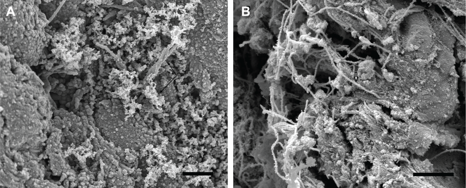
Scanning electron micrographs of specimen from stenotic segment. Scanning electron micrograph showing morphologic characteristics of bacterial biofilm on the airway stenotic segment. (A) Note the presence of bacterial structures (black single arrows). (B) Note nanofibers connecting bacterial cells (black and white arrow). (Scale bars: A = 2 µm, B = 2 µm.)
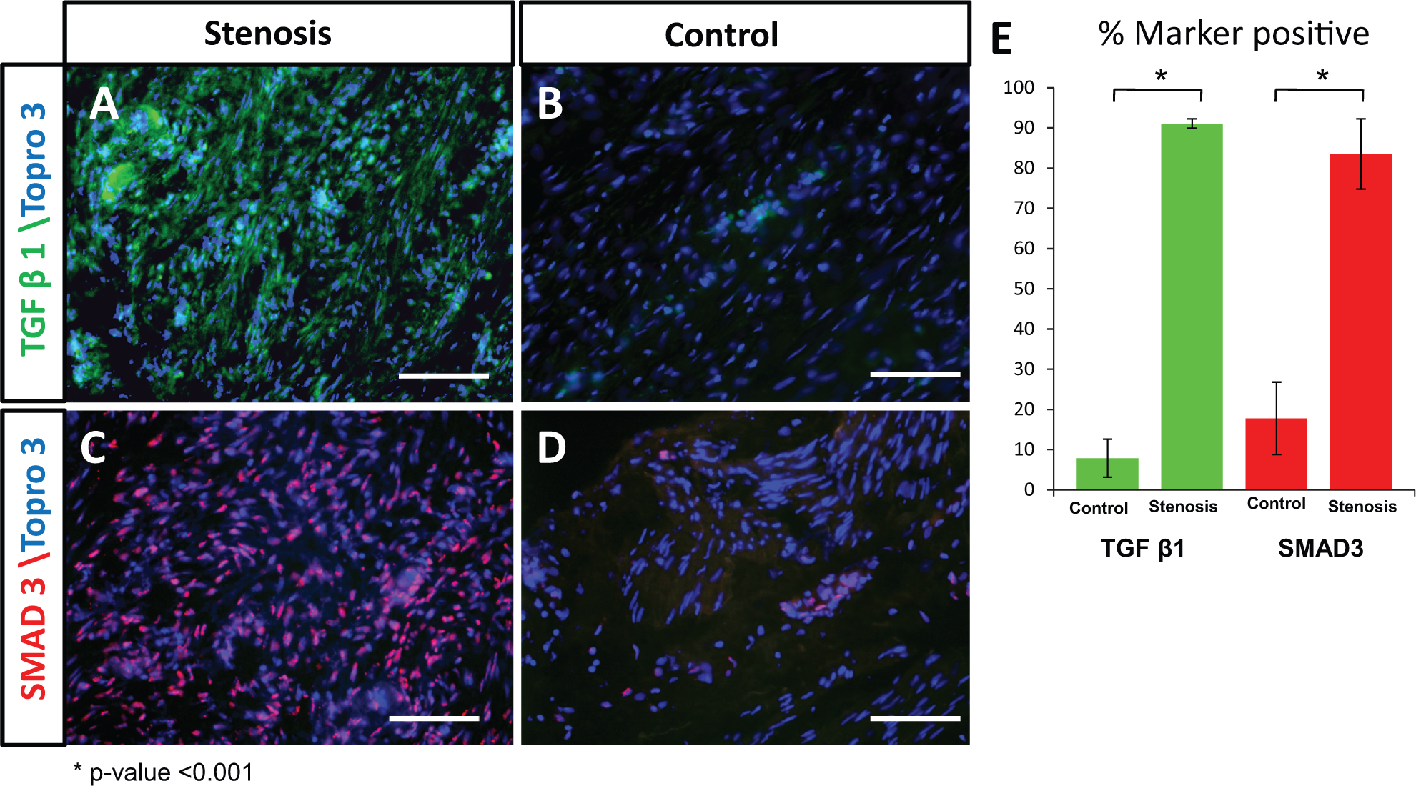
Immunohistochemistry of transforming growth factor β1 (TGF-β1) and SMAD3. TGF-β1 is expressed in a greater percentage of total cells in stenosis versus control tissue (91% vs 8%, P = .0000008) (A, B, E). SMAD3 is also expressed in a greater percentage of cells in stenotic tissue (83.5% vs 17.8%, P = .0002) (C, D, E). Scale bar = 200 µm.
Discussion
Endotracheal tubes, tracheostomy tubes, laryngotracheal reconstruction sites, and laryngotracheal stents are all associated with the formation of granulation tissue. An excess of granulation tissue can cause postoperative bleeding, airway obstruction, and excess wound contracture leading to the narrowing of the airway lumen. This may lead to respiratory distress, difficulty with decannulation, and delay of definitive treatment, which is often avoided in the presence of active inflammation.24,25 Pathophysiology of airway granulation is not completely understood. Proposed theories range from mechanical irritation, laryngopharyngeal reflux, and polymicrobial colonization.7-10,26,27
In their study on canine larynx, Sasaki et al 10 demonstrated that granulation tissue was a response to bacterial infection. They also demonstrated the beneficial effects of antimicrobial agents. 10 Previous studies have also demonstrated the association between tracheal granulation tissue and bacterial colonies.8-12 Similarly, our study showed a statistically significant higher expression of 16S rRNA gene copies in tissues from patients with airway stenosis compared with patients with no airway stenosis. Presence of a tracheostomy tube in the airway stenosis group did not affect the findings of biofilms or the 16S rRNA gene copy numbers. However, we did notice a higher expression of 16S rRNA gene copies in patients with soft stenosis and active granulation tissue compared with those with mature stenosis in the airway stenosis group. The small sample size of our study prevented us from performing a statistical subgroup analysis. Although the presence of bacteria does not prove an etiologic role, it may add to the microbial burden on the laryngotracheal mucosa, thereby affecting the host inflammatory response.
The advantages of using 16s RNA for bacterial quantification instead of microbiological culture are 2-fold: (1) it can detect nonculturable bacteria, and (2) it gives the actual bacterial count in the tissues rather than in the nutrient media. We feel that the incubation method may be selectively biased in favor of a particular organism depending on the nutrient and incubation environment. In addition, the incubation method does not take into account nonculturable bacteria. The qPCR combines the detection of a target template with quantification by recording the amplification of a PCR product via a corresponding increase in the fluorescent signal associated with product formation during each cycle in the PCR. Quantification of gene numbers is determined during the exponential phase of the PCR amplification when the numbers of amplicons detected are directly proportional to the initial numbers of target sequences present within the tissue specimen.
Biofilms are pervasive in nature and disease. The Centers for Disease Control and Prevention estimate that biofilms are related to 65% of all human infectious diseases. 28 Luminal biofilm is present in 84% to 100% of postextubation endotracheal intubation.17,18 Bacterial structures have been detected within the luminal biofilm by EM in up to 68% of specimens.16,18 There is a positive correlation between the presence of microbial structures on EM and positive microbial cultures.29,30 Our study is the first to report the presence of bacterial biofilms in the tracheal tissue specimens of patients with laryngotracheal stenosis. Our observation that higher amounts of microbes were identified in the stenotic tracheal segment compared with normal trachea and in soft and active granulation tissue compared with those with mature stenosis suggests that biofilms may contribute to airway granulation tissue by facilitating greater bacterial burden.
Karagiannidis et al 23 had reported significantly increased expression of TGF-β1 mRNA in stent-related stenosis compared with nonstenotic controls. In our study, we noticed similar findings of increased expression of TGF-β1 in tissues from the stenotic segment in the airway stenosis group compared with tracheal tissue from the group with no airway stenosis. TFG-β1 is associated with inflammation and fibrosis and is one of the strongest inducers of myofibroblasts. 31 Studies have shown that myofibroblasts have a higher collagen synthesis activity compared with normal fibroblasts. 32 Hence, myofibroblasts may be responsible for the structural changes seen in acquired airway stenosis. In addition, our study also shows increased expression of the SMAD3 gene, which is an intracellular signal transducer for TGF-β1. SMAD3 plays an important role in luminal fibrosis seen in obliterative bronchitis, and disruption of SMAD3 in experimental models inhibits extracellular matrix deposition and luminal fibrosis. 33
Our finding of biofilms and increased bacterial count in the airway stenosis segment might suggest a pathophysiological process similar to that described in chronic rhinosinusitis, in which bacterial biofilms are associated with persistent inflammation and host immune response.34,35 Bacterial biofilms may be the potential cause of persistent laryngotracheal mucosal inflammation, eliciting a host immune response in terms of overexpression of inflammatory markers, which then promotes tracheal granulation tissue formation and eventual tracheal stenosis.
Limitations of our study include a small sample size, which limits the generalizability of our findings. A study with a larger sample size should be performed to confirm or refute our findings. Since this was a preliminary study to evaluate the presence of biofilm in the airway stenotic segment, we did not perform bacterial speciation or phylogenetic study to identify the bacterial species involved in the biofilm production. We are, however, currently performing high-throughput culture-independent molecular methods to study the microbial community composition in the collected specimens. Another limitation of our study is the lack of quantitative analysis of the inflammatory markers (TGF-β1 and SMAD3) using qPCR techniques and limiting our evaluation to only TGF-β1 and SMAD3. Although the semiquantitative method used in this study has been previously described in the literature, 36 future studies should evaluate additional inflammatory markers such interleukins (IL-1, IL-6, IL-8) and fibroblast growth factors.
Conclusion
Bacterial biofilms, increased bacterial counts, and increased expression of TGF-β1 and SMAD3 are seen in stenotic segments of patients with laryngotracheal stenosis compared with normal tracheal tissue. Increased bacterial counts and development of bacterial biofilms may explain the inflammatory response of the airway epithelium and subsequent stenosis in these patients.
Author Contributions
Disclosures
Footnotes
Acknowledgements
We thank Ariel Larro-Popkin, MPH, for editorial assistance.
No sponsorships or competing interests have been disclosed for this article.
This article was presented at the 2013 AAO-HNSF Annual Meeting & OTO EXPO; September 29–October 3, 2013; Vancouver, British Columbia, Canada.