
Abstract

Keywords
Introduction
A 5-year-old girl with a 2-week history of dry cough, aversion to food, and vomiting after meals was noted to have a coin-shaped, radiopaque esophageal foreign body with central perforation on plain film ( Figure 1 ). She had a previous history of a double aortic arch repair with inadvertent esophageal injury requiring repair and subsequent esophageal dilations. The foreign body was removed and noted to be an eroded penny. This study was reviewed and determined to be exempt by the Institutional Review Board at the University of Oklahoma.
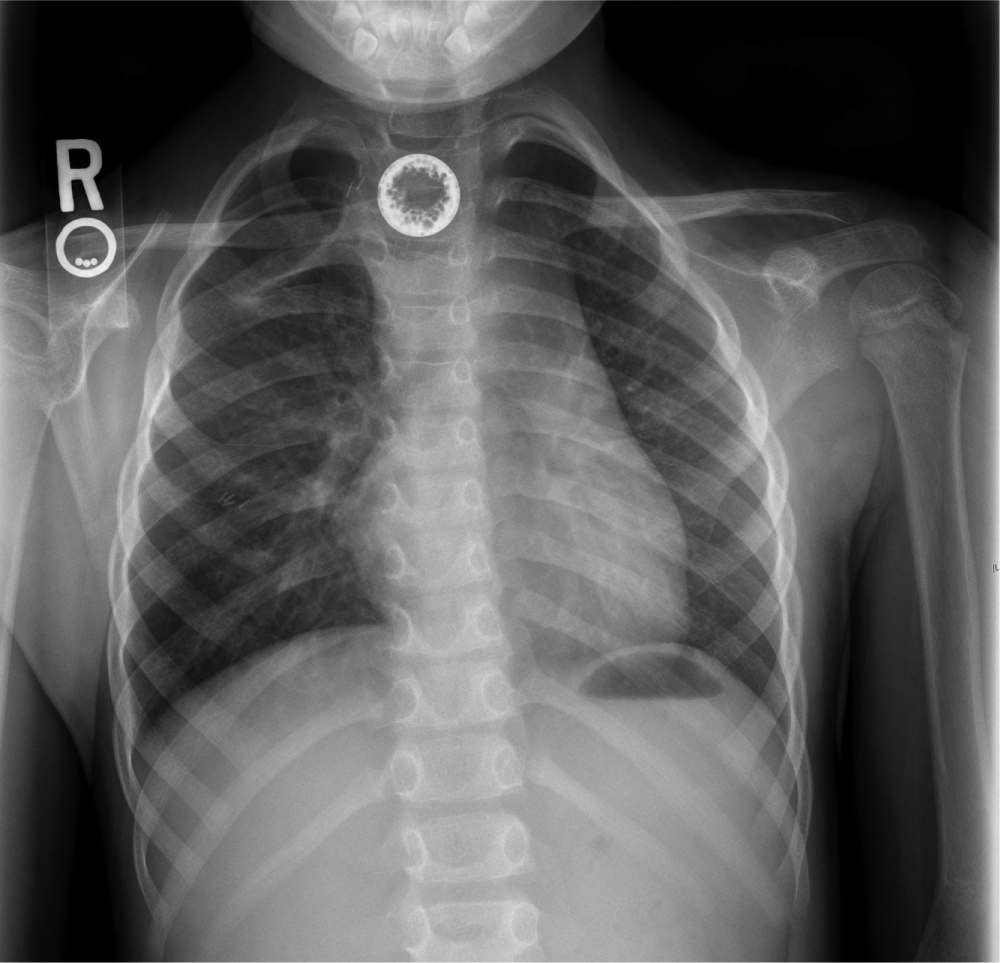
Chest x-ray showing radiopaque esophageal foreign body with irregular, central perforation.
Discussion
Esophageal foreign bodies are common in the pediatric population, with coins being the most common ingested object. 1 The penny in our case demonstrated diffuse erosive changes with central perforation ( Figure 2 ). There was, however, no mucosal injury to the esophagus. Post-1982 zinc-based pennies have been noted to have erosive changes after only 2 days of exposure to postprandial gastric acid concentration, but case reports have shown conflicting evidence of mucosal injury with chronically retained esophageal pennies.2,3 Clinicians treating esophageal foreign bodies should be aware of the potential changes to zinc-based pennies, their subsequent unique perforated appearance on plain film, and possible esophageal injury. Coupled with knowing that the diameter of a penny is 19.05 mm, more potentially dangerous foreign bodies, such as button batteries, can be ruled out. 4
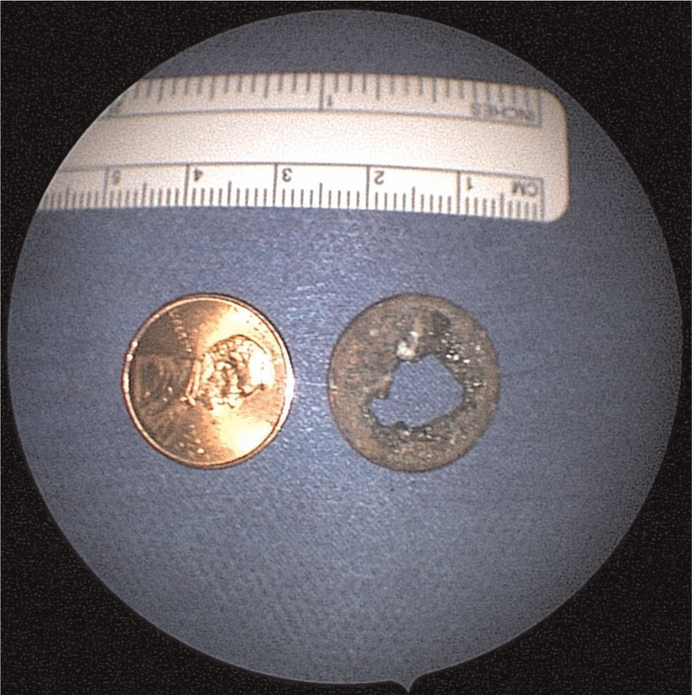
A standard penny (left) compared with the eroded penny (right) removed from the patient.
Author Contributions
Disclosures
Footnotes
No sponsorships or competing interests have been disclosed for this article.