
Abstract
Objective
Cervical schwannomas are benign tumors that commonly present as asymptomatic masses and are managed with observation, radiation, or surgery. To our knowledge, the rate of volumetric change seen on serial imaging is not currently used to determine surgical candidacy. We assess average growth rates and determine whether growth rate of cervical schwannoma predicts having undergone surgery.
Study Design
Case series with chart review.
Setting
Quaternary academic medical center.
Subjects and Methods
Patients were identified with at least 2 imaging studies and pathologic or imaging characteristics of cervical schwannoma. Volume was calculated with the formula 4/3πxyz, with x, y, and z representing the 3 orthogonal dimensions. Volume and rate of volume change were compared among observed, surgical, and gamma knife groups.
Results
Thirteen patients were identified and divided into subgroups: surgical (n = 5), observation (n = 6), and gamma knife (n = 2). Mean follow-up time was 21 months (range, 1-80 months) and not significantly different among subgroups. The average changes in volume were 3.61 cm3/mo (entire group), –2.75 cm3/mo (observation), 11.97 cm3/mo (surgery), and 1.78 cm3/mo (gamma knife). Average initial volume for the entire group was 124.4 cm3 (range, 5-608 cm3) and 142 cm3 (range 5-613) at follow-up. The surgical group had a statistically significant change in volume (P = .03). A statistically significant difference in growth rate was seen between the surgical and observation groups (P = .016) and between the surgical group and all nonsurgical patients (P = .011).
Conclusions
Rate of tumor growth can be used in the evaluation of patients with cervical schwannoma, and it may predict surgical intervention.
Neurogenic tumors of the head and neck are a rare entity and present unique challenges in management. Among the various subtypes, schwannomas—which are benign nerve sheath tumors derived from Schwann cells—can arise from peripheral, autonomic, or cranial nerves. An estimated 25% to 45% of all extracranial schwannomas occur in the head, and they are often classified according to their nerve of origin. 1 Within the head and neck, they have been reported to occur in the parapharyngeal space, nasal and oral cavities, middle ear, larynx, and orbit. As such, their symptomatology can be quite variable.
Surgical excision is the definitive treatment for head and neck schwannomas. However, the benign slow-growing nature of the tumor, combined with the potential for postoperative cranial neuropathy following resection, often renders the decision to proceed to surgery a challenging one. Depending on the nerve of origin, cervical schwannomas may present with hoarseness, dysphagia, aspiration, dysarthria, and tongue weakness, but more often they are incidentally found on imaging or present as an asymptomatic neck mass. Since surgical resection often involves sacrifice of the nerve of origin, surgery may result in significant functional deficit not experienced by the patient preoperatively. For this reason, management of these masses is not clearly defined—with some advocating immediate resection prior to development of symptoms from tumor compression and with others taking a more conservative approach by making use of serial imaging and patient symptomatology to guide timing of intervention. The use of tumor volume and volumetric change as criteria for surgery has not been previously described. The objective of our study was to calculate the volume growth rate of cervical schwannomas and determine whether it can be used as a predictor of surgical intervention.
Materials and Methods
Institutional Review Board approval was obtained from the University of California, San Francisco. A 20-year retrospective chart review was performed to identify patients with pathologic or imaging characteristics of cervical schwannoma. Patients were included if they had a histologic diagnosis of cervical schwannoma or at least 2 imaging studies consistent with a radiographic diagnosis. Patients were excluded if
they had <2 imaging studies or if films were unavailable for our review (n = 42),
the radiographic diagnosis was unclear or not consistent with schwannoma (n = 9), or
family history of neurofibromatosis was present (n = 3).
Each chart was retrospectively reviewed by 2 authors (A.S.A., C.M.H.), and information was extracted regarding patient’s age, sex, surgical pathology (if known), time between scans, and presence of symptoms. Images were separately reviewed, and dimensions of the tumors were measured and recorded. When surgical pathology was unavailable, tumor type was determined according to anatomic location and radiographic findings. Based on previous data indicating a reliable method of obtaining the volume of nonspherical shapes, cervical schwannomas were modeled as an ideal ellipsoid with the largest dimension in the anterior-posterior, medial-lateral, or craniocaudal plane measured for each lesion.2,3 The volume was calculated through the formula 4/3πxyz, with x, y, and z representing these dimensions. Tumor growth rate for each patient was calculated by dividing the change in volume (cm3) by the interval between imaging studies (months). Patients were then grouped by management strategy, and an average growth rate for each subgroup was calculated by summing all individual growth rates and dividing by the total number of patients in that subgroup. Average growth rates among observed, surgical, and gamma knife subgroups were then compared. Statistical analysis was completed with SAS 9.4 (SAS Institute, Cary, North Carolina).
Results
Thirteen patients met inclusion criteria (9 women and 4 men). The average age at the time of the initial imaging study was 58 years. Based on the previously described formula, tumor volumes on interval imaging studies were calculated. Patient demographic data, volume measurements, and individual growth rates are outlined in Table 1 .
Demographic Data of the 13 Patients Included in the Study.
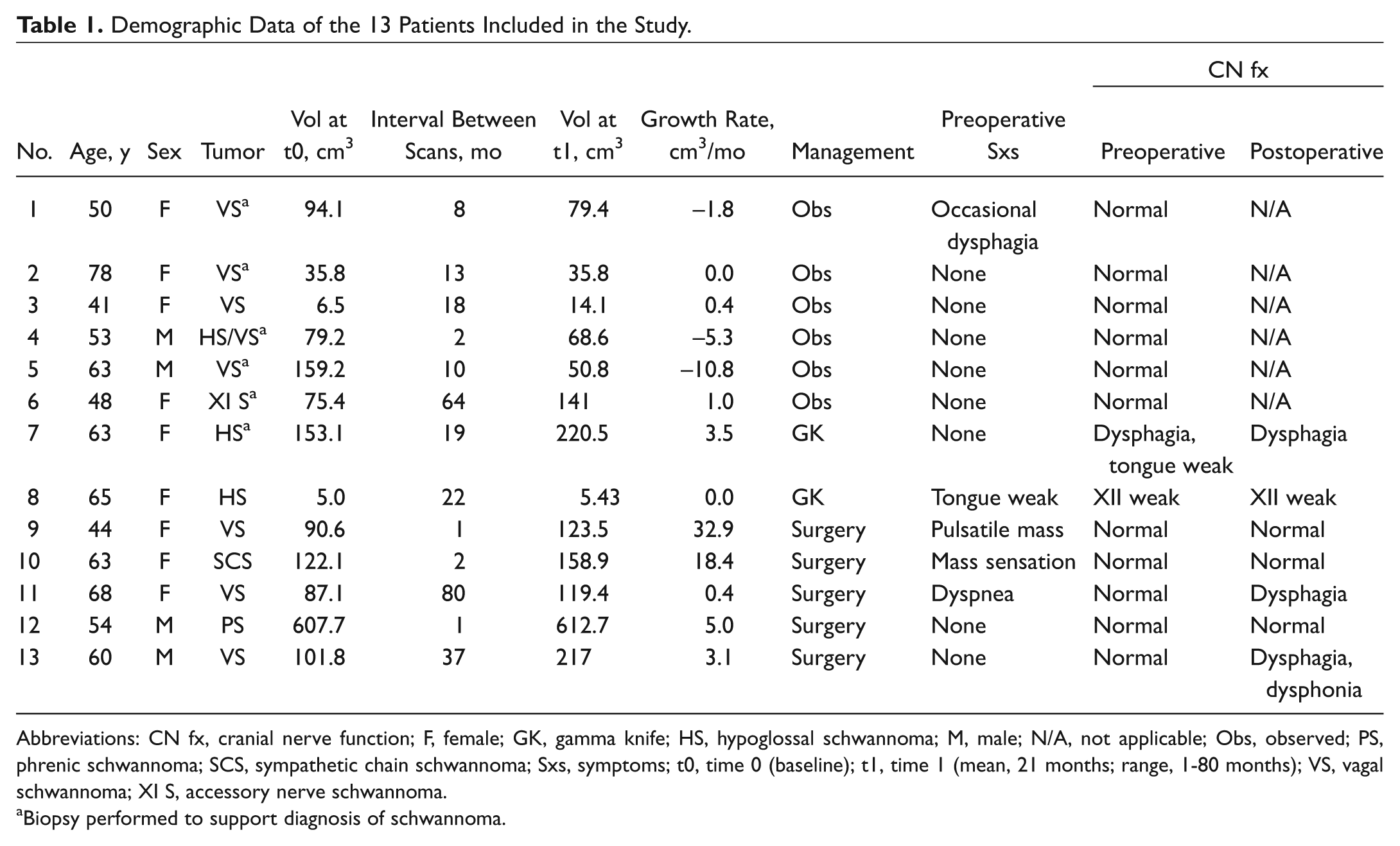
Abbreviations: CN fx, cranial nerve function; F, female; GK, gamma knife; HS, hypoglossal schwannoma; M, male; N/A, not applicable; Obs, observed; PS, phrenic schwannoma; SCS, sympathetic chain schwannoma; Sxs, symptoms; t0, time 0 (baseline); t1, time 1 (mean, 21 months; range, 1-80 months); VS, vagal schwannoma; XI S, accessory nerve schwannoma.
Biopsy performed to support diagnosis of schwannoma.
Of the 13 patients, 6 were managed with continued observation; 5 had surgical excision; and 2 had gamma knife radiation. None of the patients from the observed group had objective cranial neuropathy at any point during evaluation. Both patients from the radiated group had objective pretreatment unilateral tongue weakness; otherwise, there were no cranial nerve deficits identified. Similarly, no patients in the surgical group had preoperative cranial neuropathies. However, following surgery, 2 patients developed significant dysphagia and vocal fold paralysis due to vagal nerve sacrifice, one of whom required multiple vocal cord medialization procedures.
The average interval between initial and subsequent imaging studies for all patients was 21 months (range, 1-80 month). The average volume at the time of the first imaging study was 124 cm3 (range, 5.0-607.7 cm3) and at the time of the follow-up scan, 142 cm3 (range, 5.43-612.7 cm3). This represents a growth rate of 3.61 cm3/mo for all patients. When average follow-up intervals were classified by management strategy, analysis revealed no statistically significant difference among the subgroups: observed (19 months), gamma knife (21 months), and operative (24 months). For the observed group (n = 6), the average volume at the time of the initial scan was 75 cm3 and at follow-up, 65 cm.3 The average growth rate for the observation group was −2.75 cm3/mo (P = .34). For the surgical group (n = 5), the average volume at the time of the initial scan was 201.9 cm3 and at follow-up, 246 cm3. The average growth rate of the surgical group was 11.9 cm3/mo (P = .037). For the gamma knife group (n = 2), the average volume at the time of the initial scan was 79 cm3 and at follow-up, 113 cm3. The average growth rate of the gamma knife group was 1.78 cm3/mo (P = .25). These data, including average growth rates, are summarized in Table 2 .
Difference in Mean Volumes among Subgroups of Patients with Cervical Schwannoma.
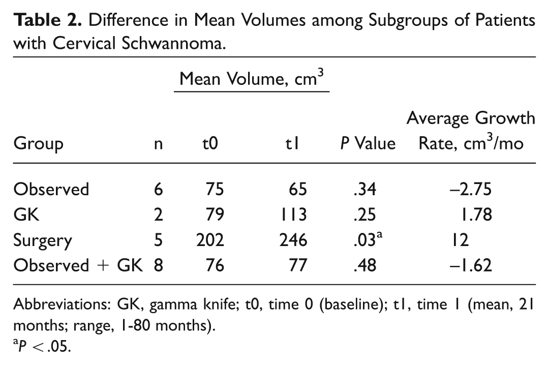
Abbreviations: GK, gamma knife; t0, time 0 (baseline); t1, time 1 (mean, 21 months; range, 1-80 months).
P < .05.
On comparison of subgroups, a statistically significant difference was found between the growth rates for patients managed with surgery (where tumors grew at an average rate of 11.9 cm3/mo) and those that were observed (which diminished at an average rate of 2.75 cm3/mo; P = .02). A Mann-Whitney test comparing the difference in volume at the 2 time points for patients managed surgically and those not managed surgically (observation + gamma knife) showed that volume was higher for the surgical group (P = .048). Similarly, a Mann-Whitney test comparing the growth rates between the surgical and nonsurgical groups (observation + gamma knife) also demonstrated a larger growth rate for tumors ultimately managed surgically (P = .023).
No statistically significant difference in growth rate was seen between patients being observed (average growth rate, –2.75 cm3/mo) and those receiving gamma knife radiation (average growth rate, 1.78 cm3/mo; P = .12).
Logistic regression modeling was performed to predict surgical outcome based on volume at the second time point and growth rate. Likelihood ratio χ2 was statistically significant (P = .0079). For a subsequent surgical excision, odds ratios were 1.011 and 1.285 for volume and growth rate and not statistically significant (P = .3959 and .406, respectively). Logistic regression with empirical standard errors was then performed, which revealed a statistically significant effect of growth rate (P = .0015). These findings are summarized in Table 3 .
Logistic Regression Modeling of Volume and Growth Rate as a Predictor of Surgical Outcome.
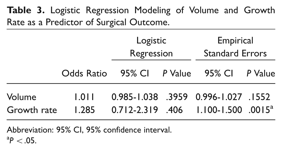
Abbreviation: 95% CI, 95% confidence interval.
P < .05.
Discussion
Schwannomas are benign slow-growing tumors that arise from all peripheral, cranial, and autonomic nerves, most commonly within the parapharyngeal space. 4 While schwannomas can occur in many subsites within the head and neck, they generally present as asymptomatic masses.5,6 However, when symptomatic, schwannomas may cause symptoms associated with the nerve of origin, including dysphagia, dysphonia, dysarthria, and facial paresis or numbness 7 or as a result of a mass effect on surrounding structures.
Given the relatively rare occurrence of these lesions, no clear guidelines exist; thus, management remains controversial. Surgical resection is the gold standard for care. However, given the benign nature of these masses and the morbidity associated with nerve resection, serial imaging is an acceptable management strategy for asymptomatic patients and those with minimal symptoms or deficits. Alternatively, patients with preoperative cranial neuropathy may be more appropriate for a surgical intervention. To our knowledge, our series presents the first use of objective volumetric data to help assess predictors for surgical management.
We found that in our cohort, the average growth rate for tumors that had multiple imaging studies was 3.61 cm3/mo. When patients were classified by management strategy, tumors that ultimately underwent surgical resection had a significantly higher growth rate than those managed with either serial imaging or gamma knife radiation. Furthermore, on regression analysis, growth rate was a statistically significant predictor of surgery as a final outcome.
On review of the medical records, patients who were managed conservatively with serial imaging were generally those who presented with asymptomatic masses. Indeed, serial imaging demonstrated that these masses changed very little over time. Those who eventually underwent surgery generally did so because of the development of cranial nerve deficit, intolerable symptoms, or tumor growth noted on surveillance imaging. Pre- and postoperative symptoms are summarized in Table 1 .
Our data indicate that at the very least, growth rate appears to be as good a predictor of surgical intervention as volume and, though underpowered, growth rate appears to be the stronger of the 2 predictors. Moreover, when taken together, tumor volume and rate of growth can be used to predict surgical intervention, beyond chance.
If applied clinically, rate of volumetric growth can be used to predict which patients will ultimately require surgical management. Patients with an average volume growth <5 cm3/mo did not receive surgical intervention in our series. Conversely, patients with an average volume growth rate >5 cm3/mo did eventually undergo surgical resection. Clinicians can add this calculation to their management algorithm to determine which patients might ultimately be appropriate for surgery.
Interestingly, in the group of patients who did not undergo surgical intervention (observed + gamma knife radiation), despite a negative average growth rate (–1.62 cm3/mo), the mean volume at the second time point was actually higher than the initial time point (77 vs 76 cm3). This is likely due to the difference in time intervals for the 2 groups and likely indicates that the volume was overall stable and that the difference noted between the 2 time points is within measurement error.
Limitations of our study include the retrospective design and low sample sizes. Given the overall scarcity of cervical schwannoma, sample sizes are small, and the possibility of variability is high. Internal validity is limited by the fact that not all patients received a pathologic diagnosis of schwannoma. In some cases, if imaging was consistent with schwannoma, the diagnosis was clinically made. There is also the possibility of selection bias, as depending on surgeon preference; some patients underwent surgical intervention after 1 imaging study and a biopsy demonstrating schwannoma. This has the potential to skew the results by erroneously eliminating those surgical patients from our cohort.
For those patients who did undergo serial imaging and routine follow-up, average volumetric change had a statistically significant difference between those who underwent surgical resection and those who did not. Multicentered aggregate or other prospective studies are required to further clarify the role of volumetric growth in the overall management of these tumors. Although it is difficult to predict surgeon behavior, we advocate that application of these objective measures can add to the clinical evaluation and counseling of these patients.
Conclusion
Rate of volume change can be calculated and serve as a predictor of patients with cervical schwannomas who will eventually undergo surgical resection. These additional data can be added to the management algorithm of cervical schwannomas.
Author Contributions
Disclosures
Footnotes
Acknowledgements
We acknowledge Alan Bostrom, PhD, for his statistical consultation.
Sponsorships or competing interests that may be relevant to content are disclosed at the end of this article.
The abstract of this article was presented at the Triologic Society Combined Sections Meeting; January 23, 2016; Miami Beach, Florida.