
Abstract

Aspiration or ingestion of foreign bodies occurs in about 2.5 million children each year in the United States, with up to 2000 deaths reported. 1 Most of these events occur in children <3 years of age. This age predilection may be due to several factors, including the tendency of younger children to place objects in their mouths, poor swallowing coordination, high respiratory rates, and immature dentition. Objects placed in the mouth while playing or at school can be inhaled or ingested when the child is distracted by talking or laughing.1-3 Most aspirated foreign bodies are successfully removed via rigid bronchoscopy using optical forceps. Larger, oddly shaped objects may not be amenable to this method.
Case Reports
Case 1
A 10-year-old male was transferred emergently to our tertiary pediatric hospital from a community hospital after aspirating a board game piece at home. The object caused complete airway obstruction, not relived by attempts with Heimlich maneuvers. The child became anoxic, lost consciousness, and suffered cardiopulmonary arrest. He was intubated in the field and was transferred to a local community hospital where attempts to remove the object via rigid endoscopy were unsuccessful. At our institution, the object was easily captured by endoscopic forceps but would not pass through the subglottis despite attempts in different orientations. A tracheotomy was performed, and the object was successfully retrieved using forceps placed through the tracheostoma. Given the uncertainty about the patient’s neurologic status, a tracheostomy tube was placed. The patient was eventually decannulated, despite suffering irreversible anoxic brain injury.
Case 2
A 9-year-old male was transferred to our institution after aspirating a pencil cap ( Figure 1 ). He was taken to an outside hospital first, where the x-ray demonstrated left lung atelectasis and leftward tracheal shift. At our hospital, oxygen saturations were 80% to 85% and improved with a non-rebreather oxygen mask. In surgery, endoscopic instruments, including several types of optical forceps, were used to easily grasp the foreign body (hollow end facing upward). The object lodged below the vocal cords on all attempts with forceps and moved into the right mainstem bronchus naturally between attempts. A tracheotomy was performed over a bronchoscope, and the object was brought into view of the incision and retrieved. The tracheotomy incision was closed in multiple layers over a drain. The patient was extubated on postoperative day 1 with no neurologic sequelae.
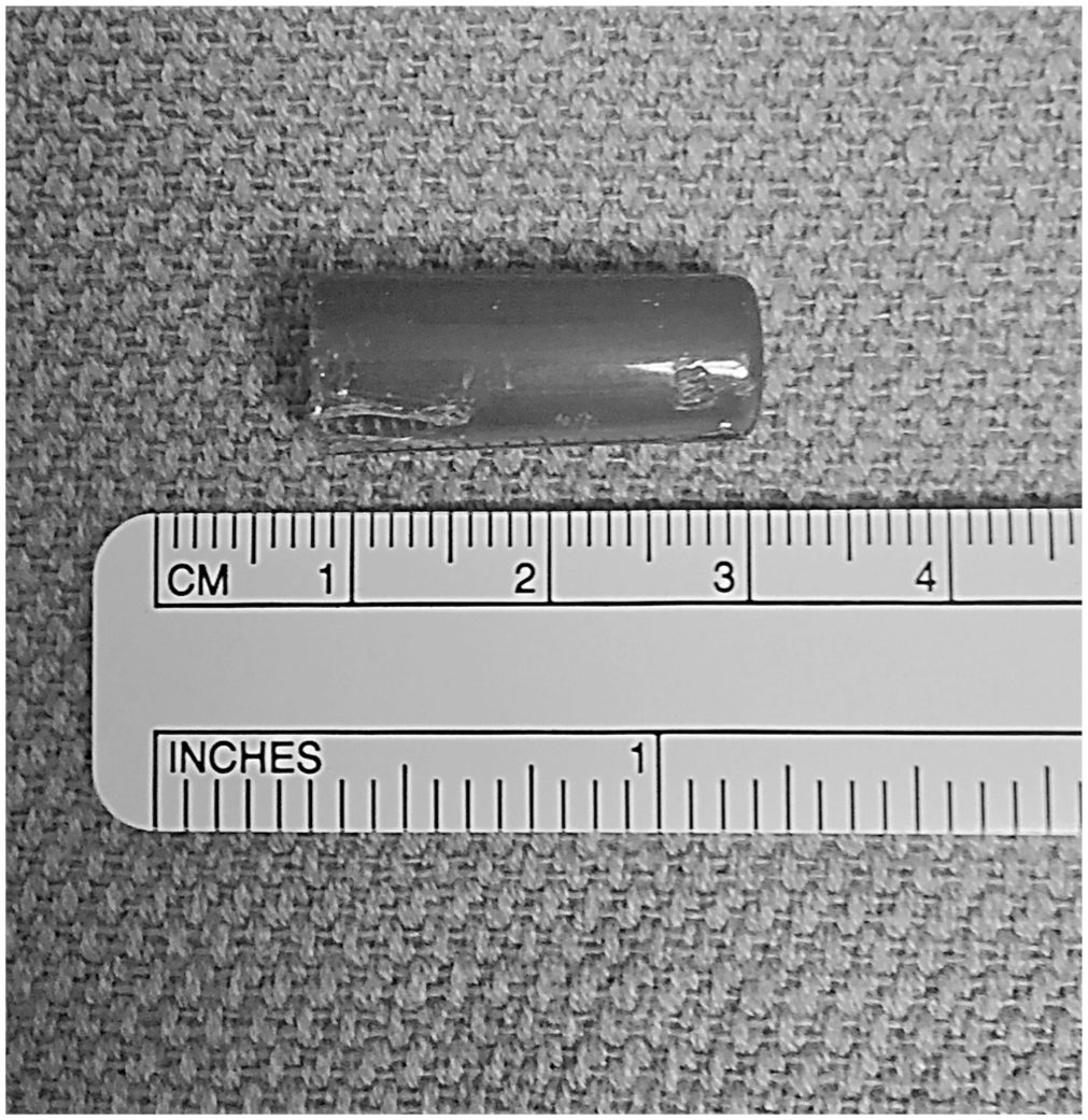
Aspirated pen cap retrieved in case 2. Despite the hollow end facing upward in the left mainstem bronchus, the cap could not pass through the glottis.
Institutional review board exemption for both cases was acquired from Ann & Robert H. Lurie Children’s Hospital of Chicago (2018-1587).
Discussion
The development of federal regulations related to manufacturing of children’s toys, games, and other objects has reduced the frequency and associated mortality due to foreign body aspirations and ingestions.1,3 Despite this, children remain at risk for life-threatening foreign body aspiration. Generally, the time of presentation after foreign body aspiration can be variable and related to the severity of airway obstruction, the presence or absence of a witnessed choking episode, patient age, and the type of management provided on arrival to the hospital. 3
Both of the airway foreign bodies described could be grasped easily with optical forceps via standard rigid bronchoscopy equipment appropriate to the age of the child. The objects lodged at the cricoid level and would not pass through the glottis. Perhaps the object had a shape that would only allow passage in one particular orientation not achieved using the optical forceps. Using standard airway endoscopy techniques assisted by a tracheotomy was the only way to remove the objects. Singh et al 4 noted, in their analysis of 342 children with airway foreign bodies, that sharp subglottic, long-duration subglottic, or objects larger than the glottis chink were most associated with tracheotomy to assist extraction.
The tracheotomy technique, after confirming the diagnosis with a flexible bronchoscope, was reported as a primary means of foreign body retrieval in a nation lacking access to rigid endoscopic equipment. 2 Flexible bronchoscopy has also been described as an alternative to rigid instrumentation. 5 Tang et al 5 cited a 91.3% success rate in extracting airway foreign bodies in a series of 1027 children. They noted wide availability, ability to reach distal foreign bodies, and ability to suction secretions as advantages. Neither of the objects encountered in our patients would have been amenable to flexible bronchoscopy given their size. Given the unpredictability of foreign body size/shape and the narrow subglottic space, clinicians tasked with foreign body removal should be prepared for the possibility of tracheotomy.
Author Contributions
Disclosures
Footnotes
No sponsorships or competing interests have been disclosed for this article.
This article was presented as a poster at the 2017 AAO-HNSF Annual Meeting and OTO Experience; September 10-13, 2017; Chicago, Illinois.