
Abstract
Objective
Transoral surgery is becoming a preferred technique because it does not leave any scar after surgery. However, transoral surgery for a dermoid cyst of the oral cavity is not standardized yet, due to the anatomic complexity of this region. The aim of this study was to evaluate the safety and efficacy of a transoral dermoid cyst excision.
Study Design
Multicenter prospective observational study.
Setting
University hospital.
Subjects and Methods
This study was designed as a 4-year prospective multicenter evaluation of dermoid cyst excisions within the floor of mouth. Clinical outcomes and complications related to procedures were evaluated among patients. The primary outcome was the efficacy of the procedure, and the secondary outcome was cosmetic satisfaction of each procedure.
Results
Twenty-one patients underwent transoral dermoid cyst excisions, and 22 underwent transcervical excisions. In the transoral surgery group, the mean size of the dermoid cyst was 5.35 cm (95% CI, 4.79-5.91), and in the transcervical surgery group, it was 6.19 cm (95% CI, 5.67-6.71). There was no significant differences with respect to overall demographic characteristics between the groups. However, the duration of the operation was shorter with the transoral group than with the transcervical group (P = .001), and cosmetic satisfaction was much better in the transoral group (P < .001).
Conclusion
Transoral dermoid cyst excision is a potentially safe and effective method that can lead to easy and quick removal of an oral cavity dermoid cyst, with excellent cosmetic outcomes.
Dermoid cysts are benign lesions that can occur throughout the body, with 7% of them occurring in the head and neck area and 1.6% within the oral cavity. 1 Dermoid cysts generally present with slow and progressive growth. Even if they are congenital, their diagnosis is usually made among young adults in their second or third decade of life. 2 They affect males more often than females, at a mean onset age of 28 years. 3 These cysts can be categorized as epidermoid cysts (when the lining refers only to the epithelium), dermoid cysts (when skin adnexa are found), and teratoid cysts (when other tissues are present, such as muscle, cartilage, and bone). 4
Various surgical techniques have been developed to excise dermoid cysts in the floor of the mouth. These procedures include transcervical approaches, such as a bilateral incision along the mandibular crest and a midline sagittal glossotomy, and they have been reported to excise dermoid cysts via intra- or extraoral routes according to the localization and size of the mass. 3 However, a large skin incision or glossotomy is unsuitable for treating a benign dermoid cyst. With recent advances in surgical techniques, an external large incision or a glossotomy is no longer needed to excise a benign dermoid cyst.5-17
In this study, we present a transoral dermoid cyst excision technique, with details of each operation. We also compare postoperative clinical outcomes of each transoral dermoid cyst excision with those of the transcervical dermoid cyst excision.
Materials and Methods
Study Design
This study was designed as a multicenter prospective observational study.
Ethics
This study was conducted after obtaining approval from the Institutional Review Board (GNUHIRB-2010-012), following the guidelines of the Declaration of Helsinki.
Patients
Of the 47 patients initially enrolled (>16 years old), 2 failed to meet our eligibility criteria: one had signs of inflammation on preoperative computed tomography, and the other had a history of a neck malignancy. Therefore, these 2 patients were excluded from this study. Surgery was performed from January 2013 to December 2016 on 45 patients for removal of a dermoid cyst from the oral cavity. Thus, transoral surgery was performed on 22 eligible patients, and the transcervical operation was performed on 23 eligible patients ( Figure 1 ). All patients gave their informed consent after being informed of the advantages and side effects of each procedure; the possibility of converting to a transcervical approach during the transoral surgery was also explained to each patient. After each cyst was excised, patients received regular checkups with clinical examinations for >1 year and ultrasonography twice a year. One patient from each group was lost to follow-up. Finally, there were 21 patients in the transoral approach group and 22 in the transcervical approach group.
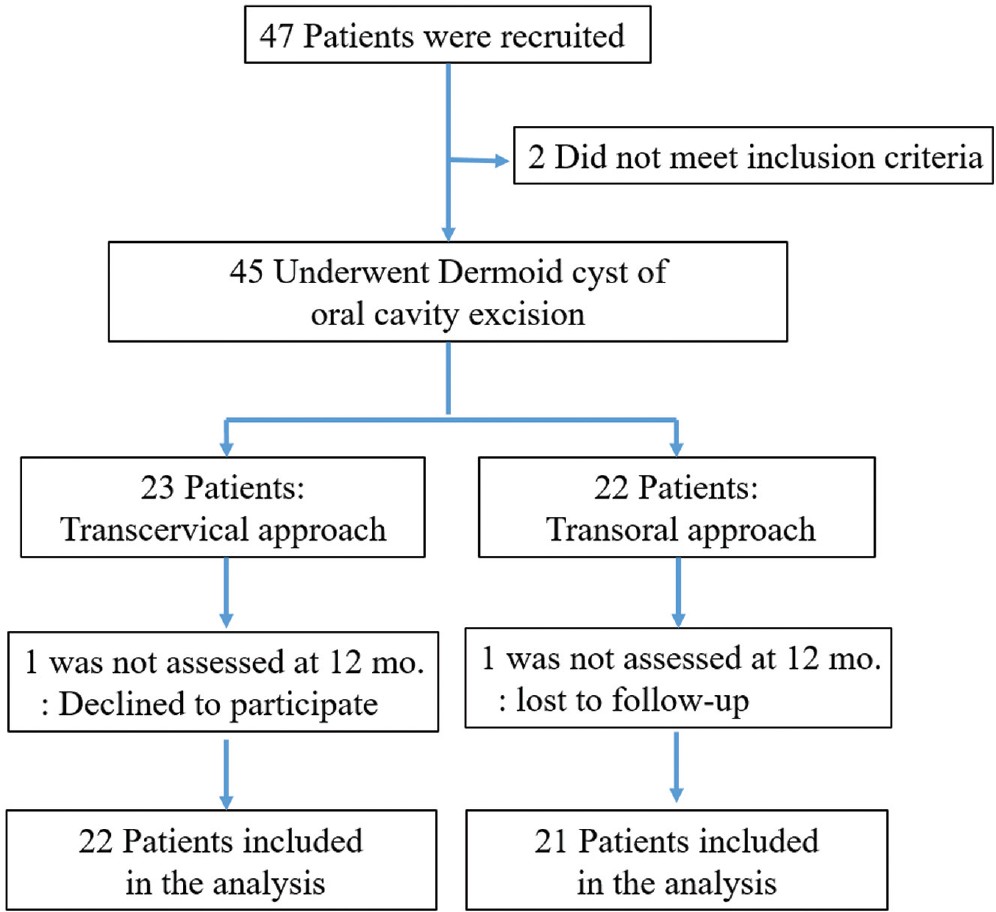
Patient enrollment.
Inclusion criteria were as follows: a diagnosis of a dermoid cyst within the floor of mouth by physical examination, neck computed tomography, and aspiration biopsy of fine needle. Exclusion criteria were as follows: abscess or inflammation suspected by preoperative tests, history of head and neck trauma, malignancy, and radiation therapy.
Outcomes
The primary outcome variable was the feasibility of the procedure. However, duration of the operation, histopathologic outcomes, recurrence, hospital stay, wound issues, and other complications were also assessed in this study. The secondary outcome was patient satisfaction after each operation, which was subjectively measured by interviewing patients after the procedure (“How satisfied are you with the surgical outcome?”). Specifically, respondents gave each satisfaction parameter a score ranging from 1 to 10 (1, extremely dissatisfied; 10, extremely satisfied) at 3 and 12 months after surgery.
Surgical Techniques
Transoral Dermoid Cyst Excision
Following general anesthesia, the mouth was opened with a Denhart oral retractor (Primestar Instruments, Sialkot, Pakistan). Each tongue was then retracted superiorly. A horizontal incision was made on the tongue’s frenulum mucosa above the submandibular gland opening ( Figure 2A ). After mucosal incision, the genioglossus muscle (GM) was identified in the operational field ( Figure 2B ). The GM is composed of 2 parts: a left side and a right side. Dissection was made between the muscles to find an avascular space under the endoscope (rigid, 10 mm, 0°; Olympus, Tokyo, Japan). After retraction of the GMs bilaterally with Sofield retractors (233-04; Zimmer Biomet, Warsaw, Indiana; Figure 2C ), the dermoid cyst was found when it was located above the mylohyoid muscle. When the dermoid cyst was located under the mylohyoid muscle, the mylohyoid was cut with an electrobovie. The dermoid cyst was then carefully dissected with an endoscopic dissector (Aesculap, Inc, Center Valley, Pennsylvania) and removed through an oral cavity incision site ( Figure 2D ). The surgical field was irrigated in each case, without any drainage. Because this procedure caused little damage to any muscle or bone (just mucosa incision and muscle retraction), there was only a small amount of drainage. Afterward, the oral mucosa was sutured vertically because a horizontal suture could result in contracture and decrease tongue movement (see Appendix: Transoral Large Dermoid Cyst Excision in the online version of the article).
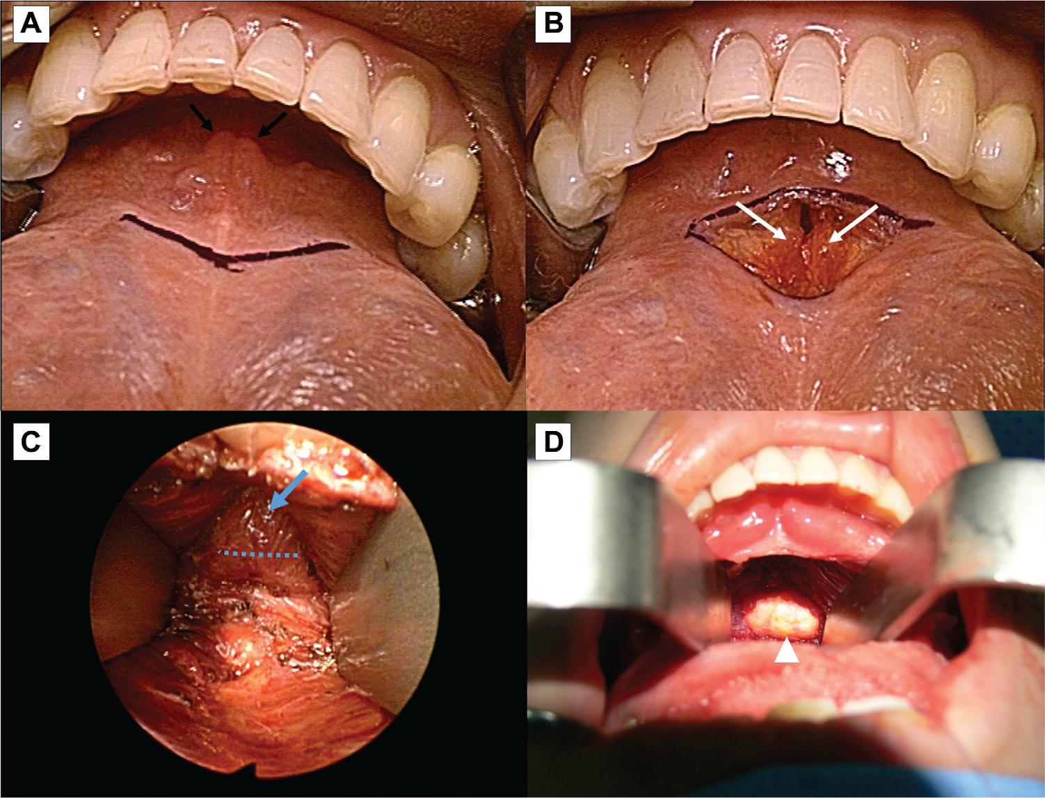
The basic concept of transoral dermoid cyst excision. (A) A horizontal incision on the tongue’s frenulum mucosa (black arrows, Wharton’s duct orifice). (B) The genioglossus muscle (white arrow) retracted bilaterally. (C, D) After the genioglossus muscle was retracted, a surgical incision (dotted line) was made on the mylohyoid muscle (blue arrow), and the dermoid cyst (arrowhead) was removed.
All patients were discharged 1 day after each operation. We encouraged frequent oral gargling with 0.02% chlorhexidine. Oral antibiotics were prescribed. Oral feeding of a normal diet was allowed at 1 day after the operation.
Transcervical Dermoid Cyst Excision
To excise each dermoid cyst, an approximately 5- to 10-cm horizontal skin incision was made in the submental or submandibular region, and a flap was created that extended above the hyoid bone and inferior to the mandible or mentum. After flap dissection, a dermoid cyst was found when the cyst was located under the mylohyoid muscle (under-mylohyoid type). It was carefully dissected and removed. When the dermoid cyst was located above the mylohyoid muscle (upper-mylohyoid type), the mylohyoid muscle was cut with an electrobovie knife. It was carefully dissected and removed through a skin incision site. Then each subcutaneous layer and skin were sutured.
Statistical Analysis
All statistical analyses were performed with SPSS 12.0 for Windows (IBM, Chicago, Illinois). Independent-sample t tests and Mann-Whitney tests were used to compared data between the groups. Statistical significance was considered when the P value was <.05.
Results
Clinical Outcomes
A total of 21 patients underwent transoral dermoid cyst excisions, while 22 patients underwent transcervical dermoid cyst excisions. All dermoid cysts were successfully excised via transoral or transcervical approaches. There was no conversion from a transoral approach to a transcervical approach during any procedure. Patient clinical data are summarized in Table 1 .
Results by Surgical Operation.
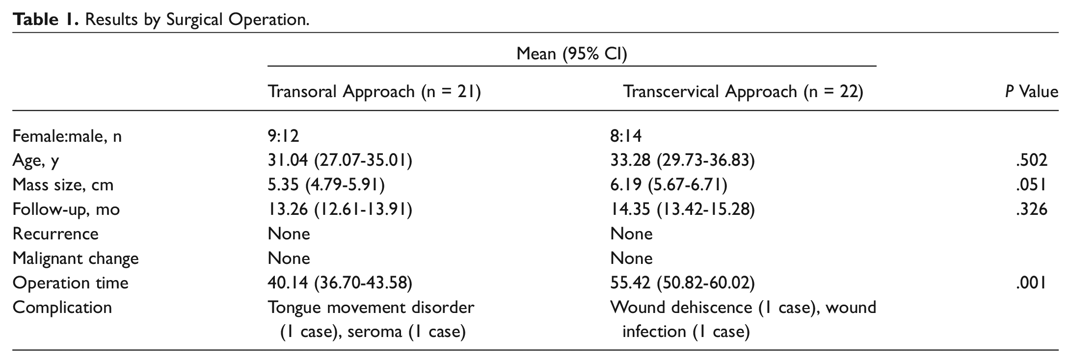
There were 12 male and 9 female patients in the transoral approach group (21 patients). Their mean age was 31.04 years (95% CI, 27.07-35.01). The mean size of the dermoid cyst was 5.35 cm (95% CI, 4.79-5.91), and the most enlarged cyst was 8.0 × 6.0 cm. Cysts could be anatomically grouped as occurring above (n = 12) or under (n = 9) the mylohyoid muscles. There were 14 males and 8 females in the transcervical approach group (22 patients). The mean age of this group was 33.28 years (95% CI, 29.73-36.83). The mean size of the dermoid cyst was 6.19 cm (95% CI, 5.67-6.71), and the most enlarged cyst was 11.0 × 6.0 cm. Cases could be anatomically grouped as occurring above (n = 12) or under (n = 11) the mylohyoid muscles.
The mean follow-up durations for the transoral and transcervical dermoid cyst excision groups were 13.26 months (95% CI, 12.61-13.91) and 14.35 months (95% CI, 13.42-15.28), respectively. During the follow-up period, we did not find recurrences or malignant changes. The transoral approach group had 1 case of a temporary tongue movement disorder and seroma, while the transcervical approach group had 3 cases of mild swelling and 2 cases of wound complications. There were no significant differences in age, size of the mass, or complication between the groups. However, operations were shorter (P = .001) in the transoral approach group (mean, 40.14 minutes; 95% CI, 36.70-43.58) versus the transcervical approach group (mean, 55.42 minutes; 95% CI, 50.82-60.02; Table 1 ).
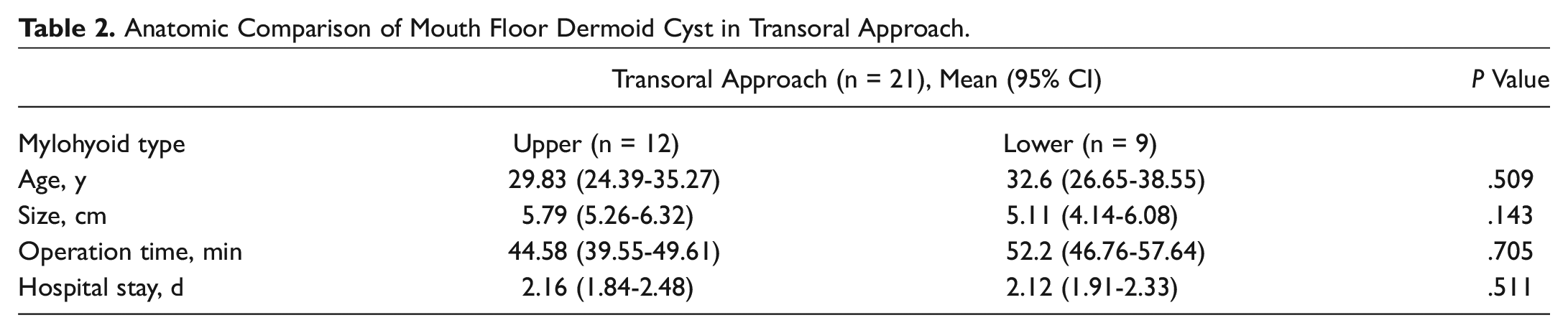
Comparisons between dermoid cyst location above and below the mylohyoid muscle showed no significant differences with respect to age, size, operation time, or hospital stay ( Table 2 ). Our results suggest that any type of oral cavity dermoid cyst (upper or lower mylohyoid muscle) can be resected through a transoral approach, thereby achieving similar results to a nontransoral approach.
Anatomic Comparison of Mouth Floor Dermoid Cyst in Transoral Approach.
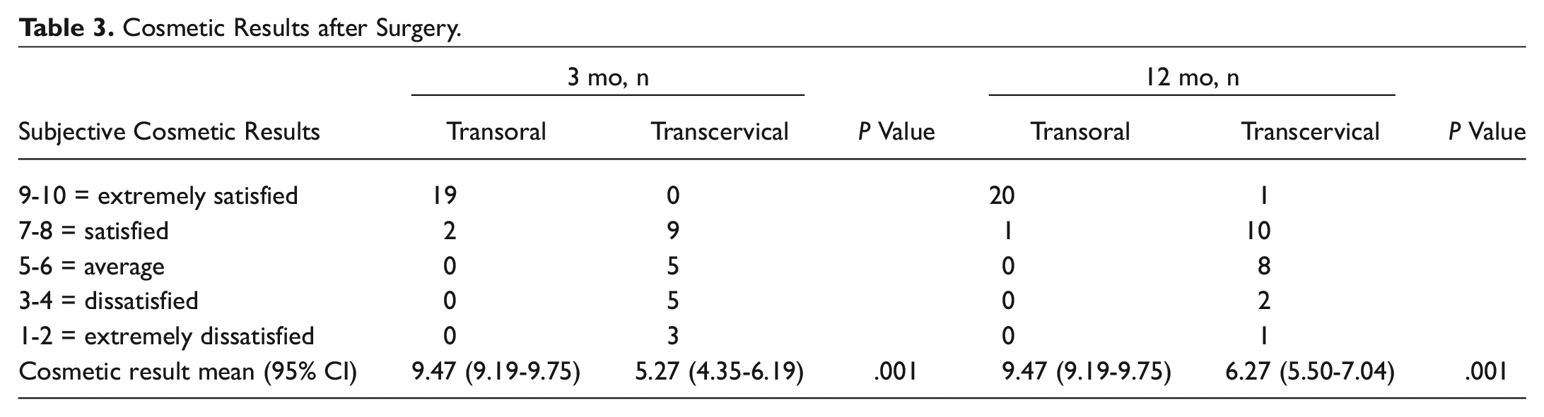
Mean satisfaction scores in the transoral group were 9.47 (95% CI, 9.19-9.75) after 3 months and 9.57 (95% CI, 9.32-9.82) after 12 months. In the transcervical group, they were 5.27 (95% CI, 4.35-6.19) after 3 months and 6.27 (95% CI, 5.50-7.04) after 12 months. Differences at 3 and 12 months between the groups were statistically significant (both P = .001; Table 3 ).
Cosmetic Results after Surgery.
Discussion
Most dermoid cysts in oral cavities are congenital developmental anomalies. They can occur where embryonic parts fuse. In the fifth week of prenatal development, a pair of lateral lingual swellings (distal tongue buds), one on the right side and the other on the left side, can form on the first pharyngeal arch. These lingual swellings can quickly expand and cover the tuberculum impar (median tongue bud). They can continue to expand through prenatal development. They form the anterior part of the tongue, making up two-thirds of its length. The line of their fusion is marked by the median sulcus. 18
A dermoid cyst in the oral cavity can arise from ectodermic elements entrapped in the tuberculum impar and covered by lateral processes of the tongue. This tuberculum impar will disappear in the adult tongue. However, entrapped ectodermic elements can remain and slowly grow, leading to dermoid cyst in the oral cavity. Thus, all congenital oral dermoid cysts are placed in the midline.
A dermoid cyst in the oral cavity is a slow-growing asymptomatic mass located in the midline, above or below the mylohyoid muscle. When it is located above, the cyst can manifest itself as a sublingual swelling. When it is located below, it will clinically manifest as a submental swelling.
Consequently, tongue elevation, speech alteration, and double-chin development are frequent complaints. Because they are almost asymptomatic, dermoid cysts are usually diagnosed only after they have reached a considerable size. 3 Such swelling on the floor of the mouth can occasionally cause serious problems for swallowing and speaking. It can also pose a critical risk to the airway.19,20
Surgical excision is the only effective treatment for a dermoid cyst in the oral cavity. Traditionally, the choice of a surgical approach depends on the size and location of the lesion. The mylohyoid muscle has been considered as a barrier to the transoral approach. Anatomically, it is possible to distinguish 2 types of dermoid cysts—an upper mylohyoid type and a lower mylohyoid type—according to the anatomic relationship between the cyst and the mylohyoid muscle of the floor of the mouth. Generally, an external approach is recommended when the cyst is present under the mylohyoid muscle. 3 An transoral approach was described for a small cyst that is located above the mylohyoid muscle. 21 This approach allows for better visualization of the surrounding structures and bleeding control, and it avoids intraoral contamination of the surgical wound. 22 Indeed, the operational field during a transoral approach is limited, and if a large incision is made to secure the operational field, it may cause injury of the lingual nerve of the Wharton’s duct. However, thanks to the development of the endoscope and other medical instrument technologies, as in our study, a mass located in the lower mylohyoid muscle could be removed by the transoral approach without complications.
This approach relies on passage through the midline incision in the floor of the mouth and utilizes the natural midline dehiscence present between the GMs; this area was shown to be a relatively avascular space (no nerve) and is composed of loose soft tissue. If the space between the GMs is spread out with a retractor, the mylohyoid muscle could be reached. Afterward, if the incision of the mylohyoid muscle is made, the lower mylohyoid space could be reached. This method is not only technically possible but could also provide an appropriate surgical field; thus, it is thought to be feasible for patients with midline neck masses manifested as a mouth floor masses.
This is a new way to excise a mass from the oral cavity, even if the dermoid cyst is large. Therefore, we believe that this method has great treatment in the treatment of dermoid cysts in oral cavities.6,14 First, the transoral approach will not lead to any scars. Most patients with dermoid cysts in their oral cavities are young adults in their second and third decades of life. Therefore, a large scar on the skin is undesirable. Second, a transoral approach leads to less tissue damage and complications. When an external approach is used, skin incision, subcutaneous dissection, and mylohyoid muscle cutting are needed. Therefore, an external approach causes damage to normal tissue (skin, subcutaneous tissue, and muscle). It also leaves a scar and can give rise to complications. However, a transoral approach involves only a mucosal incision and simple bilateral retraction of the GM. Therefore, it can minimize normal tissue damage without leaving a scar. In addition, it has fewer complications. Third, a transoral approach shortens the duration of the operation. It uses an avascular space, which is made through bilateral retraction of the GMs. Therefore, there is only very small bleeding. It reduces the number of procedures within each operation, thereby reducing its overall duration. This result is achieved without the need for subcutaneous tissue and skin suture.
However, the transoral procedure had a limitation of approach to a lateral neck mass. In our study, we used the avascular space between the GMs, which was placed in the midline of the tongue, so we could not approach the lateral part of the mouth floor. This approach was useful only for a midline neck mass.
In this study, the transoral approach group had 1 case of temporary tongue movement disorder. However, tongue movement was recovered within 4 weeks after the operation. This patient had a very large cyst. It is currently unclear whether the tongue movement disorder of this patient was due to the surgical procedure. It might be due to the large mass. Additionally, 1 patient had a seroma after the operation, which disappeared after a single needle aspiration. However, the transcervical group had 3 cases of mild swellings and 2 cases of wound issues (wound dehiscence and stich abscess).
The satisfaction levels of our patients after transoral approaches were good. Patients frequently have cosmetic concerns and make requests that require medical explanations. At present, the minimally invasive aspect and cosmetic advantage of the procedure seem to be important factors for patients. The main advantage of choosing transoral surgery is its excellent cosmetic result without scars, thus also providing an emotional benefit.
This study has some limitations. First, the number of subjects was relatively small. However, the presence of a dermoid cyst in the floor of the mouth is a rare disease. Therefore, it is very difficult to enroll enough patients to perform a randomized controlled study or case-control study. We designed a multicenter study to overcome this limitation. Future multicenter studies with larger sample sizes are needed to investigate the exact efficacy and complication of such a transoral dermoid cyst excision approach. To enhance the reliability and applicability of this approach, more data will be needed about the treatment of patients who have undergone the transoral dermoid cyst excision procedure. Second, infection is the primary concern with oral cavity surgery because of contaminated wounds. A transcervical approach is considered a “clean” procedure. Antibiotics are not indicated. However, a transoral approach is a type II clean-contaminated wound. Although the surgical field and incision site were clean, the mouth floor was not sterile. Hence, we used antibiotics and gargling for oral hygiene. Follow-up observations were carried out over 12 months. No complications were observed.
Conclusions
Transoral dermoid cyst excision is a potentially safe, quick, and effective method that can lead to easy removal of a dermoid cyst in the oral cavity, with excellent cosmetic outcomes. The importance of this report is that any type of dermoid cyst in the mouth floor can be resected through a transoral approach, even if it extends lower to the mylohyoid muscle.
Author Contributions
Disclosures
Supplemental Material
OTO791772_SupplementalMaterial_CLN – Supplemental material for Transoral Dermoid Cyst Excision: A Multicenter Prospective Observational Study
Supplemental material, OTO791772_SupplementalMaterial_CLN for Transoral Dermoid Cyst Excision: A Multicenter Prospective Observational Study by Jin Pyeong Kim, Dong Kun Lee, Jeong Hwan Moon, Jung Je Park and Seung Hoon Woo in Otolaryngology–Head and Neck Surgery
Footnotes
Sponsorships or competing interests that may be relevant to content are disclosed at the end of this article.
Supplemental Material
Additional supporting information is available in the online version of the article.
References
Supplementary Material
Please find the following supplemental material available below.
For Open Access articles published under a Creative Commons License, all supplemental material carries the same license as the article it is associated with.
For non-Open Access articles published, all supplemental material carries a non-exclusive license, and permission requests for re-use of supplemental material or any part of supplemental material shall be sent directly to the copyright owner as specified in the copyright notice associated with the article.