






Abstract
Objectives
To evaluate demographic, clinical, and polysomnographic features of children with Down syndrome suspected of having obstructive sleep apnea. To identify factors that predict severe obstructive sleep apnea among children with Down syndrome.
Study Design
Case series with chart review.
Setting
Children’s Medical Center Dallas / University of Texas Southwestern Medical Center.
Subject and Methods
Demographic, clinical, and polysomnographic data were collected for children with Down syndrome aged 2 to 18 years. Simple and multivariable regression models were used to study predictors of severe obstructive sleep apnea (apnea-hypopnea index ≥10). P≤ .05 was considered significant.
Results
A total of 106 children with Down syndrome were included, with 89 (84%) <12 years old, 56 (53%) male, 72 (68%) Hispanic, 15 (14%) African American, and 14 (13%) Caucasian. Ninety percent of children had ≥1 medical comorbidities; 95 (90%) patients had obstructive sleep apnea; and 46 (44%) had severe obstructive sleep apnea. The mean SaO2 nadir was lower among obese than nonobese children (80% vs 85%, P = .02). Obese versus nonobese patients had a higher prevalence of severe obstructive sleep apnea (56% vs 35%, P = .03). Severe OSA was associated with heavier weight (odds ratio = 1.0, 95% CI: 1.0-1.1, P = .002) and age ≥12 years (odds ratio = 1.2, 95% CI: 0.2-2.5, P = .02). The multivariable model showed that severe obstructive sleep apnea was associated only with weight (odds ratio = 1.1, 95% CI: 1.0-1.1, P = .02).
Conclusion
Obese children with DS are at a high risk for severe OSA, with weight as the sole risk factor. The results of this study show the importance of monitoring the weight of children with DS and counseling parents of children with DS about weight loss.
Down syndrome (DS) is the most prevalent chromosome abnormality seen in live-born children, currently affecting >300,000 in the United States. 1 Physical manifestations of DS include congenital cardiac anomalies, hypotonia, asthma, and facial anatomical anomalies. 2 Midfacial hypoplasia, relative macroglossia, and retrognathia of the mandible predispose children with DS to obstructive sleep apnea (OSA). 2
OSA is characterized by periodic reductions in airflow, hypoxemia, and hypercapnia, which affect up to 80% of children with DS, as opposed to just 2% to 5% of the general pediatric population.3-7 OSA is the most common reason why a child with DS has a consult with an otolaryngologist. 8 Although obesity and tonsillar hypertrophy are risk factors for developing OSA, their role among children with DS are unclear. Studies on children with DS and OSA are often limited by small sample sizes and varying definitions of OSA.2,5,9 For example, several studies reported a correlation between obesity and OSA severity among children with DS, while others reported no such association.6,7,9,10 Relationships among age, sex, tonsillar hypertrophy, and OSA severity among children with DS are also inconclusive.5,6 In addition, common disorders such as congenital cardiac anomalies, asthma, and allergies have not been shown to be predictors of OSA severity among children with DS. 9
The primary aim of this study was to use a relatively large patient population to evaluate the demographic, clinical, and polysomnographic features of children with DS suspected of having OSA. The secondary objective was to identify demographic and clinical factors that predict severe OSA among these children. Our hypothesis is that age, weight, sex, and comorbidities can predict severe OSA among children with DS.
Methods
The UT Southwestern Institutional Review Board approved this study and exempted the need for consent. A consecutive sample of children aged 2 to 18 years with DS who had a polysomnogram (PSG) at the Children’s Medical Center Dallas/University of Texas Southwestern Medical Center from January 1, 2009, to February 15, 2015, were included in a retrospective case series with chart review. Children were excluded if they had syndromic or chromosomal abnormalities other than DS; previous surgery on the pharynx, larynx, or trachea; missing data on tonsil size; or a previous tonsillectomy and adenoidectomy (T&A).
The following demographic and clinical information was obtained from the electronic medical records (Epic, Verona, Wisconsin): sex, age, ethnicity, birth status (preterm or full term), height (cm), weight (kg), body mass index (BMI) z score, tonsil size, presence of asthma, allergies, gastrointestinal reflux disease (GERD), congenital heart disease, hypothyroidism, and hearing loss. Ethnicity was self-selected by the caregiver as Caucasian, African American, Hispanic, or other. Full-term birth was defined as a gestational age ≥37 weeks, and preterm birth was defined as a gestational age ≤36 6/7 weeks, consistent with American College of Obstetricians and Gynecologists’ guidelines for birth. 11 BMI z score was calculated from the guidelines of the Centers for Disease Control and Prevention and calculated for girls and boys aged 2 to 18 years (see http://www.cdc.gov/growthcharts/zscore.htm). 12 Children were placed into weight categories based on age- and sex-adjusted BMI percentile categories via CDC classification (underweight, ≤5th percentile; normal weight, 5th-85th; overweight, 85th-95th; obese, ≥95th). 13 Children with DS were then categorized into obese (≥95th percentile) and nonobese (<95th percentile) and into age <12 or ≥12 years to compare pubescent and prepubescent children. Tonsillar size was obtained by PSG reports or prior otolaryngology clinic notes utilizing the Brodsky grading scale: 1+, occupying <25% of the oropharynx; 2+, 25% to <50% ; 3+, 50% to 75%; and 4+, >75%. 14 Tonsillar hypertrophy was defined as tonsil grade 3+ or 4+. Congenital heart disease was identified as any cardiac anomaly present at birth, including atrial septal defect, ventricular septal defect, patent ductus arteriosus, atrioventricular septal defect, and pulmonary artery stenosis. All children had treated congenital heart disease. Hearing loss, which is commonly seen among children with DS, was defined as having conductive or sensorineural hearing loss or both. Hypothyroidism, asthma, allergic rhinitis, GERD, and other medical comorbidities were treated per standard clinical care.
All children underwent a full-night in-laboratory PSG following guidelines established by the American Academy of Sleep Medicine. 15 PSG parameters included sleep efficiency, percentage of rapid eye movement (REM) sleep, arousal index, oxygen saturation nadir, peak CO2, percentage of time spent <90% O2 saturation, percentage of time spent at >50 mm Hg of end-tidal CO2 (ETCO2). AHI was defined as the number of obstructive and central apnea and hypopnea events per hour. OSA was defined as AHI ≥1. Mild OSA was defined as an AHI ≥1 to 4.99, moderate as 5 to 9.99, and severe as ≥10. Obstructive apnea was defined as at least a 90% reduction in oronasal thermal airflow signal lasting at least 2 breaths during baseline breathing despite respiratory effort. 16 Hypopnea was a decrease in airflow of at least 30% for at least 2 breaths with either an arousal or a 3% decrease in oxygen saturation. 16 Central apnea was defined by either a lack inspiratory effort for at least 20 seconds or a 3% oxygen desaturation. Sleep efficiency was defined as the percentage of time that the patient was asleep during the study. Arousal index was defined as the number of arousals from sleep per hour, including leg movements, respiratory, and spontaneous arousals. REM sleep was defined as the percentage of total sleep time spent during the REM stage of sleep. Oxygen saturation nadir (SaO2 nadir) was defined as the lowest hemoglobin oxygen saturation percentage measured by pulse oximetry. Peak CO2 was the highest carbon dioxide pressure (mm Hg) recorded by end-tidal respiratory measurement. Time spent <90% oxygen saturation and time spent at >50 mm Hg of ETCO2 were measured as percentages of the total time that the patient was asleep.
Continuous data are presented as means with standard deviations and categorical data as counts with percentages. Demographic and polysomnographic differences between obese and nonobese children with DS were compared with Pearson chi-square for categorical data and analysis of variance for continuous data.
We developed a logistic model of severe OSA (AHI ≥10) with the following strategy. First, we performed univariable analyses of the following variables: sex, age, ethnicity, birth status (preterm or full term), height (cm), weight (kg), BMI z score, tonsil size, presence of asthma, allergies, GERD, congenital heart disease, hypothyroidism, and hearing loss. Second, variables that exhibited a P value ≤.25 by the simple logistic regression were added to a multivariable logistic regression model. We then sequentially eliminated variables in the multivariable regression model that had a P value >.05. Each new regression model was compared with the previous one via the likelihood ratio test. Third, after elimination of nonsignificant variables, the remaining variables were tested for any statistical interactions. Last, the adequacy of the final regression model was tested with the Pearson chi-square test and validated with jackknife regression. P≤ .05 was considered significant. All statistics were performed with Stata Statistical Software 15 (StataCorp, College Station, Texas).
Results
A total of 152 children with DS underwent a PSG over the study period, of whom 46 were excluded, resulting in a study population of 106 ( Figure 1 ). The reasons for exclusion are as follows: 23 children underwent T&A before PSG; 15 were outside the age limits of the study; 3 had incomplete PSG; 3 had unrecorded tonsil size; and 2 had other chromosomal abnormalities (trisomy 15, chromosomal 9e22 deletion).

Study population after selection criteria. PSG, polysomnogram.
The demographics of children with DS who were referred for PSG are summarized in Table 1 . The average age was 7.3, with obese children significantly older at 9.1 years (P < .001). A total of 89 (84%) children were <12 years old, and 56 (53%) were male. A total of 72 (68%) children were Hispanic, 15 (14%) African American, and 14 (13%) Caucasian. Among African Americans, obese children were significantly overrepresented (African Americans were 14% of the study population but 23% of the obese population, P = .04). Approximately 90% of all patients had ≥1 comorbidities: 65 (61%) with congenital heart disease, 32 (30%) with allergic rhinitis, 29 (27%) with hearing loss, 26 (25%) with hypothyroidism, 25 (24%) with asthma, and 11 (10%) with GERD. Congenital heart disease was significantly more common among nonobese than obese children (71% vs 47%, P = .01). A total of 95 children (90%) were diagnosed with OSA, and 46 (44%) were diagnosed with severe OSA (AHI ≥10). Obese versus nonobese children had a significantly higher prevalence of severe OSA (56% vs 35%, P = .03). There were no significant differences between obese and nonobese children in respect to sex, Caucasian and Hispanic ethnicity, preterm birth status, allergic rhinitis, asthma, GERD, hypothyroidism, hearing loss, tonsillar hypertrophy, and no or mild to moderate OSA.
Characteristics of Children with Down Syndrome Referred for Polysomnography.

Abbreviations: AHI, apnea-hypopnea index; BMI, body mass index; CHD, congenital heart disease; GERD, gastrointestinal reflux; OSA, obstructive sleep apnea.
P values are based on analysis of variance for continuous variables and Pearson chi-square or Fisher exact test for categorical variables. Significant P values are in bold.
z score: SD score of BMI based on Centers for Disease Control and Prevention guidelines adjusted for age and sex.
Preterm: born before 37 weeks of gestation.
None, AHI < 1; mild/moderate, 1 ≤ AHI < 9.9; severe OSA, AHI ≥ 10.
Table 2 presents the polysomnographic data for obese and nonobese children with DS. The mean AHI was 16.7, and the average SaO2 nadir was 83.0, which was significantly lower for obese than nonobese children with DS (80% vs 85%, P = .02). For children with DS ≥12 years old, obese patients had an average AHI of 23, as opposed to 5.6 for nonobese patients (P = .04). For children with DS <12 years, SaO2 nadir was lower among obese versus nonobese children (80% vs 85%, P = .05). No differences were found between obese and nonobese children for apnea index, hypopnea index, central apnea index, REM, sleep efficiency, arousal index, peak CO2, SaO2 nadir, and total sleep time >50 mm Hg of ETCO2 among children aged ≥12 years. No differences were seen among children older or younger than 12 years for apnea index, hypopnea index, apnea-hypopnea index, central apnea index, peak CO2, and total sleep time >50 mm Hg of ETCO2.
Polysomnographic Characteristics of Children with Down Syndrome.
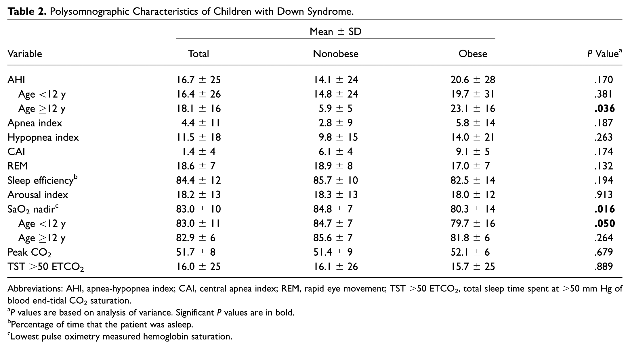
Abbreviations: AHI, apnea-hypopnea index; CAI, central apnea index; REM, rapid eye movement; TST >50 ETCO2, total sleep time spent at >50 mm Hg of blood end-tidal CO2 saturation.
P values are based on analysis of variance. Significant P values are in bold.
Percentage of time that the patient was asleep.
Lowest pulse oximetry measured hemoglobin saturation.
Table 3 summarizes the results of a univariate simple logistic regression analysis of the association of demographic and clinical risk factors and severe OSA (AHI ≥ 10). Severe OSA was associated with weight (odds ratio [OR] = 1.0, 95% CI: 1.0-1.1, P = .002), obesity (OR = 2.3, 95% CI: 1.1-5.2, P = .035), and age ≥12 years (OR = 1.2, 95% CI: 0.2-2.5, P = .020). Sex, ethnicity, BMI z score, tonsil size, and other clinical comorbidities were not associated with severe OSA.
Simple Logistic Regression of Demographic and Clinical Parameters for Predictors of Severe Obstructive Sleep Apnea (AHI ≥10).

Abbreviations: AHI, apnea-hypopnea index; CHD, congenital heart disease; Coeff, coefficient of linear regression; GERD, gastroesophageal reflux; Inter, regression intercept; OR, odds ratio.
P values are based on simple logistic regression. Significant P values are in bold.
Body mass index z score ≥95%.
Per Brodsky scale.
Table 4 is a multivariable logistic regression model to predict the likelihood of severe OSA among children with DS. All variables other than weight and age exhibited no significant changes in the corresponding coefficient when individually tested by elimination and thus were removed. The final model showed that severe OSA was affected only by increasing weight (OR = 1.1, 95% CI: 1.0-1.1, P = .020). Age (OR = 0.87, 95% CI: 0.6-1.3, P = .560) affected weight enough to be left in the final model, although it did not predict the risk of having severe OSA. Figure 2 displays the relationship between the weight of a child with DS and the probability of having severe OSA. A rise in weight correlates to a higher risk of severe OSA, as shown in the multivariable logistic regression. The predicted probability of severe OSA for a child who weighs 50 kg and 80 kg is 75% and 95%, respectively.
Multivariable Logistic Regression Model for Predictors of Severe Obstructive Sleep Apnea (Apnea-Hypopnea Index ≥10). a

Abbreviations: Coeff, coefficient of regression; OR, odds ratio.
Chi-square goodness of fit test = 103.8. Significant P value is in bold.

Risk of severe obstructive sleep apnea (OSA; apnea-hypopnea index ≥10) by weight among children with Down syndrome. Error bars indicate 95% CI
Discussion
This study evaluated demographic, clinical, and polysomnographic parameters among >100 children with DS, predominantly Hispanic, who were referred for the diagnosis and quantification of OSA. Approximately 10% had normal sleep study results, and 44% had severe OSA. Severe OSA was more likely among older obese children. Interestingly, congenital heart disease was more common among nonobese children. Tonsillar hypertrophy was present in about 50% of the study population and equally observed between obese and nonobese children. Hypoxemia, while worse among obese children, was seen in the majority of children with DS. Weight, not age, was a strong predictor of severe OSA. Sex and the treated medical comorbidities, including asthma, allergic rhinitis, and GERD, were not associated with obesity or OSA severity among children with DS.
OSA is commonly seen among children with DS. 17 In this study, 90% of children with DS had OSA. In 4 prospective studies, the prevalence of OSA among children with DS ranged from 57% to 79%.2,5,18,19 Definitions of OSA differed among these studies, but nonetheless the extent of disease throughout this population is high. According to the American Academy of Pediatrics, all children with DS should be screened with PSG by age 4 years even if asymptomatic and again afterward if they display symptoms or signs of OSA. 20 Unfortunately, there is a poor correlation between parent report of symptoms and PSG results, so children after the age of 4 years may commonly be missed. 18 This study suggests that PSG, as a screen for severe OSA, will have the highest yield among obese pubescent children with DS ( Figure 2 ).
We found a significant difference between obese and nonobese children with DS. Obese children were twice as likely to have severe OSA, with >50% of the pubescent population diagnosed with severe OSA. These findings are consistent with previous studies.2,5,21 Dyken et al prospectively analyzed 19 children with DS aged <18 years with PSG and reported that a higher BMI was positively correlated with OSA severity, as measured by AHI, and negatively with nighttime oxygen saturation levels. 2 Another study, in a retrospective review, noted increasing OSA severity with obesity among 52 children with DS aged 2 to 18 years. 21 Other studies reported no association between obesity and OSA among children with DS but were limited by small study populations, lack of PSG, and incomplete data.9,19,22 Interestingly, studies of children with DS aged <10 years with similar methods noted no association between BMI and OSA.9,22 Possible causes include the variable effects of hormones during puberty, phenotypic structural development of teens with DS, or the decreased likelihood of obesity among younger children. Kelly et al looked at this trend among children without DS, noting that BMI could not predict OSA for prepubescent children but could do so for pubescent children. 23 Based on our data and the majority of published data, weight gain is associated with OSA and a major predictor of severe OSA among children with DS.
Increasing age appears to play a role in the development of severe OSA, through its association with obesity. The majority of pubescent children with DS were obese and had severe OSA. This was reported in previous studies.5,21 Our study showed that older children with DS who were not obese showed no increased likelihood of severe OSA. This finding highlights the importance of preventing obesity among children with DS as they reach adolescence.
Tonsillar hypertrophy is also associated with OSA among children, but this relationship is less clear among children with DS. De Miguel-Díez et al reported that children with DS with tonsillar hypertrophy were nearly 4 times as likely to have OSA than those without tonsillar hypertrophy. 7 Shires et al also reported a correlation between tonsillar hypertrophy and OSA severity among 52 children with DS. 21 Most other studies of children with DS found no association between tonsillar hypertrophy and OSA.2,5,19,21,24,25 For example, in a study of 188 children with DS using home cardiorespiratory monitoring, Hill et al reported that tonsillar size did not affect any sleep parameter. 9 In our study of 106 children, we found no association between tonsillar hypertrophy and OSA. Tonsillar hypertrophy was also evenly distributed among obese and nonobese as well as prepubescent and pubescent children. This suggests that tonsillar hypertrophy is one of several factors that leads to OSA among children with DS. It is in keeping with a recent meta-analysis reporting that only 20% of children with DS are cured of OSA following T&A. 26
Asthma, GERD, and allergic rhinitis are common among children with DS.8,27 Their correlation with OSA severity, however, is unclear. A study examining the correlation between asthma and OSA severity among children with DS reported no increased likelihood of severe OSA among asthmatic children when accounting for obesity and age. 5 Similarly, GERD and allergic rhinitis have not been shown to be associated with severe OSA among children with DS.5,9,21 Our study did not show a correlation between OSA and previously identified comorbid conditions, including allergies, asthma, GERD, hypothyroidism, hearing loss, and preterm birth status among children with DS. In conjunction with other studies, the mentioned comorbidities commonly seen among children with DS should not be taken as indicators of the likelihood of OSA or factors that predict severe OSA when appropriately managed.
In this study, congenital heart disease affected 61% of children with DS and was significantly more common among nonobese than obese children. The relatively small study population of children with DS and congenital heart disease (n = 65) did not allow for a detailed study of the association between OSA and specific cardiac defects and obesity. Atrioventricular septal defect is the most congenital cardiac disorder among children with DS. 28 Studies that examined the association of weight and congenital heart disease among children with DS reported conflicting results.29,30 Increased metabolic demand and malnutrition are more common among children with congenital heart disease who do not have DS and may also play a role among children with DS.31,32 The presence of congenital heart disease is not associated with severe OSA, possibly through its inverse association with obesity.3,7,33 Of note, all patients were hemodynamically stable to undergo PSG.
Hypoxemia, measured by SaO2 nadir, was the only PSG parameter, other than AHI, affected by demographic and clinical factors. DS is a risk factor for hypoxemia, 34 as is obesity, 2 and obese children with DS had worse hypoxemic nadir when compared with nonobese children. Hypoxemia among children with DS is reported in this and multiple other studies,2,9,33-35 and yet the impacts of hypoxemia on health have not been well studied among these children. Among children without DS and OSA, hypoxemia is correlated with lower neurocognition scores and a risk of cor pulmonale.36,37
This study includes >100 children with DS who underwent PSG at a tertiary care academic children’s hospital. To our knowledge, this study is the largest to date to compare PSG in among pubescent and prepubescent children with DS. Previous studies were limited to ≤50 children.5-7,9,10 However, several limitations need to be recognized. First, the study population was from a single institution, and all children were evaluated because of suspected OSA. A community sample of children with DS may have different demographics and OSA severity. Furthermore, the predominantly Hispanic population in this study may not reflect the general population of children with DS, thus questioning its external validity. The retrospective nature of this study also leads to questions about the quality of the data and a possible selection bias in the population studied. Nonetheless, this study reports a high prevalence of OSA among children with DS who were referred for PSG and a correlation between increasing weight and severe OSA, particularly in the pubescent population. Future studies should examine the effect of nighttime hypoxemia among children with DS and the outcomes of T&A on OSA among these children.
Conclusion
Obese children with DS are at a high risk for severe OSA, with weight as the most significant risk factor. Obese children were twice as likely to have severe OSA, with >50% of pubescent children with DS diagnosed with severe OSA. The results of this study show the importance of monitoring the weight of children with DS and counseling parents of children with DS about weight loss.
Author Contributions
Disclosures
Footnotes
No sponsorships or competing interests have been disclosed for this article.