
Abstract
Objectives
Fluorodeoxyglucose positron emission tomography–computed tomography (FDG PET-CT) has been increasingly used in the past decade. Incidental FDG-avid findings are encountered in these studies, several of which with clinical significance. However, the significance of incidental FDG-avid sinonasal findings has not been studied to date.
Study Design
Retrospective cohort study.
Setting
A single tertiary medical center.
Materials and Methods
The medical records were reviewed of patients with incidental sinonasal positive FDG uptake between 2007 and 2016 who referred for further otolaryngological diagnostic workup.
Results
A total of 26 patients were identified, all of whom underwent a diagnostic surgical procedure. Histopathology revealed chronic inflammation (n = 12, 46.1%), malignancy (n = 7, 26.9%), inverted papilloma (n = 4, 15.5%), and fungal infections (n = 3, 11.5%). A unilateral maxillary sinus with FDG uptake was documented for 16 (61.5%) patients. CT evidence of bilateral disease and mucosal or sinus wall thickening correlated with inflammatory disease.
Conclusions
Incidental lesions with positive FDG uptake in the sinonasal cavities are at a high risk (40%) of being neoplastic. A diagnostic biopsy is advocated in these cases.
Since its introduction in the 1970s, fluorodeoxyglucose positron emission tomography–computed tomography (FDG PET-CT) has been increasingly used for various indications. Among them are cancer diagnosis, evaluation of regional and distant metastases, evaluation of unknown primaries, and posttreatment surveillance for residual and recurrent lesions.1-5 As FDG represents increased glucose metabolism, it was primarily linked to malignancy. However, FDG is a nonspecific tracer with increased FDG uptake demonstrated at sites of inflammatory and granulomatous lesions. Thus, FDG PET-CT has been proven to be a powerful modality for the monitoring of disease activity and response to therapy in infectious diseases. 6
Incidental PET-positive findings pose a difficult dilemma to clinicians, as FDG PET-CT is commonly performed in the setting of malignancy. The question arises whether the incidental finding is related to the primary tumor or represents a secondary tumor or an inflammatory/infectious condition.
Incidental increased FDG uptake in the sinonasal region has been scarcely discussed in the literature. The advantage of integrated FDG PET with CT images in the sinonasal cavities is the ability to gain additional information from CT findings and thus improve the diagnostic accuracy. A few studies analyzed the distribution of FDG uptake and standardized uptake values (SUVs) in different sinonasal lesions, attempting to differentiate among inflammatory, benign, and malignant lesions. Malignant lesions showed significantly higher FDG uptake than that of inflammatory lesions.7,8 Nonetheless, the treatment of patients with incidental sinonasal FDG uptake remains unclear.
The aim of our study was to investigate the implications of incidental significant FDG uptake in the sinonasal cavities of patients referred to further otolaryngological diagnostic workup.
Methods
Following approval by the Rabin Medical Center Institutional Review Board, the database of our tertiary medical center was retrospectively searched for patients with incidental suspected pathologic FDG uptake in the sinonasal area. A concomitant search was performed for all patients who underwent FDG PET-CT and surgery in the otolaryngology department from 2007 to 2016. All patients were included who had incidental significant FDG uptake in the sinonasal area and were referred to our department for further evaluation. All patients underwent biopsy of the suspected lesion under local or general anesthesia.
Patient demographic, clinical, and pathologic data were obtained from the medical chart. All relevant FDG PET-CTs were also retrospectively analyzed by 2 senior nuclear medicine specialists (H.B. and L.D.) blinded to the patients’ medical backgrounds and the final pathology reports. The presence of significant FDG uptake from the sinonasal cavities and the SUVs were measured, as well as CT parameters such as mucosal thickening, sinus wall bony thickening, and bone erosion.
Statistical analysis was performed with SPSS 21.0 (IBM, Armonk, New York). Categorical variables are described by frequency and percentage. Normally distributed variables are described by mean and standard deviation and abnormally distributed variables by median and range. Associations with nonparametric variables were compared with the Mann-Whitney test and the Kruskal-Wallis test. A P value <.05 was considered significant.
Results
A total of 26 patients were identified. Table 1 presents patients’ demographics clinical and PET CT characteristics. Mean ± SD age at endoscopic sinus surgery was 67.54 ± 14.58 years. Sixteen (61.5%) patients were female and 10 (38.5%) were male. Eleven patients (42.3%) had a history of smoking. All patients were asymptomatic.
Patients’ Demographics Clinical and PET CT Characteristics.
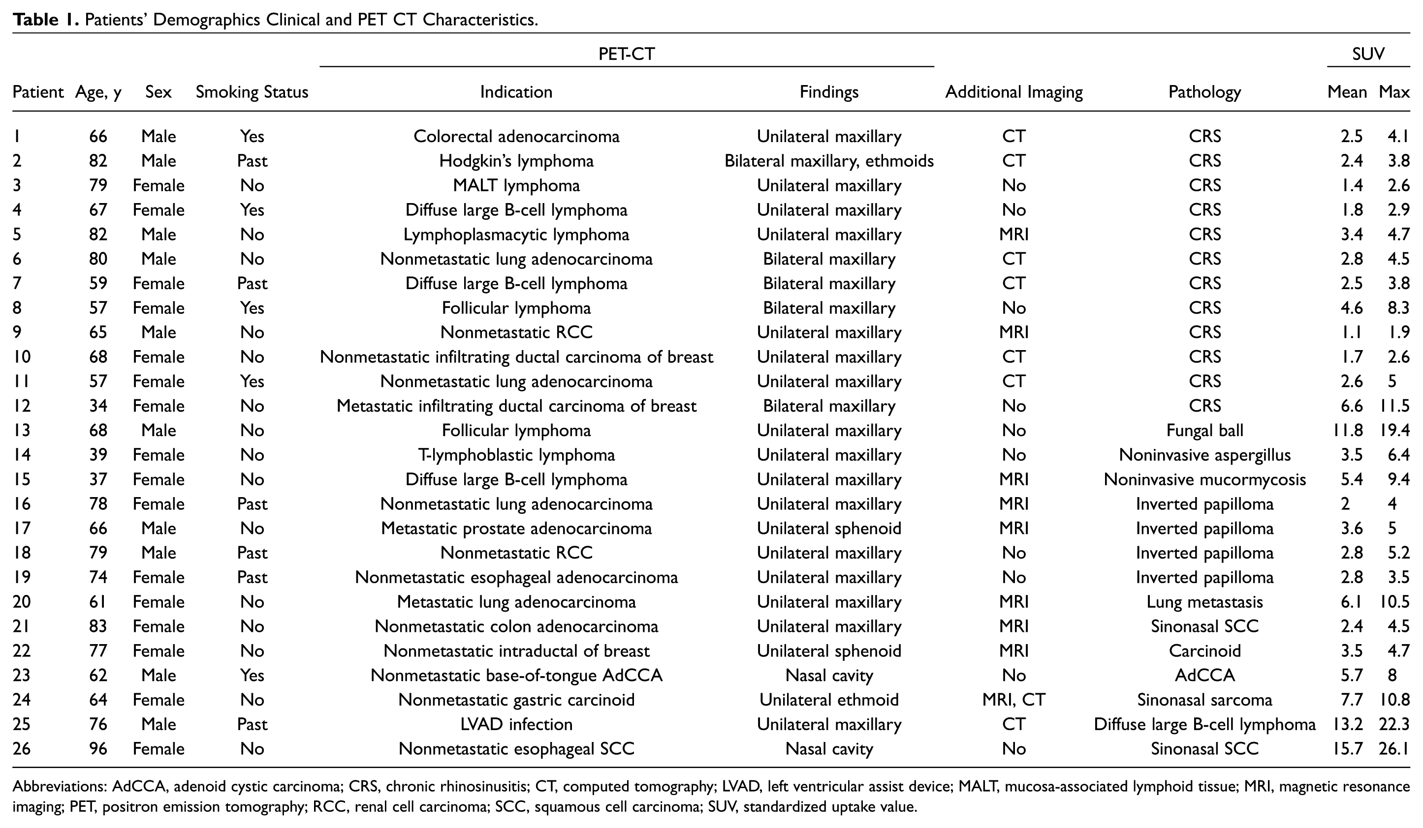
Abbreviations: AdCCA, adenoid cystic carcinoma; CRS, chronic rhinosinusitis; CT, computed tomography; LVAD, left ventricular assist device; MALT, mucosa-associated lymphoid tissue; MRI, magnetic resonance imaging; PET, positron emission tomography; RCC, renal cell carcinoma; SCC, squamous cell carcinoma; SUV, standardized uptake value.
The majority of patients (96.1%, n = 25) underwent FDG PET-CT as part of routine oncologic follow-up. Thirteen patients (50%) had lung, breast, prostate, or kidney cancer; 9 (35%) had lymphoma; and 1 had follow-up for an inflammatory event caused by a left ventricular assist device. For the remaining 3 patients (11%), 1 each had esophageal squamous cell carcinoma, esophageal adenocarcinoma, and base-of-tongue adenoid cystic carcinoma.
Twenty-one patients were diagnosed with unilateral sinonasal incidental significant FDG uptake, mainly in the maxillary sinus (16 patients, 61.5%). For 5 patients, significant FDG uptake was observed in the maxillary sinuses bilaterally.
Histopathologic analysis of targeted biopsies from these significant FDG uptake lesions revealed an inflammatory disease in 15 (58%) patients. Among them, 12 were diagnosed with chronic rhinosinusitis, and 3 were diagnosed with a fungal infection (noninvasive aspergillosis, n = 2; mucormycosis, n = 1). Remarkably, 11 patients (42%) were diagnosed with a neoplastic lesion. Of them, 7 had malignant lesions, and 4 had inverted papillomas. Malignant lesions were identified as follows: squamous cell carcinoma (n = 2) and sinonasal sarcoma, lymphoma, adenoid cystic carcinoma, carcinoid, and lung metastasis (n = 1 each).
There was no difference in smoking status between patients having neoplastic lesions and inflammatory lesions (P = .285).
Tables 2 and 3 present the correlation of SUV measurements with the histopathologic findings. There was no difference between neoplastic and inflammatory lesions in mean (P = .108) and maximal (P = .507) SUV readings. Interestingly, fungal and malignant lesions had higher mean and maximal SUVs when compared with chronic rhinosinusitis and inverted papillomas. Figure 1 depicts FDG uptakes for 3 patients with different histopathologic findings.
SUV Measurements in Neoplastic versus Inflammatory Lesions.
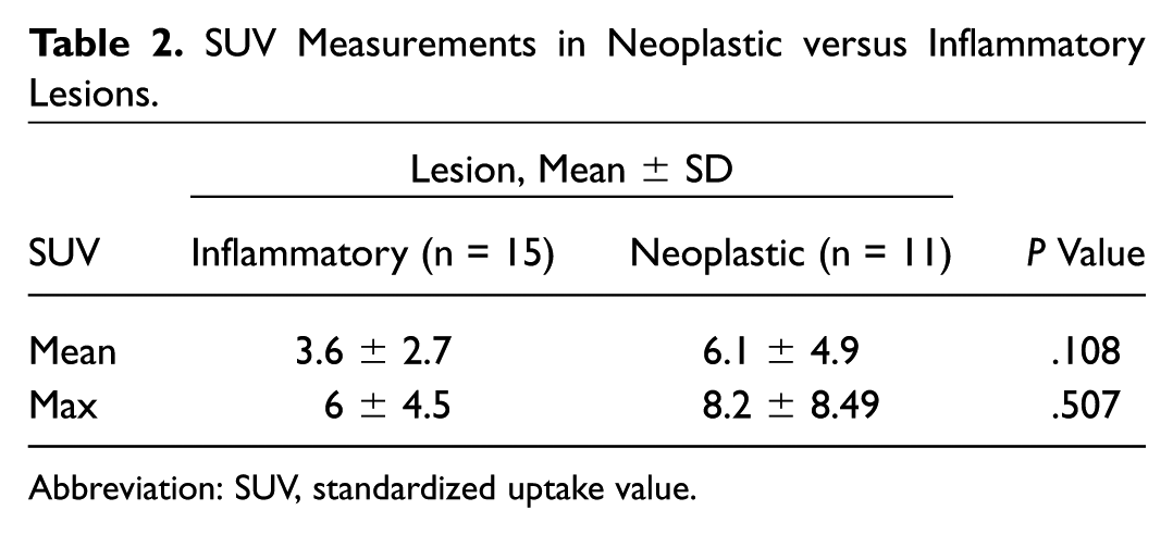
Abbreviation: SUV, standardized uptake value.
SUV Measurements Stratified to Final Histopathology. a
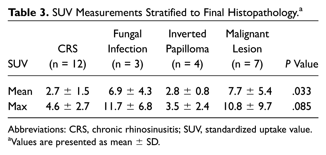
Abbreviations: CRS, chronic rhinosinusitis; SUV, standardized uptake value.
Values are presented as mean ± SD.
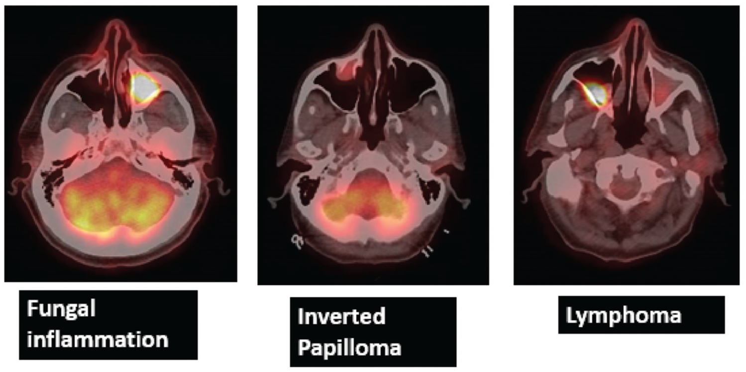
Examples of fluorodeoxyglucose uptake in histopathologic analysis.
Integration of CT findings with FDG uptake revealed that the presence of unrelated mucosal thickening and sinus wall thickening correlated 100% with inflammatory findings. Presence of bone erosion correlated with malignant lesions in 57% of cases. Table 4 depicts the correlation between FDG uptake and CT findings.
Correlation between FDG PET Uptake and Computed Tomography Findings. a
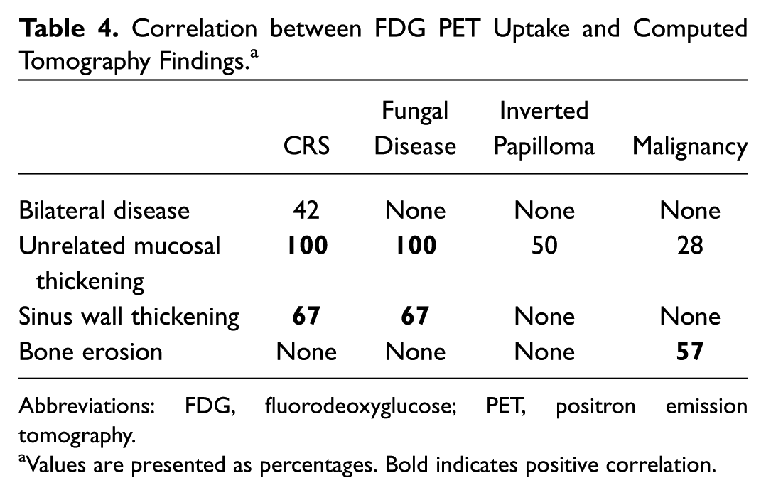
Abbreviations: FDG, fluorodeoxyglucose; PET, positron emission tomography.
Values are presented as percentages. Bold indicates positive correlation.
Discussion
In this study, we found that 40% of incidental significant FDG uptake in the sinonasal cavities, as referred to further otolaryngologic workup, represented neoplastic lesions; of them, 64% were malignant. SUVs could not differentiate between inflammatory and neoplastic lesions.
Incidental significant FDG uptake in other head and neck areas was previously discussed in the literature. In a meta-analysis by Nayan et al 9 that reviewed 31 studies, the malignancy rate of thyroid incidentalomas identified by 18-FDG PET-CT was evaluated and calculated as 19.8%. Seo et al 10 examined incidental focal FDG uptake in the parotid glands of patients with head and neck malignancies and observed a 33.3% malignancy rate. In a study by Lee et al 11 that evaluated incidental nasopharyngeal FDG uptake among patients without a history of underlying malignancy involving the head and neck, all incidental nasopharyngeal uptakes were benign on pathologic analysis. However, the authors suggested that the probability of malignancy increases if nasopharyngeal uptake is intense (SUV maximum ≥6.0) or concomitant retropharyngeal lymph node uptake is present. A recent study by Britt et al 12 examined the FDG PET-CT of 293 patients with head and neck cancer. An incidental finding was documented for 35.2% of patients—specifically in the head and neck region for 45 of them. A malignant lesion was documented for only 1 patient, in the parotid gland.
Incidental significant FDG uptake in the sinonasal areas was previously discussed in only case reports. Yılmaz et al 7 analyzed the yield of FDG PET-CT to differentiate among various histopathologic sinonasal lesions. They prospectively investigated 27 patients with a known unilateral polypoid lesion in the sinonasal cavity. Histopathology analysis of these lesions (eg, benign polyps, inverted papilloma, and squamous cell carcinoma) was correlated with SUV measurements. Higher FDG uptake was observed among patients diagnosed with inverted papilloma and squamous cell carcinoma. Moreover, FDG uptake was useful to differentiate inverted papillomas from malignant lesions, with higher uptake in the latter. All differences were statistically significant (P≤ .01). Other studies that evaluated sinonasal FDG uptake also found higher SUV measurements among patients with squamous cell carcinoma as compared with inverted papillomas.8,13-16
A study investigating the role of FDG PET-CT in fungal infections discussed the difficulty to differentiate FDG uptake of fungal infection from malignancies. In a review, Sharma et al 17 noted high FDG uptake among patients diagnosed with pulmonary aspergillosis and mucormycosis and addressed the need for histopathologic analysis for diagnosis.
In our study, we evaluated patients who underwent FDG PET-CT for a multitude of indications. Of 26 patients with incidental significant FDG uptake in the sinonasal areas, 42% were neoplastic, with 27% overall being malignant lesions. Analysis of SUV measurements showed no statistically significant difference between neoplastic lesions and inflammatory lesions. Hence, sinonasal incidental findings warrant further diagnostic biopsy to definitively assess the nature of the lesion and rule out inverted papilloma or malignancy.
Our study has several limitations. First, it is retrospective with the inherited biases of a retrospective analysis. Second, the study encompasses a low sample size, which may affect statistical significance. Note that it included only patients with incidental significant sinonasal FDG uptake who were referred to further otolaryngologic evaluation. Patients with incidental nonsuspicious FDG sinonasal uptake who were not referred for evaluation were not included in our cohort. Nonetheless, the implications of incidental significant PET-positive sinonasal uptake have not been reported previously—thus, the clinical importance of our findings.
In conclusion, incidental significant FDG uptake in the sinonasal cavities is at high risk of being neoplastic. A diagnostic biopsy is advocated in all cases for further workup.
Author Contributions
Disclosures
Footnotes
No sponsorships or competing interests have been disclosed for this article.