






Abstract
Objective
To determine adverse events after endoscopic flexible vs endoscopic rigid cricopharyngeal myotomy for treatment of Zenker’s diverticulum (ZD).
Data Sources
Systematic review of MEDLINE, Web of Science, CINAHL, Clinicaltrials.gov, and Cochrane Central Register of Controlled Trials for all years according to Preferred Reporting Items for Systematic Reviews and Meta-Analyses (PRISMA) guidelines. Additional studies were identified from review citations and a by hand search of manuscripts referencing ZD.
Review Methods
A structured literature search was conducted to identify studies for this systematic review. Methodological Index for Non-randomized Studies (MINORS) criteria were applied to assess study quality. For inclusion, each study had to provide data for at least 10 adult patients who had undergone endoscopic ZD repair reporting clear association with the postprocedure course in each case. Data extracted included all reported adverse events, recurrences, follow-up, and operative times.
Results
In total, 115 studies were included. All but 8 were retrospective case series. Sixty-one reported series of patients after rigid endoscopic stapler repair, 31 after rigid laser repair, and 13 with other rigid endoscopic instruments. Twenty-nine flexible endoscopic studies were included. Mortality, infection, and perforation were not significantly more likely in either the rigid or the flexible group, but bleeding and recurrence were more likely after flexible endoscopic techniques (20% vs <10% and 4% vs 0%, respectively). Dental injury and vocal fold palsy were reported rarely in the rigid endoscopic groups.
Conclusions
Adverse events are rare after endoscopic Zenker’s repair. The flexible approach minimizes exposure limitations and can be completed in some patients without general anesthesia. Transoral rigid approaches result in fewer revision surgeries compared with flexible diverticulotomy.
Keywords
Zenker’s diverticulum (ZD) is a hypopharyngeal pulsion diverticulum resulting from herniation of pharyngeal soft tissue through Killian’s triangle, above the horizontally oriented cricopharyngeus muscle of the upper esophageal sphincter and below the oblique inferior constrictor muscle fibers. It is a relatively rare phenomenon, typically found in the elderly population, and is characterized by dysphagia, cough, foreign body sensation, regurgitation, and aspiration. It has been treated primarily with surgical repair since the late 19th century. Surgical diverticulectomy through a cervical incision was the mainstay of treatment into the 20th century and continues to be at some institutions. Although endoscopic procedures were introduced to divide the esophageal-diverticular septum in 1917, 1 they were largely abandoned due to high surgical morbidity and mortality until Dohlman reintroduced this approach in the 1940s with the modification of diathermic coagulation.2,3 Since that time, endoscopic repair using rigid instrumentation has gained popularity, especially with the introduction of the endoscopic stapler, supplanting the open surgical approach in many centers.4-7
With the advent of interventional gastroenterology, flexible endoscopic treatment of Zenker’s diverticula was described in 1995 and has now been espoused by increasing numbers of providers around the world.8,9 A major advantage of the flexible endoscopic approach is its applicability in cases when neck extension is limited or cervical anatomy is unfavorable, leading to challenges in rigid transoral exposure or transcervical access. In addition, in some instances, the procedures can be done under sedation, an advantage for patients who are poor candidates for general anesthesia.
Rigid and flexible endoscopic Zenker’s diverticulotomy both possess advantages, although the results of these 2 approaches have yet to be compared in a large prospective trial. Published series do allow for comparison by systematic review. The purpose of this investigation was to compile and assess all reported adverse events, including recurrence rates, of patients who had undergone either rigid or flexible endoscopic Zenker’s diverticulotomy.
Methods
This study is reported as a systematic review of endoscopic methods for the treatment of Zenker’s diverticula based on the statements from the Preferred Reporting Items for Systematic Reviews and Meta-Analyses (PRISMA) guidelines.10,11 The target population of this study was adult patients with ZD. The interventions that were compared were endoscopic rigid treatment and endoscopic flexible treatment. Outcomes of both procedures were compiled with the primary goal of comparing the adverse events associated with each endoscopic procedure.
Study Identification
Studies were identified by performing searches of the following databases: Medline, the Cumulated Index to Nursing and Allied Health Literature (CINAHL), Cochrane, and Web of Science. The last search was completed on January 10, 2018. No date limitations were placed within any of the included databases. English, Romanian, and Turkish languages were filtered in Medline based on reviewer proficiency, but no language filters were used for the additional databases. The Medline search terms are as follows for English results. This was altered and repeated for additional languages:
(((“zenker diverticulum”[MeSH Terms] OR (“zenker diverticulum”[tiab] OR “zenker’s diverticulum”[tiab] OR “zenker’s diverticula”[tiab] OR “zenker diverticula”[tiab])) OR “pharyngeal pouch”[tiab]) OR (“pharyngeal diverticulum”[tiab] OR ((“hypopharynx”[MeSH Terms] OR “hypopharynx” [tiab] OR “hypopharyngeal”[tiab]) AND (“diverticulum”[MeSH Terms] OR “diverticulum” [tiab] OR “diverticula”[tiab])))) AND (“endoscopy”[MeSH Terms] OR endoscopic[tiab] OR microscopic[tiab] OR “Esophagoscopy”[Mesh]) AND English[lang] AND “adult” [MeSH Terms].
Broader search terminology was applied to other databases as follows:
CINAHL: (MH “Zenker Diverticulum”) OR AB “zenker diverticulum” OR AB “zenker’s diverticulum”
Web of Science: ((“zenker diverticulum” OR “zenker’s diverticulum” OR “zenker’s diverticula” OR “zenker diverticula”) OR ((“pharyngeal pouch” OR “pharyngeal diverticulum” OR “hypopharynx” OR “hypopharyngeal”) AND (“diverticulum” OR “diverticulum” OR “diverticula”))) AND (“endoscopy” OR endoscopic OR microscopic OR Esophagoscopy)
Cochrane: zenker
Results were compiled and deduplicated. The results were checked against a larger Medline search that included only Zenker or Zenker’s diverticulum as well as the reference lists of each of the reviews that were encountered in our search and those studies registered on clinicaltrials.gov.
Eligibility Criteria and Study Selection
Studies were compiled from all sources. The titles and abstracts of all potentially relevant studies were screened by the first author for inclusion. All study designs were considered, and they were included if they presented at least 10 adult cases of Zenker’s diverticulum (by eponymous name or anatomic description) with treatment by endoscopic rigid or flexible techniques. Two authors then independently performed full-text screening with disparities settled by consensus. Studies were excluded if adverse events were not disclosed, if outcomes were not clearly associated with a particular surgical treatment (in the case of studies that described multiple surgical approaches), if procedures did not at least transect the septum or cricopharyngeus (eg, dilation alone), or if procedures involved an external incision (eg, robot-assisted, axillary approach). A specific follow-up period threshold was not required, although discussion of the immediate postoperative course was. Studies were also excluded if the patient data presented were also presented in a subsequent work, to eliminate duplicate patient records.
Data Extraction
Data extraction was undertaken through the use of a worksheet that detailed the interventions reported in each manuscript, the number of patients involved, and the length of time during which follow-up data were collected. Each index procedure was compiled for calculation of adverse events. Recurrent procedures were treated separately as most studies reported them separately. As reporting differed, each study was carefully read for these details, and rates were calculated as reported by total for each study. For each type of intervention, adverse events were tabulated. These outcomes included infection, bleeding (controlled at the time of the procedure or requiring takeback to the operating room), perforation (treated conservatively or treated by immediate conversion to open surgery), subcutaneous emphysema, mortality, conversion to an open approach (for complication or inadequate exposure), the need for further surgery within 14 days (and the description), fever, length of hospital stay, readmission, dental damage, vocal fold palsy, and other. Recurrence of Zenker’s-related symptoms was also included with a description of how it was defined in each study, if appropriate. Length of hospital stay, length of procedure, instruments employed, inclusion of patients who previously underwent surgery for Zenker’s diverticulotomy, description of success and how it was measured, number of unsuccessful procedures, and number of aborted procedures were also extracted from each study. A uniform recurrence definition was applied to all studies for the purpose of this review. For this result, any patient who underwent at least 1 procedure at any time after the index procedure reported in the study was counted. In the event that a patient required more than 2 procedures for ZD treatment, these events were not included as additional recurrences.
Assessment of Bias
The articles were examined for bias. Selection bias was limited by extending our search through the described databases. Quality bias was limited by using the Methodological Index for Non-randomized Studies (MINORS) criteria, 12 a validated bias assessment instrument for observational studies, including those detailing surgical interventions. For noncomparative studies, the first 8 items were scored with a global ideal of 16. For studies comparing more than 1 intervention, all 12 items were scored with a global ideal of 24. Each item was assigned a score of 0 to 2 (0: not reported, 1: inadequately reported, 2: adequately reported), and a consensus was reached for a final total score between 2 independent reviewers. Studies were then grouped into high (<6 of 16 or <11 of 24), moderate (6-10 of 16 or 11-18 of 24), and low bias (>10 of 16 or >18 of 24) groups to facilitate comparisons.
Statistical Analysis
Random-effect models were used to estimate the pooled adverse events proportion, with 95% confidence interval (CI). Metaprop procedure was used to allow for Freeman-Tukey double arcsine transformation in addition to the application of the continuity correction to avoid excluding studies with 0% or 100% prevalence of side effects. Heterogeneity between the studies was evaluated and quantified. Furthermore, subgroup analyses by the intervention type were done for some of the adverse effect outcomes. Statistical analyses were conducted using STATA, version 15.1 (StataCorp LP, College Station, Texas).
Results
In total, 115 studies met all criteria and were included for analysis ( Figure 1 ; see Supplemental Table S1, available in the online version of the article). Sixty-one studies reported outcomes after rigid endoscopic stapler Zenker’s diverticulotomy (2858 procedures), 31 after rigid endoscopic laser (1999 procedures), and 13 other studies used the endoscopic rigid approach to apply other tools (other modalities), including the harmonic scalpel, ligasure, or electrocoagulation (708 procedures). Flexible endoscopic Zenker’s diverticulotomy outcomes were reported in 29 studies (1299 procedures) achieving inclusion criteria. Nineteen studies reported on more than 1 technique. Figure 2 highlights the year of publication by technique described. Bias outcomes are reported in Table 1 .

Preferred Reporting Items for Systematic Reviews and Meta-Analyses (PRISMA) flow diagram.

Number of studies by endoscopic technique and publication year.
Proportion of Low, Moderate, and High Biases Based on MINORS Scores by Surgical Technique.

Abbreviation: MINORS, Methodological Index for Non-randomized Studies.
Mortality after Zenker’s diverticulotomy was extremely low in all groups ( Figure 3 ). In the stapler group, 9 total deaths of 2858 cases were reported, although only 1 was due to mediastinitis that developed related to surgery. The other 8 deaths were attributable to pneumonia present prior to surgery, myocardial infarction, or (3) unknown or unrelated causes. In the laser group, there were 4 reported perioperative deaths of 1999 cases, none of which were directly related to the procedure. One patient in the other modalities group died of heart failure 2 days after surgery. The flexible group of studies reported 3 deaths of 1299 cases, 1 due to a pulmonary embolism on postoperative day 27 and 2 of unrelated causes.

Meta-analysis of mortality and 95% confidence interval (CI) by surgical technique for endoscopic repair of Zenker’s diverticula. ES, effect size.
Infection following surgery was noted with all approaches, although also with low incidence. The other modalities group reported an infection proportion of 2% (95% CI, 1%-4%, with a low level of heterogeneity between studies, I2 = 23.1%; P = .21), significantly higher than all other groups ( Figure 4 ). In the stapler group, 5 of 2858 cases of mediastinitis were reported while 18 of 1999 mediastinitis cases were reported in the laser group and 15 of 708 cases were found in the other modalities group. Two cases of mediastinitis in 1319 cases were noted in the flexible group. Pneumonia and cervical or retropharyngeal abscess were also reported in all groups in a small proportion of patients. In many cases, the occurrence of infection prompted surgery within 14 days ( Table 2 ).

Meta-analysis of infection proportion and 95% confidence interval (CI) by surgical technique for endoscopic repair of Zenker’s diverticula. ES, effect size.
Incidence and Reason for Additional Procedure within 14 Days of Index Endoscopic Zenker’s Diverticulotomy.
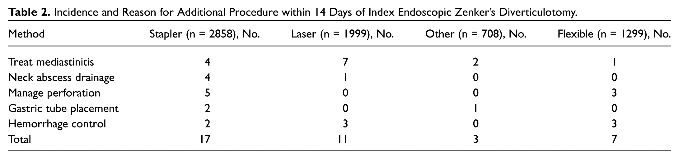
Intraoperative bleeding was reported in all 4 groups ( Figure 5 ), although at a slightly higher proportion in the flexible group ( Figure 6 ). Bleeding occurred in 4% of cases (95% CI, 2%-7%) with a high level of heterogeneity: I2 = 74.3%; P < .001. Similar proportions of operating room (OR) takeback were noted ( Table 2 ), but in the flexible group, bleeding during the surgery occasionally resulted in an incomplete or unsuccessful surgery. 13 One flexible study 14 was excluded from this analysis as intraoperative bleeding was reported to occur “usually” during the course of the procedure.

Meta-analysis of intraoperative bleeding and 95% confidence interval (CI) by surgical technique for endoscopic repair of Zenker’s diverticula. ES, effect size.

Meta-analysis of intraoperative bleeding proportion and 95% confidence interval (CI) in the flexible endoscopic group for endoscopic repair of Zenker’s diverticula. ES, effect size.
These studies reported subcutaneous or mediastinal emphysema separately from perforation. The former was reported after physical exam or imaging, while the latter was reported due to visualization at the time of surgery, appearance on contrasted swallow evaluation, or sequelae such as fistula or abscess. In the stapler group, reports of subcutaneous emphysema were lower than all other groups, with the lowest heterogeneity (I2 = 0%; P = .78) ( Figure 7 ). Perforation was noted to occur in 1% to 2% of cases completed with stapler, laser, or flexible instruments ( Figure 8 ). The procedure was converted to open due to perforation in only a few cases of the stapler (15), laser (6), and other modalities (2) groups.

Meta-analysis of subcutaneous or mediastinal emphysema proportion and 95% confidence interval (CI) by surgical technique for endoscopic repair of Zenker’s diverticula. ES, effect size.

Meta-analysis of perforation and 95% confidence interval (CI) by surgical technique for endoscopic repair of Zenker’s diverticula. ES, effect size.
Descriptions of symptom recurrence or procedure failure could not be included in our analysis due to vast methodological heterogeneity, lack of standardized symptom descriptions, differences in follow-up periods, and loss to follow-up or death. To compare between studies, recurrence was defined as the requirement of at least 1 revision surgery or procedure for a recurrent or persistent symptomatic Zenker’s diverticulum. Only patients who submitted to or were scheduled for at least 1 additional surgery were included, as recurrent symptoms could not be quantified or compared across studies ( Figure 9 ). The flexible approach resulted in a significantly higher proportion of patients undergoing revision procedures, with some studies reporting multiple procedures within a single hospitalization, separated by 1 or 2 days.8,15

Meta-analysis of the need for more than 1 procedure expressed as recurrence by approach and 95% confidence interval (CI) by surgical technique for endoscopic repair of Zenker’s diverticula. ES, effect size.
Laser and stapler studies typically reported a number of procedures that were aborted or converted to an open approach at the time of initial surgery for inability to expose the diverticulum. Laser procedures were aborted in 1% (95% CI, 1%-3%) and converted in fewer (0%; 95% CI, 0%-2%). Stapler procedures were aborted in 3% (95% CI, 1%-5%) and converted in 1% (95% CI, 0%-2%). Conversion or abandonment was not reported in flexible studies with the exception of Jones et al 16 ; rather, a proportion of unsuccessful procedures, either due to inability to expose or inability to completely incise the septum, was reported in 7 studies, with an overall proportion of 10% (95% CI, 3%-19% with a high level of heterogeneity; I2 = 77.3%; P < .001) ( Figure 10 ). The high outlier reported inability to divide the septum completely in 94% of cases at the initial procedure, although subjective dysphagia was improved. 14

Meta-analysis of the proportion of unsuccessful procedures and 95% confidence intervals (CIs) reported in flexible endoscopic Zenker’s diverticulotomy studies. ES, effect size.
Fever was reported often but not in all studies. When fever was reported, it was rarely defined, although occasionally a study reported low or high pyresis.17,18 Only 6 (21%) studies in the endoscopic flexible group and 9 (15%) of the stapler group reported fevers. One study 15 (3%) in the flexible endoscopic group reported a 17% fever incidence, with the remaining 5 (17%) studies ranging from 2% to 10%. The high outlier kept patients in the hospital and repeated the diverticulotomy procedure as necessary at intervals. In the stapler group, fever was detected in 0% to 4% of patients in 59 (97%) studies and 8% 19 and 10% 20 in the remaining 2 studies. Dissard et al 19 was the only study in this stapler group to describe the occasional use of the laser to complete a stapler division. The only 2 other studies to use additional cutting methods at the limit of the staple line did not report any fever events. Fever events were reported at high rates in the laser and other modalities groups, as shown in Figure 11 .

Meta-analysis of fever proportion and 95% confidence interval (CI) reported by publication in other and laser groups. ES, effect size.
Dental trauma was reported in 25.8% of studies in the rigid laser group and 27.9% in the rigid stapler group. Most studies reported only 1 or 2 instances of dental trauma in each group with an overall proportion of 1% (95% CI, 0%-1%) in the stapler group and 0% (95% CI, 0%-1%) in the laser group. However, 1 high outlier 21 reported 11 cases of dental damage after rigid stapler repair in a total of 150 procedures. Temporary recurrent laryngeal nerve palsy was reported only in the rigid stapler and rigid laser groups, with 8 cases (of 2858) in the former and 4 cases (of 1999) in the latter. No permanent cases of recurrent laryngeal nerve palsy were reported in any study after endoscopic Zenker’s diverticulotomy.
Discussion
Endoscopic Zenker’s diverticulotomy has achieved greater acceptance as technology has advanced during the past century. Flexible endoscopic esophagogastroscopes have permitted this procedure to be completed in patients who are not candidates for general anesthesia or who have unfavorable cervical anatomy. However, the rarity of this condition presents challenges for rigorous scientific examination to determine the most effective, safe, and efficient technique to apply. The purpose of this investigation was to compile and assess all reported adverse events, including recurrence rates, of patients who had undergone either rigid or flexible endoscopic Zenker’s diverticulotomy.
This systematic review and meta-analysis were originally undertaken with the purpose of analyzing the results of studies that compared rigid with flexible approaches, but even after more than 20 years of experience with the newer approach, only 2 of these studies were encountered.16,22,23 A comparison of 30 patients after rigid endoscopic diverticulotomy vs 28 after flexible endoscopic diverticulotomy was presented by Repici et al 23 in 2011. Their groups had similar complication rates (1 perforation per group) and length of hospital stay. Three of the rigid and 2 of the flexible group had persistent dysphagia. Two of the rigid and 1 of the flexible group had recurrent dysphagia in less than 2 years. In Jones et al, 16 6 of 15 rigid cases were converted to open with an additional case due to a perforation compared with 5 of 20 in the flexible group. There were no other adverse events reported except “minor” events (2 rigid, 1 flexible). These studies did not clearly support one approach over the other: both approaches were found to be safe and efficacious, although one might be preferred over another in individual cases. The former study advanced surgeon preference as the determining factor in a decision between rigid and flexible approaches while commenting that air leakage was more common in the flexible approach, although that approach minimized exposure difficulties. The latter study concluded that the flexible approach could be applied to large diverticula with fewer exposure problems than the rigid approach.
As direct comparisons between rigid and endoscopic techniques were limited, our investigation was broadened to include case and cohort studies that did not compare techniques. In reviewing these studies, some features became immediately apparent: most studies were small, retrospective, and not standardized. Many studies did not exclude patients who had had previous surgery for their Zenker’s diverticulum, and most reported insufficient follow-up to truly monitor for recurrence in all patients.
Reporting in the surgical literature (rigid endoscopic) differed from that of the medical literature (flexible endoscopic). Two examples were procedure times and bleeding. In the rigid approach, the surgical time was usually measured from the moment before the surgeon touched the patient to when the patient’s care was returned to anesthesia, while the procedure time in gastroenterology was counted in some instances as the time the procedure took after placement of the overtube, excluding initial endoscopic examination and overtube placement and removal. As times were defined differently, they could not be compared between rigid and flexible approaches. Bleeding was reported significantly more frequently in the medical literature after flexible procedures, whereas it was rarely reported in surgical cases. This may be due to the expectations of surgeons working in this area and the multitude of tools available for endoscopic hemorrhage control, including a 2-handed, 2-instrument working approach not feasible with a flexible endoscope. Operative takeback for hemorrhage was noted after both rigid and flexible approaches, although the only reports of bleeding preventing the completion of the procedure were using the flexible approach. 13
Inconsistencies in reporting among studies were noted for emphysema and perforation events. Both were extracted from the text directly as reported. The stapler and flexible groups had slightly higher rates of perforation than the laser group, while the laser, flexible, and other modalities groups had significantly higher emphysema rates than the stapler group. Unique among the instruments is the stapler, with its higher risk of trauma and subsequent perforation during placement due to poor visualization relating to its thick shaft and cumbersome head. Despite this, the staple line provides complete sealing of the incision, likely accounting for the lower incidence of emphysema.
The size of the diverticulum was inconsistently reported, and when it was, measurement techniques differed with questionable reproducibility. For this reason, size of diverticula was not collected or analyzed. It is important to note that diverticular size may create its own selection bias as some practices do not approach very small or very large diverticula endoscopically. 24 This increases the likelihood that many small-diverticula patients have already been eliminated from participation in endoscopic research cohorts. However, many surgeons do complete rigid endoscopic cricopharyngeal myotomies in the absence of a diverticulum, supporting the application of a rigid endoscopic approach. Very small and very large diverticula also present difficulties in the endoscopic flexible approach, preventing overtube placement in the former and requiring multiple procedures in the latter. The size of the diverticulum does not seem to favor the flexible or the rigid endoscopic approach, although further investigation is certainly warranted.
Advantages of the flexible endoscopic approach (avoidance of general anesthesia and exposure difficulties) are not universal. Although avoidance of general anesthesia is possible, most series of flexible endoscopic cases include patients who received either sedation or general anesthesia.9,15,25-30 Few studies report the absolute avoidance of general anesthesia, and most fail to detail decision making when deciding between forms of anesthesia. One important consideration is that intubation protects the airway when food and debris from the pouch are removed prior to septum division or in cases of severe gastroesophageal reflux disease. In considering access, the flexible approach may provide a means of circumventing a difficult exposure, although it is uncommon for exposure to entirely fail with rigid techniques, especially if there are several different rigid scopes and division instruments available. However, in the case of limited cervical extension, the flexible approach may be the only appropriate endoscopic option.
It is difficult to directly compare the flexible with the rigid group as a whole because of the differences in instrumentation. The laser and flexible groups may be most comparable as the laser most closely resembles the flexible devices. However, the placement of a clip in many cases at the distal aspect of the wound during the flexible approach may account for a lower fever rate, although this could also be due to an overall more conservative approach resulting in a higher recurrence rate in the flexible group or a higher same-day discharge rate. When the single flexible stapler group is compared with the rigid stapler studies, the incomplete rates are much higher for the flexible approach (25%) and neck extension is still required. 31
The rigid endoscopic stapler is the most studied and perhaps used tool when otolaryngologists approach endoscopic ZD repair. However, in practice, most are prepared to convert to another method (endoscopic or open) when the larger endoscope required for stapler use cannot be safely placed. In the same manner, if no rigid exposure is possible, a flexible endoscopic approach may be attempted under the same anesthetic. This approach minimizes the risk of dental or laryngeal nerve damage. Its most powerful application is in the case of a diverticulum that is difficult to expose with rigid instruments in a patient who cannot be converted to a transcervical approach because of laryngeal anatomy or patient preference.
There are a number of significant limitations of this study. The most obvious limitation stems from the heterogeneous nature of included citations. Although every effort was made to standardize data collection and assignment, differences in reporting occurred in all cases. Moderate and high bias scores were noted in the majority of studies. In most, the retrospective nature of the studies reduced the value of their conclusions, especially as pertaining to success, with almost universal incomplete patient follow-up. Very few studies excluded patients with multifactorial dysphagia relating to neurologic disease, esophageal dysmotility, or other factors that would confound swallow outcomes after Zenker’s diverticulotomy. 32 Likewise, only a minority of studies expressly accounted for patients who underwent a previous repair prior to the study period or afterward at another institution. Time to feeding and hospitalization time varied widely between studies and ultimately were not included in our analysis due to the algorithmic preferred practices applied to patient management in uncontrolled retrospective studies. Included studies occurred over the course of decades as technology, practice patterns, and experience evolved, limiting our ability to draw strong conclusions. Finally, these studies were subject to significant selection bias as most were retrospective, and initial choices regarding the decision to operate and the best method of approach for each patient situation were not accessible for analysis.
The safety of these procedures does not recommend one over another. Both rigid and flexible endoscopic techniques can be performed on an outpatient basis in under an hour with rapid return to oral intake. Exposure may be easier for small diverticula using a rigid approach, but exposure is limited when the cervical spine cannot be extended, when the maxilla or dentition is prominent, and when the hyomental distance is short. In these cases, flexible endoscopic approaches hold a potential advantage compared to the rigid approach, and the risk of damage to dentition or vocal fold palsy is virtually nil. However, this study demonstrated that rigid endoscopic diverticulotomy is associated with fewer revision procedures compared with the flexible approach. For surgeons familiar with a rigid endoscopic procedure, a flexible approach might be considered when rigid exposure is not possible, with the expectation of similar results.
Conclusions
Adverse events are rare after endoscopic Zenker’s repair. Flexible approaches minimize exposure difficulties and may be completed under sedation, while rigid approaches result in fewer revision surgeries. Endoscopic diverticulotomy is safe and effective through either endoscopic approach.
Author Contributions
Disclosures
Supplemental Material
OTO839991_suppl_mat – Supplemental material for Adverse Events after Rigid and Flexible Endoscopic Repair of Zenker’s Diverticula: A Systematic Review and Meta-analysis
Supplemental material, OTO839991_suppl_mat for Adverse Events after Rigid and Flexible Endoscopic Repair of Zenker’s Diverticula: A Systematic Review and Meta-analysis by Brianna Crawley, Salem Dehom, Shanalee Tamares, Abdullah Marghalani, Julina Ongkasuwan, Lindsay Reder, Chandra Ivey, Milan Amin, Mark Fritz, Michael Pitman, Ozlem Tulunay-Ugur and Philip Weissbrod in Otolaryngology–Head and Neck Surgery
Footnotes
Sponsorships or competing interests that may be relevant to content are disclosed at the end of this article.
Supplemental Material
Additional supporting information is available in the online version of the article.
References
Supplementary Material
Please find the following supplemental material available below.
For Open Access articles published under a Creative Commons License, all supplemental material carries the same license as the article it is associated with.
For non-Open Access articles published, all supplemental material carries a non-exclusive license, and permission requests for re-use of supplemental material or any part of supplemental material shall be sent directly to the copyright owner as specified in the copyright notice associated with the article.
