






Abstract
Objective
Ballistic injuries to the temporal bone are uncommon but devastating injuries that damage critical neurovascular structures. This review describes outcomes after ballistic injuries to the temporal bone and offers initiatives for standardized high-quality future research.
Data Sources
A systematic search of PubMed, Embase, and Cochrane.
Review Methods
Studies in the review included adults who experienced temporal bone fractures due to gunshot wounds and survived longer than 48 hours. Individual case reports were excluded. The various searches returned 139 results, of which 6 met inclusion criteria.
Results
Most of the included studies are case series with low-level evidence that report a wide range of outcomes and follow-up. Outcomes include demographic patient information, audiologic outcomes, vascular injuries, intracranial complications, facial nerve function, and surgical indications.
Conclusion
This review is the first to characterize the nature and progression of patients who experienced gunshot wounds to the temporal bone. Although all patients share an etiology of injury, they often have vastly different hospital courses and outcomes. This review provides a basis for future studies to guide care for these injuries, as most of the existing literature includes small dated case series.
Temporal bone fractures derive from a multitude of traumatic etiologies, but none is more catastrophic than the ballistic projectile, which represents the greatest threat to the function of the vestibulocochlear system and facial nerve. Temporal bone fractures caused by gunshot wounds are rare and represent only 3% of all temporal bone fractures. 1 Temporal bone fractures occur in about 5% of all head injuries. 2
The 2 main types of trauma to the temporal bone include blunt and penetrating. Blunt injuries are more common, but penetrating injuries cause more significant damage. 3 Penetrating injuries are largely typified by gunshot wounds. Approximately 20% of gunshot wounds to the head cause temporal bone injuries. 4 A massive quantity of kinetic energy (KE) is required to induce fractures within the temporal bone, as it is the most dense bone in the human body. 5 Bullets carry this requisite KE, as revealed by the equation KE = 0.5 mv2, where the high velocity of the bullet is the main determinant of KE. 6
Gunshot wounds can be categorized into low- and high-velocity projectiles. Low-velocity projectiles travel <1000 m/s, and high-velocity projectiles travel >1000 m/s. 7 Historically, head injuries were characterized during times of military conflict where many injuries were from high-velocity projectiles and often resulted in death. In the past 50 years, these injuries have transitioned to low-velocity projectiles related to .22- to .38-gauge civilian-owned handguns. 8 This transition has allowed for review of outcomes because patients can survive low-velocity projectiles despite catastrophic injury. 4
Given the uncommon nature of gunshot injuries to the temporal bone, there is a paucity of literature describing patient evaluation and outcomes. This review of literature describes the outcomes of patients who experienced temporal bone fractures caused by gunshot wounds. The purpose of this article is to review the literature and report the evaluation, treatment, and outcomes of these patients. There has been a significant number of studies published on temporal bone fractures, but only a small number have isolated ballistic injury as the underlying etiology. Few studies have compared their results with existing literature, and those studies that do review the literature do not consistently report the same outcomes. This review utilizes the existing body of literature to report patient demographics and clinically relevant outcomes, which include hearing loss, facial nerve injury, cerebrospinal fluid (CSF) leak, intracranial injury, and vascular injuries. Additionally, the last 50 years of literature are reviewed to describe changes in injury patterns, diagnostic methods, operative interventions, and operative outcomes over time.
Review Methods
The review protocol was created in accordance with PRISMA-P (Preferred Reporting Items for Systematic Review and Meta-analysis Protocols), and Figure 1 demonstrates the PRISMA flowchart. This study was a systematic review, so registration with our institutional review board was not required.
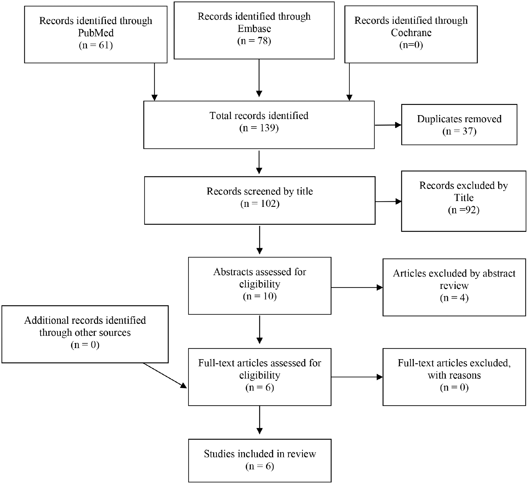
PRISMA flowchart.
The primary purpose of this study is to provide a literature-based description of patients who experienced temporal bone fractures resulting from gunshot wounds. We sought to answer the following questions: What are the presenting findings and clinical course of patients with these injuries? What are the major outcomes and complications? When is surgery indicated, and what are the outcomes of patients who undergo surgical procedures related to the injury?
In May 2019, systematic searches were conducted in PubMed, Embase, and the Cochrane Library with a combination of MeSH terminology and keywords in the Title/Abstract fields relating to “gunshot,”“wounds,” and “temporal bone” but without publication date restrictions. By limiting publications to the English language, 61 citations were retrieved in PubMed, 78 in Embase, and none in Cochrane (see Supplemental Table S1 for full search strategies). Removal of 37 duplicates left 102 distinct citations for screening.
Specific criteria were used to narrow the search to the appropriate topic. Only articles written in English were considered, which limited the results to 47 citations. Individual case reports were excluded, as these added unnecessary confounding factors to the review. The list of citations was then limited to subjects related to temporal bone injuries caused by bullets. One article was excluded because it comprised only patients who had facial nerve injuries, and another was excluded because its analysis considered patients who had immediately died.3,9 Only patients who had temporal bone fractures caused by gunshot wounds and survived for at least 48 hours were included in this review.
Based on these criteria, 6 studies were included on the subject of temporal bone fractures caused by gunshot wounds, ranging from 1972 to 1998 in publication date. These studies were cross-referenced to ensure that no relevant study was excluded by the search method. All studies were retrospective, and 1 was a multicenter study, 6 which provides a low-quality level of evidence. Furthermore, the studies are subject to publication bias. Additionally, some studies utilized representative cases without providing information on all patients for comparison.6,8 Other studies failed to provide objective information, such as audiometry and the severity and timing of facial nerve paresis. Poor follow-up has the possibility of introducing bias. Patients with better outcomes may not follow up, causing the study outcomes to appear worse than the actual patient outcomes.
Statistical synthesis of data was limited because the studies included in the review did not provide sufficient data for rigorous statistical analysis. Descriptive statistics were instead calculated, and these are shown in Table 1 . Weighted averages were calculated when studies did not include the relevant information.
Outcomes of Patients Included in the Review.
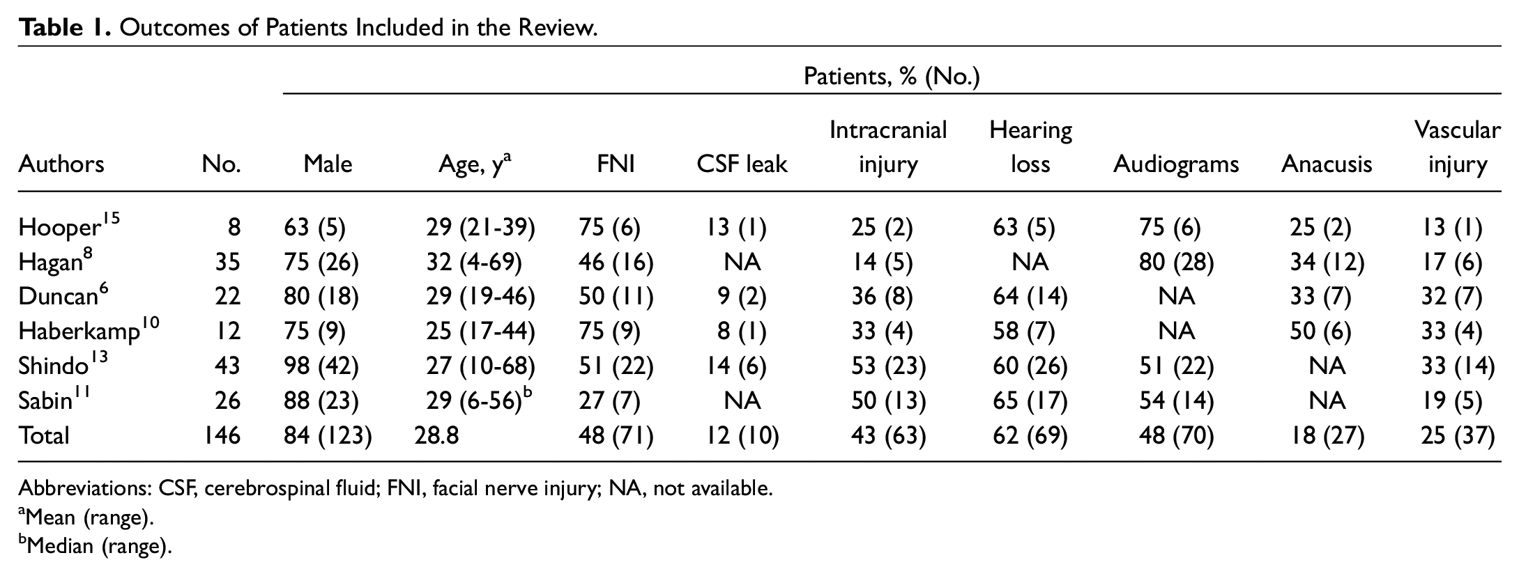
Abbreviations: CSF, cerebrospinal fluid; FNI, facial nerve injury; NA, not available.
Mean (range).
Median (range).
Results
Patient Demographics
Patient demographics were remarkably similar across most of the studies. In every study, there was an overwhelming majority of young males involved: 84% of the 146 total patients included in the review were male, with an average age of 28.8 years. Unfortunately, demographic information beyond this level of detail is unavailable for most patients due to the use of representative cases for subcategories of injuries, as seen in Hagan et al and Duncan et al.6,8 Interestingly, Haberkamp et al noted that the right side was injured 75% of the time; however, the laterality of each patient was not noted in any other studies. 10 Though speculative, this could be related to handedness of the shooter and would be an interesting hypothesis to test in a future study. 10 Hagan et al excluded shotgun blasts, but other authors chose to include them. 8
Hearing Loss
Hearing loss was also frequently reported in the studies included. About 62% of patients exhibited some type of hearing loss, but just 5 of the 6 studies included the hearing status of all patients within the study. Only 48% of the patients had preoperative audiograms. Some studies utilized tuning fork tests, whereas others reported audiometric data obtained prior to surgery. The tuning fork examination can be performed to distinguish conductive and sensorineural hearing loss (SNHL). Obtaining audiograms of patients who are critically ill can be technically difficult from a systems perspective but still valuable for guiding care. Sabin et al described 14 of 22 patients who had audiograms, and 86% of these patients showed SNHL or mixed hearing loss. 11 Furthermore, 18% of the patients included in their review had anacusis, but only half of the other studies reported this finding. Ideally, all future studies would report the presence of subjective hearing loss, hemotympanum, and tuning fork findings. This would help establish the type and severity of hearing loss. Patients should be provided with follow-up information to obtain an audiogram once discharged from the hospital. Audiometric outcomes are used to guide patients to appropriate interventions, such as amplification for SNHL or surgical exploration for persistent conductive hearing loss. 12 Additionally, documentation of pre- and postoperative audiograms will allow a more robust study of outcomes.
Facial Nerve Injury and Rehabilitation
Facial nerve injury was universally mentioned in every study, with facial nerve weakness reported in 48% of all patients in this review. This rate included total, incomplete, immediate, and delayed paralysis. The inconsistent reporting of onset and severity of facial nerve function makes it difficult to generalize the outcomes across studies. Overall, 92% of reported facial nerve injuries were immediate. Only 1 of 6 studies reported House-Brackmann facial nerve grading scale, with the remaining studies describing facial nerve function as complete or incomplete.13,14
Transection of the facial nerve can occur because it is tethered within the temporal bone and blast injuries will tear or crush the nerve. In most studies, facial nerve injury was the most common indication for intervention, though patients often had multiple indications for surgery. 10 The use of corneal reflex or grimace as an extension of the physical examination can elicit facial nerve function in patients who are unable to follow commands. This allows the timely diagnosis and assessment of the severity of facial nerve injury. Immediate and total facial nerve paralysis with concern for facial nerve transection is a clear indication for surgical exploration. Furthermore, most authors considered a comminuted fracture of the facial nerve canal on computed tomography as a consistent indication for exploration and repair. 10 Two-thirds of the studies reported the location of the facial nerve injury and the corresponding method of surgical intervention, including facial reanimation. The vertical portion of the facial nerve was the most common site of injury and was seen in 48% of patients with confirmed facial nerve injury. The tympanic portion of the nerve was the second-most common site of injury. Many patients had multiple segments of the facial nerve injured, making classification difficult. There were various facial nerve reanimation techniques utilized, but the most frequently used was an interpositional cable graft with the great auricular or sural nerve. When anastomosis was indicated, 59% of grafts involved an interpositional cable graft. The interpositional cable graft is frequently used. Tension-free primary neurorrhaphy is rarely possible because the injury frequently induces thermal injury to the frayed ends of the nerve, which have to be trimmed back to create a healthy anastomosis. 13 Alternatively there may be a complete transection of the nerve without thermal injury. Variability in practice setting and geographic location adds to the various possibilities of facial nerve injury. Future studies should specify immediate versus delayed facial weakness. They could characterize the degree of injury by describing function of the individual branches of the nerve. At this time, the House-Brackmann scale remains the standard assessment tool.
CSF Leak
Overall, 12% of patients developed a CSF leak, with a range of 8% to 14%. This was either at the time of presentation or after surgical exploration, but estimates may be inaccurate as some studies did not report CSF leak rate. The leak rate may be underreported as some leaks resolve spontaneously. This makes the reported low incidence of CSF leaks difficult to interpret. Postoperative CSF leak was sometimes included with preoperative CSF leaks, which confounds the study of outcomes. Postoperative CSF leaks could be related to the timing, duration, and extent of the procedure as well as the experience of the surgeon involved. Future studies should separate initial CSF leaks from postoperative CSF leaks and report laboratory confirmation with beta-2 transferrin where possible to facilitate differentiation.
Intracranial Complications
Intracranial injury was present in 43% of patients and included stroke, hematoma, abscess, carotid cavernous fistula, and carotid artery injury. Two of the 6 studies lack detail in the types of intracranial injuries.8,10 Hooper et al described 2 intracranial injuries from the bullet itself, but no intervention was required. 15 Duncan et al used representative cases to describe bullets within the temporal lobe and a carotid cavernous fistula, but details were not provided for the other 6 patients with intracranial injuries. 6 Shindo et al provided the most detail, including subtypes of intracranial hemorrhage (eg, subdural vs subarachnoid) as well as descriptions of the neurologic complications: cortical injuries, stroke, pneumocephalus, and meningitis. 13 Meningitis occurred in 9% of their patients. CSF leak is a well-known risk factor for development of meningitis; however, 1 of the patients in their study developed meningitis despite no evidence of a CSF leak.
Vascular Injury
Vascular injury was reported in every study and occurred in 25% of patients. This is likely because vascular injuries direct the patient’s treatment priorities. In accordance with Advance Trauma Life Support protocol, patients are not evaluated for otologic intervention until hemodynamically stabilized. Vascular injury typically implied intracranial vascular injury, such as jugular foramen or carotid artery injury, but could also imply extracranial carotid artery injury. Because the most recent study in this review was 21 years old, most vascular injuries were discovered by angiography, which was not performed on every patient. 15 With the evolution of technology, computed tomography angiography is now more commonly utilized. Computed tomography angiography is helpful because it assists modern interventional radiologists or neurosurgeons with planning for embolization if necessary.
Surgical Considerations
Haberkamp et al described 5 major indications for surgical intervention: facial paralysis, vascular injury, CSF leak, external auditory canal destruction or cholesteatoma prevention, and intracranial injury. 10 Treatment algorithms are difficult to create, as each patient and pattern of injury are unique. Figure 2 and 3 demonstrate the significant comminution seen with these types of injuries. Subsequently, surgical interventions are carefully individualized. The greatest variation among the studies included in this review originates from the type of procedures and the timing in which they were performed. Urgent surgical indications include facial nerve paralysis and CSF leaks. Immediate facial paralysis with confirmation of a bony spicule abutting the facial nerve is a widely accepted indication for immediate exploration and facial nerve reanimation. If a patient has immediate complete facial paralysis, then an electroneuronography will typically be performed at least 72 hours after the injury. If the electroneuronography demonstrates 95% degeneration, then facial nerve decompression is recommended. Electromyography can be used several weeks following a traumatic injury to confirm complete nerve injury when there are no fibrillation potentials or if there is an absence of compound muscle action potentials. Electromyography can also be used to detect polyphasic potentials several months after an injury, which suggests nerve regeneration. 16 CSF leaks are typically managed first with CSF precautions, and if these fail, then a lumbar drain is placed. If the leak continues despite drain placement, then surgical intervention is recommended during the same admission. CSF leaks are managed aggressively because of the risk of developing meningitis. Vascular injuries are often initially managed by neurosurgery. Another important consideration is the risk of infection and cholesteatoma relating to the entry of skin into the middle ear and mastoid space. Gunshot wounds to the temporal bone universally introduce skin and mucosa into the middle ear and mastoid space, which portends eventual infection and cholesteatoma if not managed properly. These patients will eventually require surgery, but delayed surgical intervention as an outpatient is acceptable and sometimes recommended. Performing delayed outpatient surgery is recommended when patients remain critically ill and the delay will not reduce the risk of a positive outcome. In 1 study, the authors typically performed a canal wall down mastoidectomy, removed all visible bullet fragments, and obliterated the mastoid space to prevent future infection and cholesteatoma formation. 11 Figure 4 demonstrates removal of bullet fragments embedded in a temporal bone. If a nerve anastomosis has been performed, closure of the external auditory canal may be considered, as this procedure will help to protect the nerve graft.
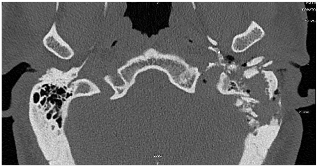
Axial view of a computed tomography scan demonstrating a highly comminuted temporal bone fracture caused by a gunshot wound.
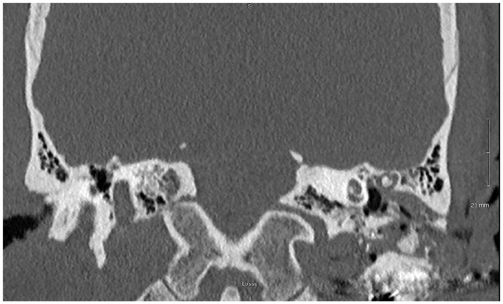
Coronal view of a computed tomography scan demonstrating multiple bullet fragments within the temporal bone.
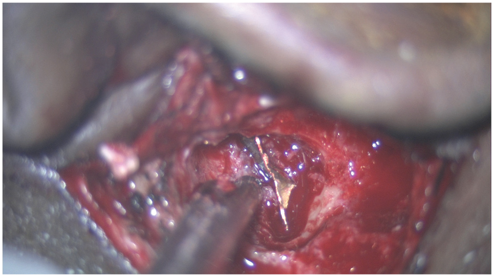
Bullet fragment embedded in the mastoid cavity of a patient.
This review excluded non–English language articles, which may result in some missed studies. Several studies were excluded because they described patients who immediately died after injury. Numerous single-case reports of gunshot wounds to the temporal bone were excluded as these add unnecessary heterogeneity to the systematic review. Finally, follow-up is incompletely reported among the studies discussed. Selection bias may arise from poor follow-up of patients with positive outcomes, which would cause the results of a retrospective study to appear worse than actual patient outcomes.
Conclusion
This review ascertains the most up-to-date outcomes of temporal bone fractures caused by gunshot wounds and compares previous studies of these injuries. Patients may share a common cause of injury, but their outcomes vary greatly. Which variables have the greatest influence on the pattern of injury? Only future prospective studies with more rigorous reporting of data can help address that question.
Hearing loss was universal among patients, and 18% had anacusis. Facial nerve paralysis was expected among most patients, though outcomes varied greatly. CSF leaks are uncommon but usually resolve without surgical intervention. Forty-three percent of patients had intracranial complications; therefore, otolaryngologists should recognize the severity of the injury and carefully consider surgical intervention at the appropriate level of recovery. Vascular injuries are typically managed prior to evaluation by otolaryngology, but these injuries should be recognized and treated if missed on the initial survey. The English language restriction, while necessary due to the fluency limitations of researchers and the lack of translation resources available, may have excluded valuable data from publications in other languages. The synthesis and analysis of the review were limited by the heterogeneity and the outcomes assessed. Although there is variation on how studies are conducted, a few broad suggestions for future research can be made. Future research should include detailed demographics, audiograms, rate of complications, surgical procedures, and ideally 1- to 2-year follow-up. Inclusion of this information would add tremendous value to the body of knowledge on the subject and would help formulate study questions for prospective cohort studies to create treatment algorithms for patients with these injuries. Furthermore, existing studies are dated, and as technology evolves, an update would be beneficial to provide relevant data to physicians. Prospective cohort studies and multi-institutional retrospective reviews would help facilitate the study and treatment of patients with these injuries.
Supplemental Material
Supplemental_Table_1_1 – Supplemental material for Temporal Bone Fractures Caused by Ballistic Projectiles: A Systematic Review
Supplemental material, Supplemental_Table_1_1 for Temporal Bone Fractures Caused by Ballistic Projectiles: A Systematic Review by Kenneth L. Kennedy, Elizabeth Cash, Jessica Petrey and Jerry W. Lin in Otolaryngology–Head and Neck Surgery
Footnotes
Author Contributions
Disclosures
Supplemental Material
Additional supporting information is available in the online version of the article.
References
Supplementary Material
Please find the following supplemental material available below.
For Open Access articles published under a Creative Commons License, all supplemental material carries the same license as the article it is associated with.
For non-Open Access articles published, all supplemental material carries a non-exclusive license, and permission requests for re-use of supplemental material or any part of supplemental material shall be sent directly to the copyright owner as specified in the copyright notice associated with the article.
