
Abstract
To explore the effect of lingual artery ligation on tongue vascularity, we performed an analysis of 25 patients who underwent transoral robotic surgery for base of tongue cancers (May 2011 to December 2019). Hounsfield units of the intrinsic muscles (IMs) and genioglossus muscles (GGs) were measured in postoperative imaging (mean 4 months) as a surrogate for vascularity. In ligated patients (n = 15), the values from the ligated/resected side of the tongue were compared with the contralateral side and the nonligated side of resection. Individually, IMs and GGs on the ligated side demonstrated no significant difference to the contralateral side (P = .662 and .618, respectively). Ligation produced a significant decrease in IM measurements but no difference between GG values vs nonligated patients (P = .050 and .818, respectively). No difference was appreciated in mean values for combined IMs and GGs between cohorts (P = .212). No gross tongue atrophy or complications were incurred. Future studies are warranted to delineate long-term effects.
The increasing incidence of oropharyngeal squamous cell carcinoma (OPSCC) has stimulated advances in the management of OPSCC in recent years. 1 One of these major technological developments is transoral robotic surgery (TORS) for resection of OPSCC.2-4
A feared complication, postoperative oropharyngeal hemorrhage (OPH), is reported in 5% to 10% of TORS patients. Severe hemorrhage can portend acute airway compromise, particularly in patients with impaired airway protection mechanisms.5-7 Ipsilateral lingual artery ligation (iLAL) is a reasonable prophylactic measure against OPH, decreasing the incidence of life-threatening hemorrhage.5-8 While animal models have demonstrated collateral vascular compensation, studies exploring this relationship in human subjects are lacking.9,10 Therefore, we sought to examine effects of iLAL on tongue vascularity.
Methods
Patient Selection
The UAMS institutional review board (#260316) approved a retrospective review on 25 patients (21 male, 4 female) who underwent TORS for base of tongue (BoT) OPSCC at our tertiary academic institution (May 2011 to December 2019). Hounsfield units (HU) of the intrinsic muscles (IMs) and genioglossus muscles (GGs) were measured by a staff radiologist as a surrogate marker for tongue vascularity in postoperative contrasted computed tomography (CT) images (range, 2-9 months; mean, 4 months).11-14 In iLAL patients (n = 15), the values from the ligated/resected side were compared with the nonligated, contralateral hemi-tongue ( Figure 1 ), as well as with patients with nonligated vessels (nLAL, n = 10) ( Figure 2 ). Comparisons were also performed in nLAL patients between the side of resection and the contralateral hemi-tongue and between individual GG and IM values as well as averaged IM and GG values, using independent samples t test and paired t test. Categorical variables were analyzed using χ2 and paired t test.
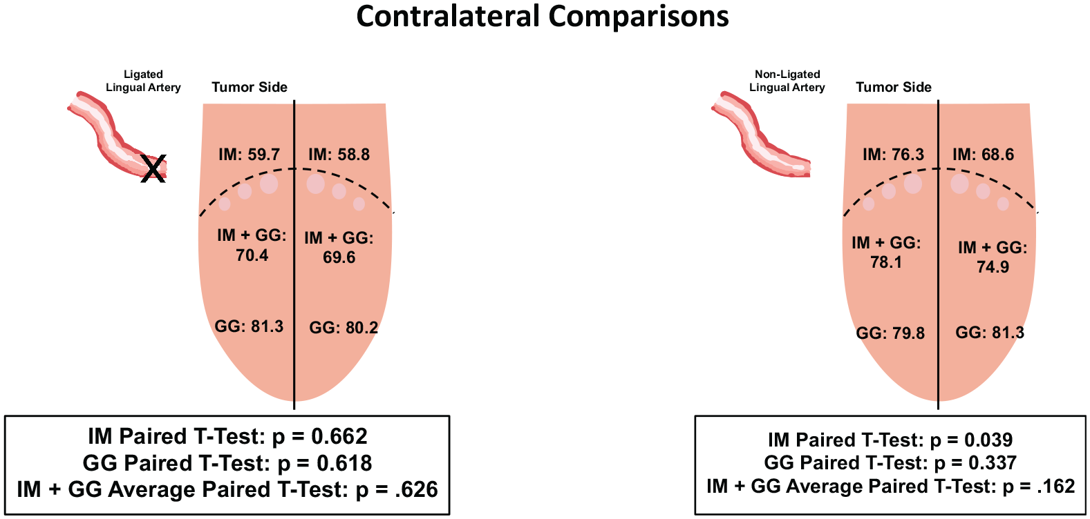
Comparison of side of tumor resection to contralateral hemisphere.
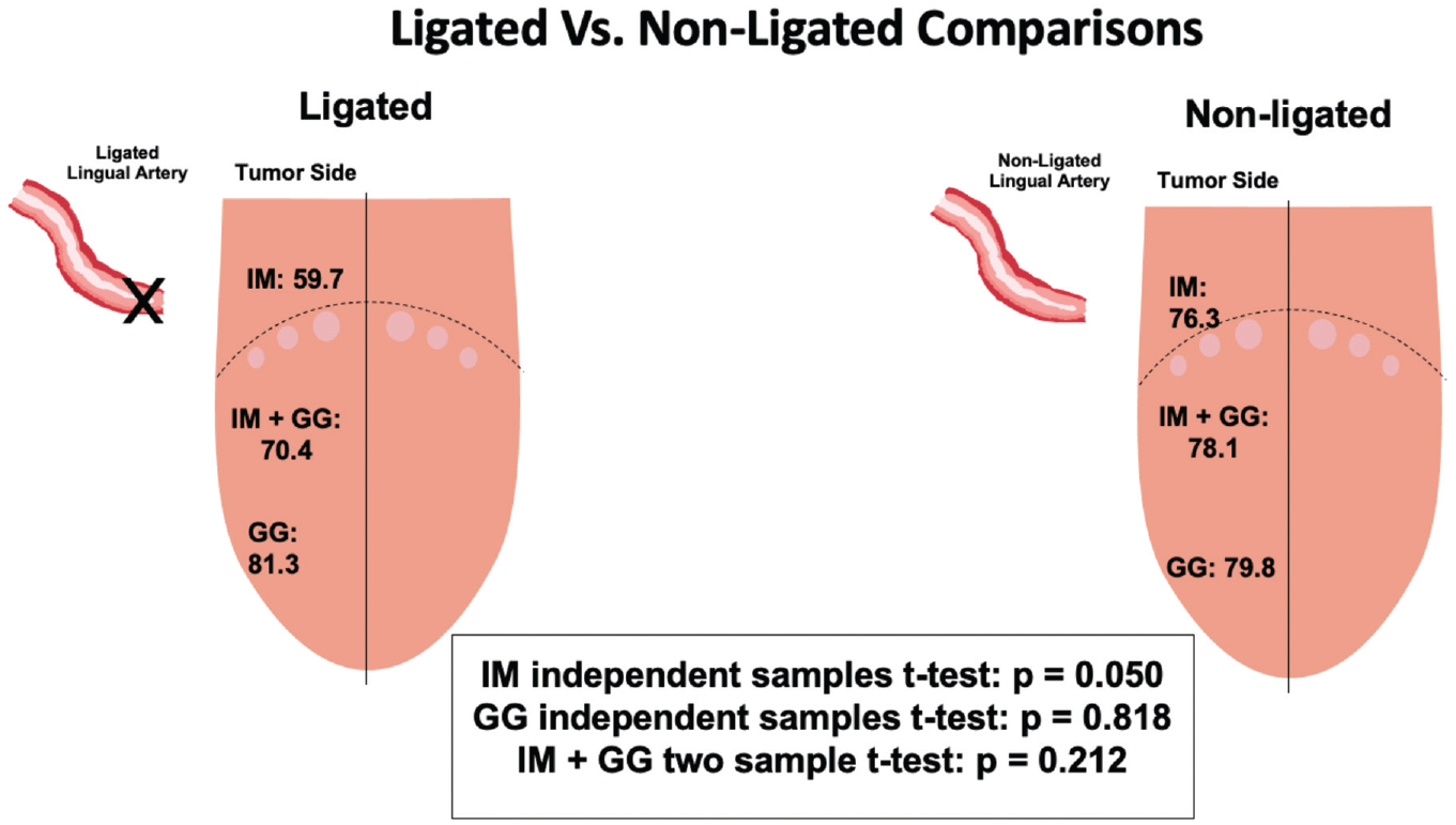
Comparison of side of tumor resection between ligated and nonligated patients.
Results
No significant differences were found in demographic, pathologic, or preoperative variables among cohorts ( Table 1 ). One significant, nonfatal bleeding event occurred intraoperatively in the nonligation group.
Patient Demographics and Clinicopathologic Data Comparison.
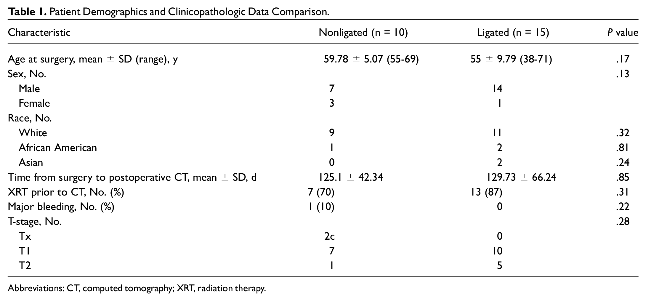
Abbreviations: CT, computed tomography; XRT, radiation therapy.
Comparison of the IMs and GGs on the side of ligation/resection to the contralateral side revealed no significant difference in HU (P = .662 and .618, respectively; Figure 1 ). Comparison of the side of resection between cohorts demonstrated significantly higher HU in nonligated patients on IM measurements but no difference between GG values (P = .050 and .818, respectively; Figure 2 ). No significant difference was appreciated, however, upon comparison of combined muscle groups (average of IM + GG) between cohorts with respect to laterality (P = .212; Figure 2 ). Neither gross tongue atrophy nor complications in the neck were incurred as a result of the performance of iLAL during the study.
Discussion
As utilization of TORS for OPSCC increases, optimization of the surgical technique to minimize complications is paramount. iLAL has been shown to significantly decrease the severity of OPH in several studies.5-8 Our investigation adds to the literature as reported on the effect of iLAL on human tongue vascularity is not reported.
Using HU, we quantitatively examined the effects of iLAL on tongue vascularity following intervention (mean 4 months). The nLAL IM on the side of resection demonstrated significantly higher HU measurements and thus vascularity than all other IM groups. While this may be an expected finding compared to the iLAL/resected IM (
Considering the iLAL/resection IM showed no significant difference from the contralateral IM (P = .662) and significantly lower vascularity in comparison to the nLAL/resection IM (P = .050), it is reasonably inferred that performance of iLAL was effective. This finding correlates well with other studies demonstrating higher incidence of severe OPH in nLAL patients. Further studies are needed to explore the above hypotheses concerning the etiology and consequence of observed trends in vascularity.
Although we did find significant differences in vascularity between IM muscle groups, none were appreciated when combining muscle groups (IM + GG) with respect to laterality in contralateral comparison of iLAL patients or iLAL vs nLAL comparisons, respectively (P = .626, P = .212;
While this investigation serves well as a pilot study, it is not without important limitations. A post hoc power analysis was performed for all significant findings, demonstrating inadequate study power to definitively reject the null hypothesis. Due to small cohort size and limited power, the findings of this study should be interpreted with caution. Further limitations exist in the relatively short-term follow-up (~4 months), preventing conclusive assessment of long-term vascular changes. Large-volume studies with longer follow-up are warranted to characterize long-term outcomes in this patient population.
Conclusion
iLAL may have no significant effect on tongue vascularity with respect to laterality in the early postoperative period. While these novel findings are encouraging and support the iLAL performance, further studies are warranted to corroborate our findings as well as demonstrate safety and efficacy, particularly with regard to long-term effects.
Footnotes
Abstract was presented virtually at the AAO-HNSF 2020 Virtual Annual Meeting & OTO Experience, September 13-16, 2020.