
Abstract
Objective
To examine the changes in measures of sleep apnea severity and hypoxemia on the first postoperative night following implantation of the hypoglossal nerve stimulator.
Study Design
This was a single-arm prospective cohort study.
Setting
A single academic sleep surgical practice.
Methods
Subjects with moderate to severe obstructive sleep apnea underwent implantation of the hypoglossal nerve stimulator (HGNS) and were discharged to home the same day as surgery. A single-night WatchPAT study was performed on the night immediately following surgery (PON 1) and was compared to baseline sleep testing.
Results
Twenty subjects who were an average of 58.6 ± 2.5 years old, were 25% female, and had a mean body mass index of 28.1 ± 0.9 kg/m2 completed the study. Mean O2 nadir at baseline was 79.6% ± 1.1% compared to 82.7% ± 0.9% (P = .013) on PON 1. One patient demonstrated a >10% worsening in O2 nadir. Only 2 additional patients demonstrated a worsening in O2 nadir on PON 1, each by only 1 percentage point. Neither mean time spent below SpO2 88% nor oxygen desaturation index (ODI) worsened postoperatively (mean time spent below oxygen saturation of 88%, 27.8 ± 7.85 vs 11.2 ± 5.2, P = .03; mean ODI, 29.6 ± 5.2/h vs 21.0 ± 5.4/h, P = .10). Mean obstructive apnea hypopnea index (AHI) was no worse (40.6 ± 4.7/h to 28.7 ± 4.2/h, P = .02), with only 2 patients experiencing an obstructive AHI >20% more severe than baseline. Only 1 patient demonstrated a clinically meaningful increase in central AHI on PON 1.
Conclusions
Overall, AHI and measures of nocturnal hypoxemia are stable, if not improved, on PON 1 following HGNS implantation. These findings support the safety of same-day discharge following implantation of the hypoglossal nerve stimulator.
Patients with obstructive sleep apnea (OSA) undergoing surgery have been shown to be at an increased risk of perioperative complications in a wide range of surgical specialties.1-4 This heightened risk has been demonstrated in a number of outcome measures, including cardiopulmonary and neurologic complications, intensive care unit utilization, increased length of stay, readmissions, and perhaps even increased mortality.5-23 This added risk is attributed to a multitude of factors, including worsening in OSA metrics, such as apnea-hypopnea index (AHI) and hypoxemia, effects of opiates and general anesthesia, sleep disruption, and changes in inflammatory markers.24-29
Perioperative management of patients undergoing traditional OSA surgery is of particular concern given the addition of postoperative upper airway edema, significant narcotic requirements, and frequent inability to tolerate positive airway pressure (PAP) therapy on top of the abovementioned risks.30,31 Nonsurgical treatment options such as positive airway pressure therapy, oral appliance therapy, and positional therapy are clearly advantageous in terms of safety. However, a significant number of patients are intolerant of or do not derive benefit from these therapies and seek surgical treatment options.
Hypoglossal nerve stimulation (HGNS) is a hybrid treatment option in that it is surgically implanted but medically titratable. 32 HGNS also differs from traditional upper airway surgery in that there is typically minimal postoperative pain and no mucosal surface swelling due to the minimally invasive, transcutaneous means by which the device is implanted. It therefore stands to reason that HGNS has a superior perioperative safety profile, and indeed many surgeons perform this on an outpatient basis, including at surgical centers without admitting capabilities. However, there are others who feel that any surgical patient with moderate to severe OSA requires overnight observation in the hospital. The current best evidence on perioperative management of patients undergoing OSA surgery, including HGNS, is largely based on expert opinion. 31
The objective of this study was to objectively examine the changes in sleep study metrics, including hypoxemia, on the first postoperative night following implantation of the HGNS as a means to assess the safety of same-day discharge.
Methods
This was a prospective, single-arm cohort study of adult patients undergoing implantation of the Inspire Medical Systems HGNS. All implants were placed by the author using the standard 3-incision or modified 2-incision approach. All patients were discharged to home from the postanesthesia care unit. Patients who were prescribed acetaminophen-hydrocodone (5/325 mg every 6 hours as needed for pain) were assumed to have used it as directed on the first postoperative night. Subjects were instructed to wear the WatchPAT 300 with central apnea monitor (Itamar Medical Ltd) on the first postoperative night (PON 1). Otherwise, no changes to the standard postoperative instructions were made. No patients were actively using other sleep apnea treatments such as positive airway pressure or oral appliance therapy.
Demographic data, including age, sex, and body mass index (BMI), were extracted from the medical record. Baseline sleep study data originated from a mixture of type 1 or type 3 studies, some of which were interpreted by the author. Surgical data such as the time of day of surgery (morning/evening) and duration of general anesthesia were noted in the anesthesia record. Time under general anesthesia was defined as the number of minutes from induction to extubation, not surgical time. WatchPAT data from postoperative night 1 reflect the automated scoring of the signals to reduce potential bias introduced by the author. Rule 1a (3%) and rule 1b (4%) scoring was matched to the baseline study scoring.
Data were compiled in an Excel file (Microsoft) and subsequently analyzed using SPSS version 24 (SPSS, Inc). Comparisons were made between baseline sleep testing and the PON 1 WatchPAT results using the paired samples t test. To assess for associations between patient (sex, BMI, age), surgical factors (number of incisions, length of general anesthesia, timing of surgery, nasal packing, opiate prescription), or baseline sleep study findings (OSA severity, nocturnal hypoxemia, positional OSA) and worsening PON 1 sleep apnea measures (>20% worsening in obstructive AHI [oAHI], central AHI [cAHI], time spent below oxygen saturation of 88% [T88], and oxygen desaturation index [ODI] or >10% worsening in O2 nadir), Fisher exact test was used. This study was approved by the Partners Institutional Review Board, and all patients signed informed consent.
Results
Of 25 subjects enrolled in the study, 5 experienced technical issues, resulting in a failed WatchPAT recording, and were excluded from further analysis. Final study participants (n = 20) were an average of 58.6 ± 2.5 years old (range, 30-77 years), were 25% female, and had a mean BMI of 28.1 ± 0.9 kg/m2 (range, 19.5-34.6 kg/m2; 5 patients >32 kg/m2). Overall, subjects demonstrated severe obstructive sleep apnea with moderate nocturnal hypoxemia on baseline sleep testing ( Table 1 ).
Comparison of Sleep Study Metrics Between Baseline and Postoperative Night 1 After Implantation of the Hypoglossal Nerve Stimulator.
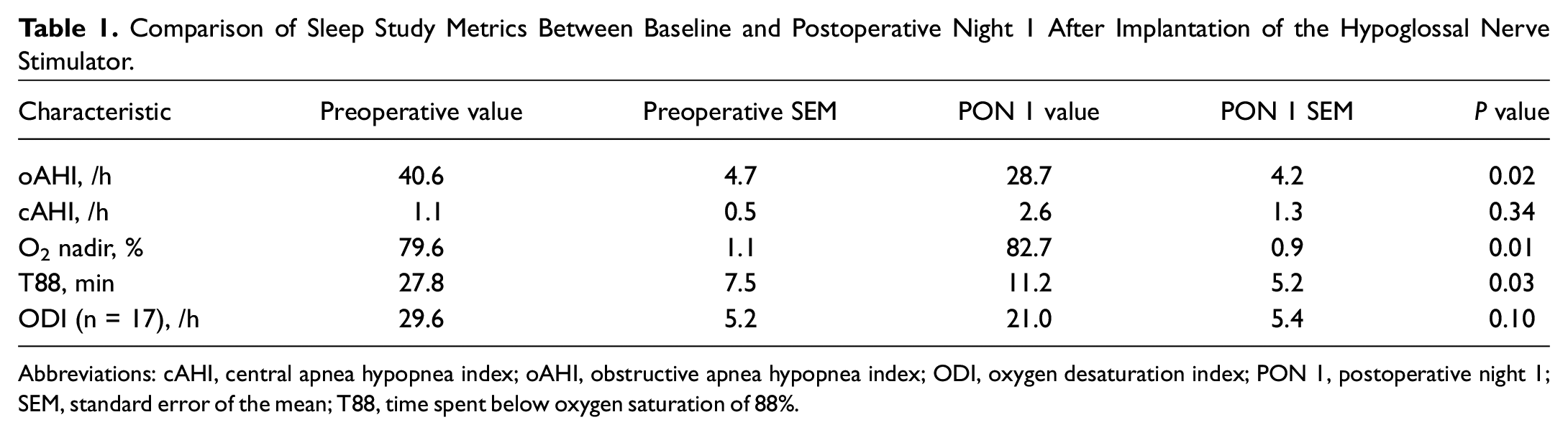
Abbreviations: cAHI, central apnea hypopnea index; oAHI, obstructive apnea hypopnea index; ODI, oxygen desaturation index; PON 1, postoperative night 1; SEM, standard error of the mean; T88, time spent below oxygen saturation of 88%.
Three subjects had functional nasal surgery at the time of HGNS implantation, each with Doyle splints placed. Average time under general anesthesia was 123.5 minutes (range, 86-188 minutes), and most surgeries were performed in the morning (14/20). Just over half of cases (12/20) were 2-incision approaches. Prescriptions for hydrocodone-acetaminophen were ordered for 16 of 20 patients. One subject had mild, transient postoperative tongue weakness; otherwise, there were no intraoperative or postoperative complications.
Mean estimated total sleep time on the PON 1 WatchPAT study was 408.6 minutes with 16.0% detected as rapid eye movement (REM) sleep. Table 1 displays the PON 1 mean oAHI, cAHI, O2 nadir, T88, and ODI, which all showed a lack of consistent worsening, if not statistically significantly improved, values when compared with baseline sleep study values. oAHI was only >20% worse in 2 individuals, 1 with an increase from 22.9/h to 35.0/h and the other from 39.3/h to 57.4/h ( Figure 1 ). Therefore, only 1 patient was reclassified in terms of severity of OSA (moderate to severe). In both of these cases, the patient spent 100% of the WatchPAT study night in the supine position vs 40.4% and 42.9% on their baseline study nights. cAHI was only increased above 5 events per hour in a single patient (who was prescribed acetaminophen-hydrocodone); no others showed development of central sleep apnea.
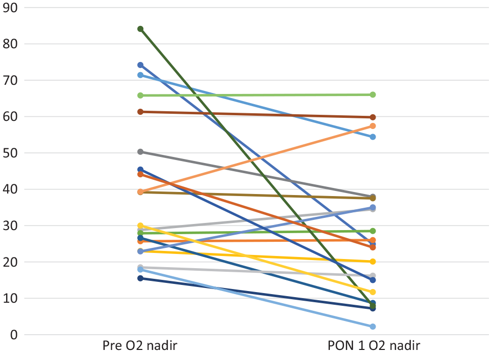
Plot of change in obstructive apnea-hypopnea index by individual subject. PON 1, postoperative night 1.
O2 nadir demonstrated a slight but statistically significant improvement overall on PON 1, and only a single participant had a clinically relevant worsening (88% to 79%). The other 2 subjects who had a lower O2 nadir on PON 1 were by a single percentage point (
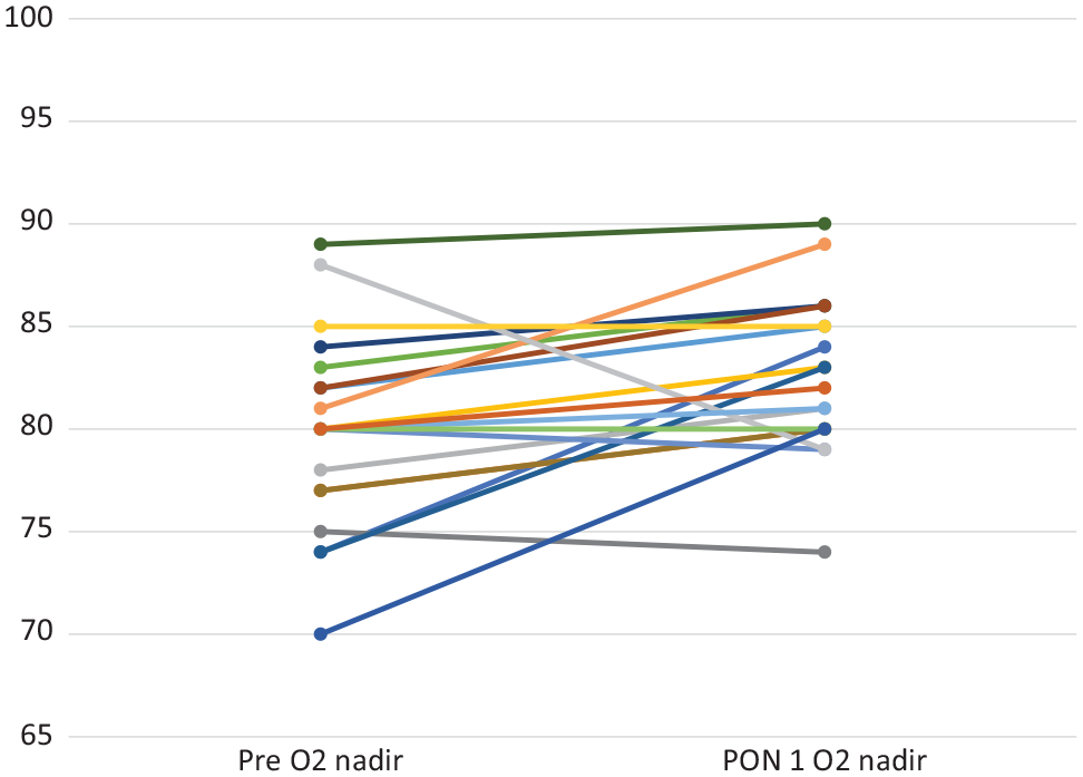
Plot of change in O2 nadir by individual subject. PON 1, postoperative night 1.
No statistically significant associations were found between any of these sleep apnea or oximetry findings (>20% worsening in oAHI, cAHI, T88, or ODI and >10% worsening in O2 nadir) and variables such as age (±60 years), sex, BMI (±30 kg/m2), concomitant nasal surgery/packing, general anesthesia time (±120 minutes), surgical timing (morning vs evening), surgical approach (2 vs 3 incisions), opiate prescription, baseline AHI (±30/h), baseline nocturnal hypoxemia (T88 ±5 minutes), or baseline positional OSA. None of the patients with a BMI over 32 kg/m2 (n = 5) or an AHI >60/h (n = 5) demonstrated worsening oximetry or AHI on PON 1.
Discussion
This is the first study examining immediate postoperative changes in sleep apnea measures, including AHI and oximetry, following implantation of the HGNS. The PON 1 WatchPAT studies demonstrated remarkable consistency with baseline diagnostic sleep studies, especially when one considers normal night-to-night changes in OSA severity reflected in the test-retest variability of sleep testing.33-38 Despite using different testing modalities, the consistency in AHI between baseline and PON 1 in this study was better than in studies with no intervention between nights of testing.
Ultimately, the true value in these findings lies in the safety of this procedure, specifically the safety of same-day discharge. From the perspective of apneas, hypopneas, and hypoxemia, patients do not consistently experience significant worsening in disease severity, and therefore in the absence of other indications for admission, same-day discharge is justified. During times of strained inpatient resources, such as the current coronavirus disease 2019 (COVID-19) pandemic, strategies to safely conserve beds and staff are more important than ever.
The lack of changes in sleep study measures may be fairly intuitive. The HGNS device is not activated until 4 weeks postoperatively and, when turned off, would not be expected to influence OSA severity. Furthermore, the implant surgery itself is minimally invasive in that it is performed transcutaneously, does not involve significant alteration in anatomy or edema, and is largely performed with blunt dissection, resulting in mild pain.
However, what is not necessarily intuitive, but is reassuring, is that the effects of general anesthesia and any opiate pain medications received in the recovery room or at home do not seem to have major, detectable effects on OSA and hypoxemia on PON 1. The stability of oximetry (O2 nadir, T88, and ODI) is especially compelling as SpO2 is the only measure that would otherwise be followed overnight if patients were admitted (and at a lower sampling rate). Only a single subject had a clinically meaningful worsening in O2 nadir and T88 that was directly attributable to supine sleep-incorporating positional therapy into discharge instructions may represent a simple prevention strategy. Positional therapy would have likely prevented the 2 cases with worsening oAHI as well given that both slept supine for the entirety of PON 1.
This study also provides some insight into the risk of OSA and non-OSA surgery in OSA patients. It appears that general anesthesia and mild to moderate opiate use do not contribute to changes in OSA severity that are thought to occur after OSA surgery (including central apneas). OSA patients undergoing non-OSA, nonairway surgeries such as hemithyroidectomy or otologic surgery likely do not require routine admission on PON 1 for the purposes of OSA monitoring.
With that said, these findings should not be generalized to all OSA patients undergoing all procedures. By definition, the HGNS population tends to be less obese and often has less severe OSA. The study cohort presented here had a mean BMI below the obese level and mean AHI in the lower severe range. These PON 1 findings likely suggest that it is the upper airway edema and more significant opiate use, and perhaps obesity, associated with traditional upper airway surgery are more likely to drive this presumed worsening in OSA measures. This study protocol can be applied to those undergoing palate and tongue base surgery to address the long-held belief that upper airway surgery causes a transient worsening in OSA postoperatively.
Changes in OSA may not be reflected until postoperative days 3 to 5 when swelling, REM rebound, and opiate consumption are often at peak values. While it is not practical to admit patients for this length of time or readmit patients during this window, it may be worth investigating the changes in OSA severity over the first postoperative week. Chung et al 26 performed polysomnography on PON 1, 3, 5, and 7 in predominantly orthopedic patients and found that AHI was increased for controls and OSA patients on all nights but peaked on PON 3, and sleep architecture was most affected on PON 1. In another study, preoperatively high AHI, age, and 72-hour opioid use were positively correlated with postoperative increases in AHI. 39 This is of concern given that by definition, patients undergoing HGNS have an elevated AHI and are often of advanced age, but fortunately, this increase in AHI was not seen on PON 1 following HGNS implantation.
This study is limited by several factors. Ideally, WatchPAT studies would have been performed before and after surgery for improved internal consistency. This study was intended to be a pilot study to assess if any major differences in OSA measures would be detected compared to existing baseline testing. Most, but not all, of the literature on peripheral arterial tonometry shows high correlations in AHI with traditional polysomnography.40-42 A recent large study of 500 patients undergoing simultaneous polysomnography and WatchPAT found that overall WatchPAT tended to overestimate the AHI. Furthermore, most patients in this study had home sleep apnea tests serve as the baseline sleep study, which inherently underestimate AHI. Taken together, this only adds confidence that that the stability of AHI seen in this study is real.
It could also be assumed that the AHI and measures of hypoxemia were falsely low because of poor sleep quality after surgery. This does not seem to be the case as WatchPAT provides an estimated total sleep time and percentage of time spent in REM, and both of these measures indicate a fairly normal sleep period: nearly 7 hours of sleep and 15.7% REM sleep. If subjects had been admitted, sleep in the hospital setting would have likely been of shorter duration and more fragmentated. 43
Finally, the sample size is relatively small. The research protocol originally planned for further recruitment, but given the lack of consistent worsening in sleep apnea and oximetry measures, enrollment was stopped. As mentioned, the intent of this study was as an initial investigation to ensure that the practice of same-day discharge was safe, which the data have fairly convincingly validated. A larger sample size may have aided in identifying associations between variables such as nasal packing, general anesthesia time, opiate use, and timing of surgery (morning/evening), but it stands to reason that optimizing/minimizing these variables whenever possible is a sound practice. A larger sample size may have also allowed for the capture of clinically relevant postoperative complications such as readmission or reintubation, which are fortunately uncommon (there were none in this study). Instead, more granular measurements of AHI and hypoxemia were used as surrogates for complications/safety. While it is not known if changes in these values necessarily correspond to a clinically detectable complication, the stability in the values is certainly reassuring.
In conclusion, this study finds that there is no consistent, significant worsening in OSA severity or hypoxemia on the first postoperative night following implantation of the HGNS. This supports the safety of same-day discharge of these higher-risk patients with moderate to severe OSA following HGNS implantation.