
Abstract
Objective
To determine the range of incidental mucosal changes in a general sinonasally asymptomatic population on radiology.
Data Sources
Medline (1996-present) and Embase (1974-present) were searched on March 14, 2020, to identify articles that reported radiological sinus mucosal findings in asymptomatic population groups. Bibliographic search of included studies was conducted to identify additional articles.
Review Methods
The review followed the Preferred Reporting Items for Systematic Reviews and Meta-Analyses (PRISMA) guidelines and Cochrane Handbook for Systematic Reviews of Interventions. A comprehensive search strategy was formulated and articles screened to extract data reporting Lund-Mackay (LM) score, presence of mucous retention cysts, and maxillary mucosal thickening. A random-effects model was used in meta-analysis.
Results
A total of 950 articles were identified, of which 33 manuscripts met the inclusion criteria. The included studies involved 16,966 sinonasally asymptomatic subjects. The mean LM score was 2.24 (95% CI, 1.61-2.87), and an LM score of ≥4 in 14.71% (95% CI, 6.86-24.82%) was present across all general asymptomatic population groups. Mucous retention cysts were noted in 13% (95% CI, 8.33-18.55%) and maxillary mucosal thickening of ≥2 mm in 17.73% (95% CI, 8.67-29.08%).
Conclusion
The prevalence of incidental mucosal changes in a general asymptomatic population on radiology needs to be considered when making a diagnosis of chronic rhinosinusitis.
Chronic rhinosinusitis (CRS) is a common condition with an estimated global prevalence of 3% (clinical CRS based on symptoms and computed tomography [CT] findings), 1 which has a profound impact on quality of life and burden on health care resources. Four cardinal symptoms of CRS have been described: nasal congestion/obstruction, nasal discharge, facial pain/pressure, and hyposmia/anosmia. According to the European Paper on Rhinosinusitis and Nasal Polyps (EPOS) 2020, CRS is defined as inflammation of the nasal cavity and paranasal sinus with 2 or more symptoms, with at least 1 being nasal congestion/obstruction or discharge, lasting over 12 weeks with evidence of inflammation on either endoscopy or radiology. 1 CRS is also further classified based on the anatomical distribution of the disease and underlying endotype. 2
A clinical diagnosis of CRS, however, is not straightforward, due to the range of symptoms overlapping with other sinonasal conditions, such as rhinitis. The recommendation to ensure that sinus imaging, with or without nasal endoscopy, confirms diagnosis of CRS has been in place since 2005. 3 Contemporary assessment often includes CT of the sinuses, which has become the gold standard for diagnosis. CT is relatively inexpensive and readily available, and imaging of the sinuses provides additional information such as the degree of disease, as well as sinonasal anatomy, which is essential in the planning of endoscopic sinus surgery. However, CT is very sensitive to any mucosal thickening, and not all mucosal changes represent active inflammation.
Previous studies have reported on various incidental sinus abnormalities such as mucosal thickening or sinus opacification, with an estimated prevalence of such findings between 16% and 60%. 4 However, many of these studies were not strictly comparable, as they had heterogenous populations, symptom screening, and methodologies, especially in their definitions of abnormality. Furthermore, there is a lack of correlation between the presence of CT sinus abnormality and severity of symptoms. 5
The Lund-Mackay (LM) score 6 is a simple, validated, and reliable scoring system created to consistently quantify sinus imaging findings. The LM score grades the degree of opacification of the maxillary, frontal, sphenoid, anterior ethmoidal and posterior ethmoidal sinuses (0, none; 1, partial; 2, complete) and ostiomeatal complex (0 or 2), for a total score of 24. However, the range of “normal” or expected LM scores in a general asymptomatic population is still debated. Attempts to address this report a “normal” LM score of anywhere between 1 and 4.4,7-12 In addition to the LM score, other key aspects on sinus imaging are the presence of mucous retention cysts and degree of sinus mucosal thickening.
Currently, there is limited literature exploring the range of normal or incidental radiological findings (such as LM score, prevalence of mucous retention cysts, and sinus mucosal thickening) in a general population that is asymptomatic for sinonasal complaints. Establishing a range of normative values would allow clinicians to use a standard frame of reference to assess radiological findings in their practice. Thus, this systematic review of the literature aimed to evaluate studies that investigated the sinuses using CT or magnetic resonance imaging (MRI) in a general asymptomatic population for sinonasal complaints. Within this cohort, a meta-analysis was conducted to determine the average LM score, prevalence of mucous retention cysts (MRCs), and degree of maxillary sinus mucosal thickening.
Methods
Protocol and Registration
A systematic review to identify studies that reported sinus mucosal radiological findings in a general population, without sinonasal symptoms, was retrospectively registered in Research Review Registry (https://www.researchregistry.com/; ID: reviewregistry1009). This review was reported as per recommendations from the Preferred Reporting Items for Systematic Reviews and Meta-Analyses statement (PRISMA). 13 The Cochrane Handbook for Systematic Reviews of Interventions 14 was also used where applicable.
Eligibility Criteria
The inclusion criteria consisted of studies with participants of all ages, investigating sinus mucosal radiological findings in a general population asymptomatic for sinonasal complaints, defined as patients without any history of sinus disease, previous sinus surgery, or facial trauma. Only studies in English were included. Animal studies, case reports, reviews, letters, and technical reports were excluded. Three main outcomes were reviewed: LM score (0-24), the presence of MRCs, and maxillary sinus mucosal thickening ≥2 mm.
Search Strategy and Information Sources
A comprehensive search strategy (see Suppl. Tables S1 and S2 in the online version of the article) was used to conduct an electronic systematic search on both Medline (1996-present) and Embase (1974-present) databases on March 14, 2020.
Study Selection
The results from the literature search were reviewed by 2 authors (B.R. and A.P.). After removal of duplicates, studies were selected based on the inclusion and exclusion criteria by screening titles, abstracts, and full texts. Discrepancies in study selection were resolved by discussion between the 2 reviewers.
Data Extraction
Following final selection, eligible studies were further subcategorized into 3 general asymptomatic population groups: (1) specific symptom screening with endoscopy, (2) specific symptom screening without endoscopy, and (3) no specific screening. Studies allocated to group 3 reported their patient cohort as being sinonasally asymptomatic, but they did not describe their screening methodology. Data from each of these studies were extracted from text, graphs, tables, and figures and entered into a table of characteristics. The table documented study type, objective, population size, patient selection criteria, imaging modality, and outcome. When not provided, the standard deviation was imputed in accordance with the Cochrane handbook. 14 A risk of bias assessment of the studies was conducted based on a modified Newcastle-Ottawa scale, using a traffic light system based on population recruitment, population screening, and outcome assessment (see Suppl. Table S3 in the online version of the article).
Statistical Analysis
All data and statistical analyses were performed using Excel 2019 (Microsoft) with the statistical add-on application, MIX2.0 version 2.016 (BiostatXL) 15 by 2 authors (B.R. and A.S.). Two main statistical analyses were performed using a random-effects model, a meta-analysis of studies with sufficient continuous data, and a meta-analysis of single proportions for dichotomous data. A subgroup analysis was performed by grouping studies by their population age; those with a mean age <18 years were placed in the pediatric group; all other studies were named “mixed age-group populations.” Meta-analysis was performed using a continuity correction (ie, 0.5) in studies that reported “zero events” as per Ma et al. 16 Heterogeneity was assessed in all statistical analyses using the I2 test. This was conducted to assess the variability and discrepancies of studies for each outcome explored. Differences between the study cohort mean age and groups (as defined earlier) were explored.
Results
The systematic search strategy resulted in a total of 1117 studies, 3 of which were identified outside of the database search, through a review of reference lists ( Figure 1 ). In total, 950 remained after duplicate removal, 127 after title screening, and 85 following abstract review. Evaluation of these full texts based on the eligibility criteria resulted in 48 studies being included in the qualitative synthesis and 33 in the meta-analysis (see Suppl. Table S4 in the online version of the article). Fifteen studies did not have sufficient or complete data that could be analyzed statistically and thus were excluded from the meta-analyses. The standard deviation was imputed for 2 studies.7,10
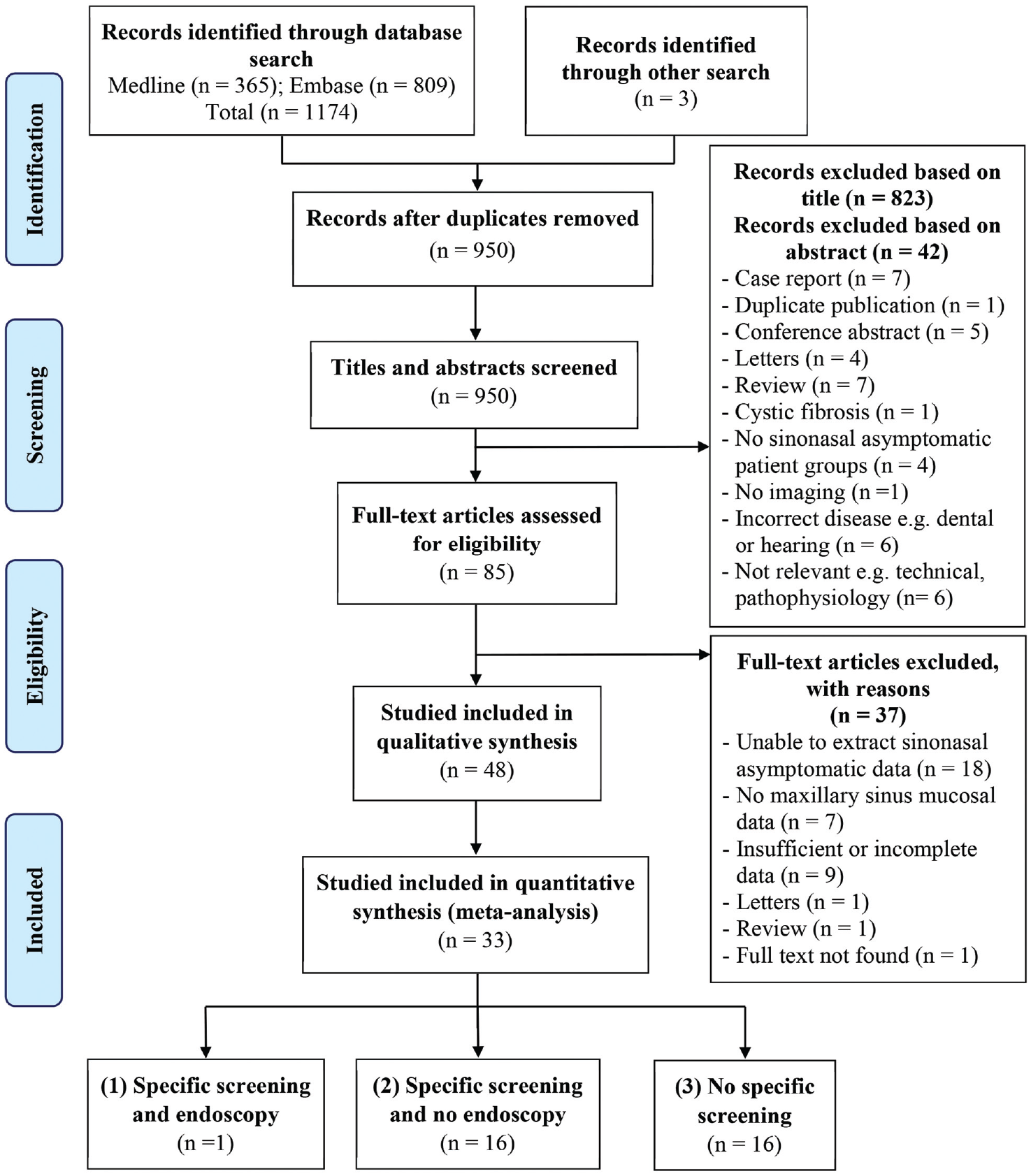
Preferred Reporting Items for Systematic Reviews and Meta-Analyses flowchart.
Study Characteristics
Most of the studies included in the meta-analysis were conducted in adults, 6 studies were conducted in pediatric cohorts,4,10,17-20 and 10 studies8,21-29 were in a mixed patient cohort. The breakdown of the 33 studies into population groups was as follows: 1 study in group 1 (specific symptom screening with endoscopy), 16 in group 2 (specific symptom screening without endoscopy), and 16 in group 3 (no specific screening). In addition, of the 33 studies, 8 reported LM score data, 20 studies described MRC findings, and 13 studies reported maxillary sinus mucosal thickening.
LM Score as Continuous Variable
LM score was used as an outcome in 8 of the 33 studies included in the meta-analysis, of which only 5 articles reported this as continuous data ( Figure 2 ). The total sample size across these 5 articles was 532 patients, one of which was conducted in a pediatric population. 10 All studies used the LM scoring system out of 24. Across all general asymptomatic populations, the overall LM score was 2.24 (95% CI, 1.61-2.87). These studies were heterogenous with an I2 of 93.67% (95% CI, 86.97%-96.92%). In mixed age-group populations, the LM score was significantly different from the LM score of the pediatric population (1.74 [95% CI, 1.56-1.91] vs 2.12 [95% CI, 1.82-2.41], P < .01). 10 After removing a possible outlier, 8 the LM score of a general asymptomatic population reduced to 1.80 (1.42-2.18) and I2 reduced to 78.88% (43.45%-92.11%).
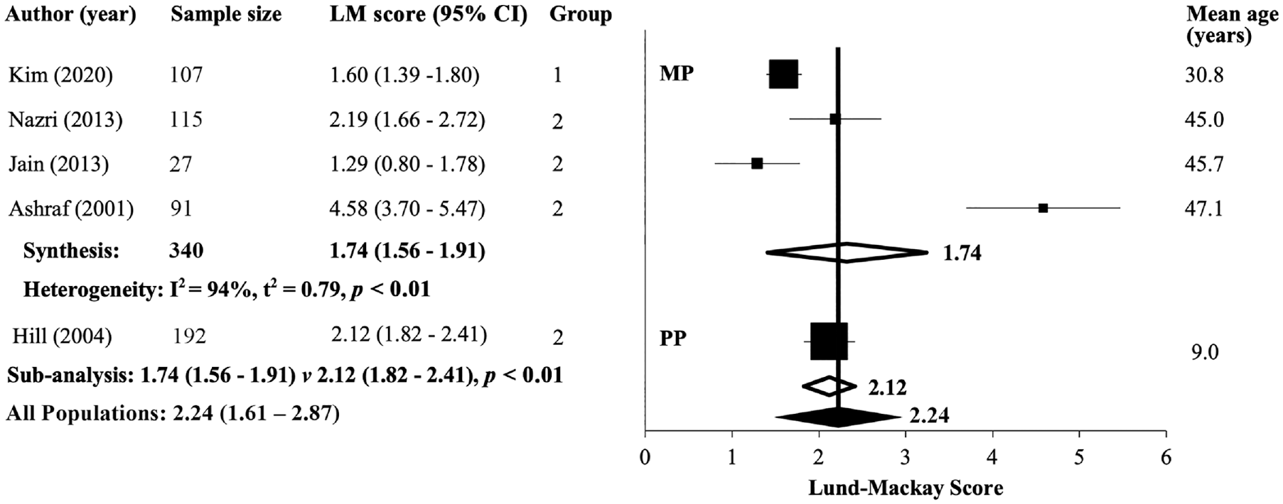
Forest plot of radiologic Lund-Mackay scores in all asymptomatic populations. LM, Lund-Mackay; MP, mixed age-group population; PP, pediatric population.
A meta-analysis of single proportions was also conducted on the remaining 3 studies, which revealed that an LM score ≥4 was present in 14.71% (95% CI, 6.86%-24.82%) of the general asymptomatic population ( Figure 3 ). The I2 of this group of studies was 95.79% (95% CI, 90.78%-98.05%). Three additional studies reported incomplete LM score data and thus were not included in the meta-analyses. In these 3 studies reporting incidental sinus abnormalities, a mean LM score of 1.23 was found in 43 pediatric patients, 4 while the 2 adult cohorts found mean LM scores of 1.24 (N = 114) 9 and 0.22 (N = 50). 30
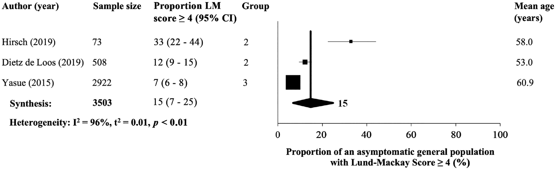
Forest plot illustrating the proportion of patients with a radiologic Lund-Mackay score of ≥4 in all asymptomatic populations. LM, Lund-Mackay.
Mucous Retention Cyst Prevalence
Studies included for the meta-analysis of proportions documented either a concise definition of an MRC or a maxillary cyst. All other studies reporting on polyps or polypoid mucosal lesions were not included in the meta-analysis. The combined sample size of the 20 studies was 7277 patients. Overall, an MRC was found in the sinuses of 13.01% (95% CI, 8.33%-18.55%) asymptomatic patients. The heterogeneity of this group of studies was 98.00% (95% CI, 97.54%-98.37%). No significant difference was found between the presence of MRCs in mixed-age and pediatric populations (14.36% [95% CI, 8.81%-20.97%] vs 8.10% [95% CI, 2.14%-17.21%], P = .39) ( Figure 4 ). Representative images of incidental MRCs are shown in Figure 5A (CT) and Figure 5B (MRI).
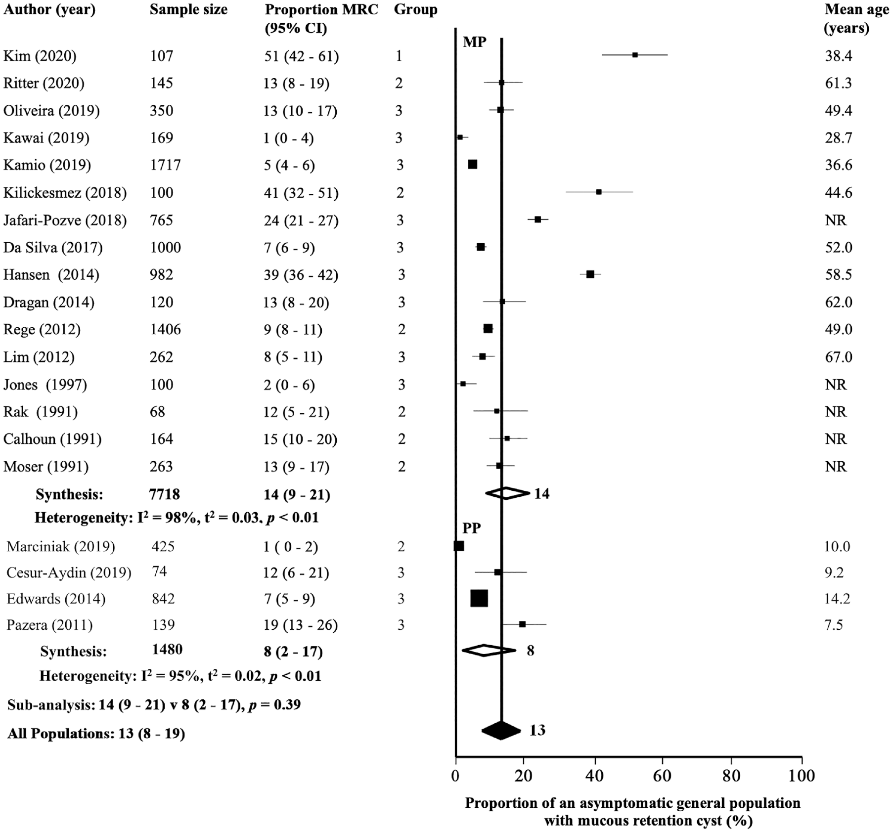
Forest plot illustrating the proportion of patients with the presence of mucous retention cysts in all asymptomatic populations. MP, mixed age-group population; MRC, mucous retention cysts; PP, pediatric population.
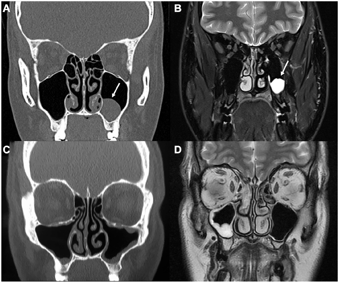
Representative images of incidental sinus mucosal findings. Mucous retention cysts (arrow) on computed tomography (A) and T2 magnetic resonance imaging (B). Maxillary sinus mucosal thickening on computed tomography (C) and T2 magnetic resonance imaging (D).
Maxillary Sinus Mucosal Thickening ≥2 mm
All studies had varying definitions and cutoffs of “normal” mucosal thickening. Most studies referred to the largest measurable mucosal thickening in any wall of the maxillary sinus, and the most common cutoff used was ≥2 mm. The combined sample size of the 14 studies was 8386 patients. Studies were excluded from this analysis if they did not state an exact definition of pathological mucosal thickening. Overall, in this general asymptomatic population, 17.73% (95% CI, 8.67%-29.08%) presented maxillary mucosal thickening of ≥2 mm. The heterogeneity of this group of studies was 99.15% (95% CI, 98.98%-99.29%). There was no significant difference in the prevalence of pathological thickening between mixed-aged and pediatric populations (19.87% [95% CI, 19.03%-20.73%] vs 10.56% [95% CI, 7.97%-13.46%], P = .25) ( Figure 6 ). Representative images of incidental maxillary sinus thickening are shown in Figure 5C (CT) and Figure 5D (MRI).
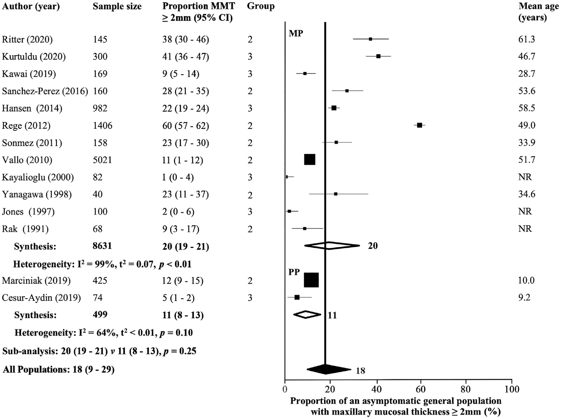
Forest plot illustrating the proportion of patients with maxillary mucosal thickness ≥2 mm in all asymptomatic populations. MMT, maxillary mucosal thickness; MP, mixed age-group population; PP, pediatric population.
Six of the 33 studies21,23,31-34 included in the meta-analyses elsewhere did not state their cutoff for classifying significant mucosal thickening but reported a combined prevalence of 21.3% of some degree of thickening. Nine of the studies excluded from the meta-analysis due to incomplete data found a combined 12.8% prevalence of some degree of maxillary sinus mucosal thickening. However, they did not describe the criteria used to classify pathological thickening of the maxillary sinus mucosa.19,24,35-41 Two additional studies reported maxillary thickening >3 mm combined with additional findings, but individual data could not be extracted to be included in the meta-analyses performed here.20,25 Finally, 1 study reported a 37.5% prevalence of maxillary sinus mucosal thickening ≥1 mm. 26
Discussion
EPOS 2020 reported that an LM score of >5 has a positive predictive value of 96% for CRS.1,10 This study found a mean LM score of 2.24 (95% CI, 1.61-2.87) across all general asymptomatic population groups, and approximately 15% of an asymptomatic population would be expected to have an LM score ≥4. Therefore, less than 5% with LM >5.01 will not have CRS. Ashraf and Bhattacharyya 8 appeared to be a significant outlier, finding a mean LM score of 4.58 in adults, while the other studies ranged between 1.2 and 2.2 despite having a similar study cohort. The same group repeated a similar study in a pediatric cohort and reported a lower mean LM score 2.12 (95% CI, 1.82-2.41). 10 In contrast, this study found a lower mean LM score in adults (1.74) compared to children (2.12). Previous studies in seemingly asymptomatic pediatric populations have found a prevalence of 16% to 45% incidental paranasal sinus findings.10,17 It is unclear why a pediatric population would have greater “incidental” abnormalities or a higher LM score, but repeated viral upper respiratory exacerbations during childhood or dental eruption may account for this observation42,43 or technical difficulties in evaluating and applying the LM score to the developing sinuses of children, seasonal effect on sinus imaging, differing study designs, and inclusion/exclusion criteria.
MRCs are usually asymptomatic and discovered incidentally on imaging, with a reported incidence between 1.4% and 9.6% in the general population.44,45 MRCs have been shown to either stay the same size or regress in size spontaneously in the long term. 44 One study found an MRC prevalence of 22% in symptomatic CRS patients. 46 This meta-analysis found an expected overall prevalence of maxillary retention cysts of 13.01% (95% CI, 8.33%-18.55%) across all general asymptomatic population groups with no difference between adult and pediatric populations. A sample of rhinologists was asked to quantify the prevalence of MRCs in rhinology patients. They universally estimated a 33% prevalence of MRCs in this patient population, which is significantly higher than the general asymptomatic population. There is no clear reference as to the percentage of MRCs in a general population, which is what this study sought to clarify. This difference could suggest an underlying significance of MRCs in an ear, nose, and throat/rhinology patient cohort. Rhinology patients may have previously experienced sinusitis or allergic symptoms, which has been postulated as a potential factor in the pathogenies of MRCs. 47 There was a wide range of MRC prevalence reported by the studies included in this analysis (1%-51%). Reasons for this variation may be differing diagnostic criteria, imaging interpretation, and the difficulty in distinguishing MRCs from polyps or polypoid thickening. Another reason for the discrepancy could be related to environmental factors such as the location and season during which the scans were undertaken. MRCs have been postulated to be closely associated with seasons, particularly winter, during which there is a higher incidence of viral upper respiratory tract infections in winter 48 and environmental changes, with reported increased incidence with higher air pollution and humidity.49-51 Last, the definition of an MRC was unfortunately not consistent (eg, “round hypodense lesions with a sharp edge” 17 ; “well-defined, non-corticated opacity, being dome shaped and homogenous mass” 18 ; and “semispheric thickening of the Schneiderian membrane” 22 ), which could also have resulted in some discrepancies between studies.
Maxillary sinus mucosal thickening and its significance has been extensively studied in the literature. This study found that 17.73% (95% CI, 8.67%-29.08%) of a general asymptomatic population would be expected to have mucosal thickening ≥2 mm, with no difference between adults and children. Discrepancy in results between studies could be due to the cutoff used to define pathological thickening, which ranged from 1 to 5 mm, and the grouping of thickening reported. The most common cutoff reported was ≥2 mm and thus allowed for meta-analysis. There is some evidence of periodic physiological mucosal thickening of up to 2 to 3 mm. 29 Several studies were conducted in dental patient cohorts,18,22,27,28,32,52-57 and while some did screen for sinonasal symptoms and CRS, periodontal disease could have contributed to mucosal thickening. Other potential confounding factors include the season during which imaging was performed, imaging protocols, and the method of determining the maximal thickness of a maxillary sinus. Studies rarely described which wall of the sinus was used and exactly how they measured maximal mucosal thickening.
In conducting this systematic review and meta-analysis, several limitations of the literature were apparent. First, while attempts were made by studies (groups 1 and 2) to exclude symptomatic patients, there was no consistency in the exclusion criteria. Some studies used formal symptom survey scores while others only examined patient histories. Group 3 studies made no mention of the criteria used to classify their patients as truly asymptomatic. In all of the analyses conducted in this study, a high heterogeneity was found. A number of subgroup analyses between the study cohort mean ages and groups were conducted, but this did not reveal any significant improvement in the I2 statistic. This is likely to be due to differences in patient selection, imaging protocols, and outcome definitions (such as MRC and mucosal thickening). Another limitation of the studies included in this meta-analysis was inadequate reporting of data on patient age, which made subanalysis based on study age groups difficult. There were few studies exploring incidental sinus findings in a pediatric population; many studies in the “mixed age group” did not report patient age ranges, and some used a mixture of children and adults (but had an overall mean age >18).
Conclusion
This meta-analysis of current literature reporting incidental CT imaging of a general asymptomatic population lacking significant sinonasal symptoms revealed an LM score of 2.24 (95% CI, 1.61-2.87), 14.71% (95% CI, 6.86%-24.82%) of patients had an LM score ≥4, 13.01% (95% CI, 8.33%-18.55%) exhibited MRCs, and 17.73% (95% CI, 8.67%-29.08%) had maxillary mucosal thickening ≥2 mm. A clinician needs to be aware of the prevalence of such mucosal changes when making the diagnosis of CRS over other conditions that produce sinonasal symptoms.
Supplemental Material
sj-docx-1-oto-10.1177_01945998211035097 – Supplemental material for Sinus Radiological Findings in General Asymptomatic Populations: A Systematic Review of Incidental Mucosal Changes
Supplemental material, sj-docx-1-oto-10.1177_01945998211035097 for Sinus Radiological Findings in General Asymptomatic Populations: A Systematic Review of Incidental Mucosal Changes by Basil Razi, Adam Perkovic, Raquel Alvarado, Anna Stroud, Jacqueline Ho, Larry H. Kalish, Raewyn G. Campbell, Raymond Sacks and Richard J. Harvey in Otolaryngology–Head and Neck Surgery
Supplemental Material
sj-docx-2-oto-10.1177_01945998211035097 – Supplemental material for Sinus Radiological Findings in General Asymptomatic Populations: A Systematic Review of Incidental Mucosal Changes
Supplemental material, sj-docx-2-oto-10.1177_01945998211035097 for Sinus Radiological Findings in General Asymptomatic Populations: A Systematic Review of Incidental Mucosal Changes by Basil Razi, Adam Perkovic, Raquel Alvarado, Anna Stroud, Jacqueline Ho, Larry H. Kalish, Raewyn G. Campbell, Raymond Sacks and Richard J. Harvey in Otolaryngology–Head and Neck Surgery
Supplemental Material
sj-docx-3-oto-10.1177_01945998211035097 – Supplemental material for Sinus Radiological Findings in General Asymptomatic Populations: A Systematic Review of Incidental Mucosal Changes
Supplemental material, sj-docx-3-oto-10.1177_01945998211035097 for Sinus Radiological Findings in General Asymptomatic Populations: A Systematic Review of Incidental Mucosal Changes by Basil Razi, Adam Perkovic, Raquel Alvarado, Anna Stroud, Jacqueline Ho, Larry H. Kalish, Raewyn G. Campbell, Raymond Sacks and Richard J. Harvey in Otolaryngology–Head and Neck Surgery
Footnotes
Author Contributions
Disclosures
Supplemental Material
Additional supporting information is available in the online version of the article.
References
Supplementary Material
Please find the following supplemental material available below.
For Open Access articles published under a Creative Commons License, all supplemental material carries the same license as the article it is associated with.
For non-Open Access articles published, all supplemental material carries a non-exclusive license, and permission requests for re-use of supplemental material or any part of supplemental material shall be sent directly to the copyright owner as specified in the copyright notice associated with the article.