
Abstract
Objective
Inadvertently ingested grill brush bristles can lodge in various locations and lead to a variety of injuries. They can also be difficult to identify and remove. Our primary objective was to perform a systematic review of cases reported in the literature, with analysis of trends in clinical presentation and success of diagnostic modalities and treatment approaches.
Data Sources
Cases of reported grill brush bristle ingestion reported in PubMed, PubMed Central, and Google Scholar databases through April 30, 2021.
Review Methods
Databases were searched for the following terms: (“ingestion” OR “injury” OR “barbeque” OR “BBQ” OR “grill” OR “foreign body” OR “brush” AND “wire” OR “bristle”). Data were collected on patient demographics, clinical presentation, and treatment course. Statistical analysis was performed on characteristics with low risk of confounding.
Results
An overall 57 studies involving 91 patients were included. Grill brush bristles presented most commonly in the upper aerodigestive tract (48/91), followed by the abdomen (26/91) and deep neck (17/91). Computed tomography was the most accurate imaging modality for initial diagnosis, identifying 92.8% of bristles. Less invasive or adjunctive techniques such as endoscopy, intraoperative imaging, or minimally invasive surgery may be useful particularly for bristles located in the head and neck given the low rate of success of transoral surgery (66.7%).
Conclusion
Although this review of retained bristle may be biased toward complex cases, retained grill brush bristles represent an underrecognized and difficult-to-manage hazard. When cases are suspected, clinicians should obtain computed tomography imaging based on presentation and tailor management appropriately.
Outdoor barbequing is an integral part of American culture with a predominance for the summertime months. A sometimes-overlooked safety concern related to best practices of barbequing is the use of wire bristle grill brushes. Dislodged grill bristles have a predilection for adhering to the food being cooked. Consumption of these bristles is relatively common and was the underlying reason for nearly 1700 emergency department visits in America between 2002 and 2014. 1 Bristles can lodge in a variety of locations, such as the upper aerodigestive tract (UADT), deep neck (DN), and abdomen. Previous case reports have identified serious complications from these foreign bodies, including abscess formation,2-6 vascular injury, 7 intestinal perforation, 8 and even death. 9
In 2017, Health Canada performed a risk assessment on wire bristle grill brushes following calls from physicians and patients to have them recalled. 10 Ultimately, recall was deemed impractical, but the Canadian Standards Association took steps forward in 2020 to raise awareness through the implementation of a warning label that will accompany new grill brushes. 11 Despite this ongoing international debate regarding wire bristle grill brushes, a systematic review has yet to be performed to analyze the published literature for trends in presentation, diagnosis, and treatment.
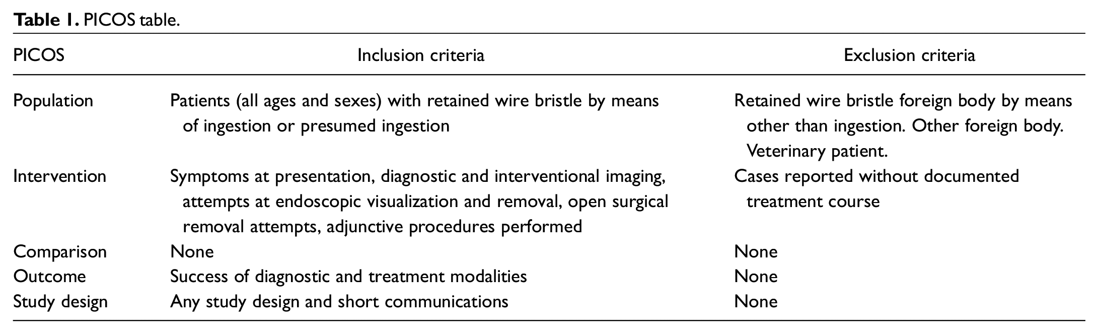
The primary objective of this study was to systematically review the literature on wire bristle ingestion and evaluate the different presentations, diagnostics techniques, and interventional techniques for this condition. Our secondary aim was to assimilate an optimal treatment algorithm. Utilizing the PICOS acronym, we identified the following study inclusion criteria:
Patients: any individual (all ages and sexes) with retained wire bristle by means of ingestion or presumed ingestion
Interventions: symptom presentation, diagnostic and interventional radiographic imaging, endoscopic technique, open surgical removal technique, adjunctive procedures
Comparison: different symptom presentation, other types of imaging, other types of surgical intervention, other adjunctive procedures
Outcome: success of different diagnostic and treatment modalities, complications rates, symptoms based on wire location
Study Design: any study design and short communications
This is summarized in Table 1 .
PICOS table.
Methods
Protocol
Walter Reed National Military Medical Center reviewed and approved the initial Institutional Review Board application (protocol WRNMMC-EDO-2021-0716).
Study Eligibility Criteria
Studies were included without any limitation based on year of publication. Exclusion criteria were other type of foreign body ingestion, inadequate documentation of bristle location, nonhuman patient, inadequate documentation of treatment course, and non–English language literature.
Information Sources
A comprehensive review of the English-language literature was performed through April 30, 2021, from the PubMed, PubMed Central, and Google Scholar databases through the Ovid portal.
Search Strategy
To identify all reported cases of bristle ingestion, databases were queried for the following terms: (“ingestion” OR “injury” OR “barbeque” OR “BBQ” OR “grill” OR “foreign body” OR “brush” AND “wire” OR “bristle”).
Study Selection and Data Collection
A systematic search was performed per the PRISMA literature selection process ( Figure 1 ).2-9,12-75 Two authors (N.M. and Y.M.) independently searched the literature for relevant articles. The title and abstract of every article were screened for relevance to wire bristle ingestion. The full text of potentially relevant studies was then downloaded and reviewed. Data from articles meeting inclusion criteria were then documented in a table for later analysis.
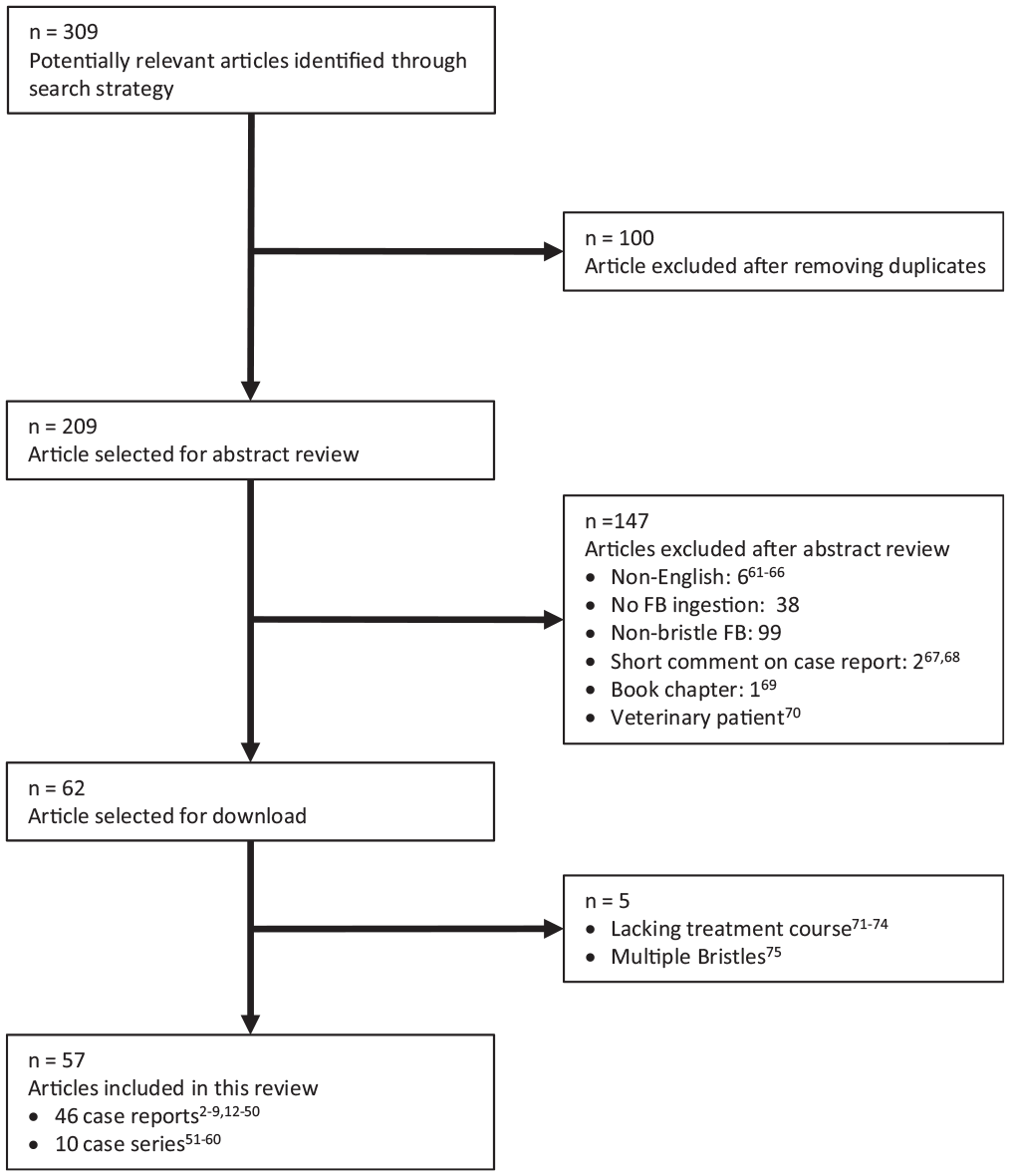
PRISMA flowchart (Preferred Reporting Items for Systematic Reviews and Meta-analyses). FB, foreign body.
Data Items
The anatomic sites in this study were divided into the UADT, DN, and abdomen. UADT was used to describe the following locations and all their subsites: oral cavity, oropharynx, supraglottis, hypopharynx, and cervical esophagus. DN was used to describe the following locations: parapharyngeal space, retropharyngeal space, prevertebral space, submental space, infrahyoid neck, and thyroid. The mediastinum was included with the DN as both cases described the superior mediastinum (level VII of the neck). Cases were classified as abdominal if they were in the distal esophagus or alimentary tract or within the abdominal cavity or its viscera. The subsites described within each article were used to characterize each grill bristle location. If overlapping locations for the same grill bristle were noted in an article, the least morbidly accessible location was used. Migration was defined as clinical verification of a bristle’s movement from its primary presenting location to another subsite.
Transoral surgery included floor of mouth exploration, pharyngotomy, lingual tonsillectomy, or palatine tonsillectomy. Minimally invasive surgery was defined as a percutaneous or permucocutaneous approach for bristle removal. Direct laryngoscopy included suspension microlaryngoscopy or cervical esophagoscopy when employed.
Head and neck symptoms were defined as odynophagia, globus, throat pain, otalgia, cough, and halitosis. Abdominal symptoms were defined as abdominal pain, nausea and vomiting. Systemic symptoms were defined as fever and dyspnea. Delayed presentation was defined among all cases by time course as a report ≥48 hours from ingestion to presentation.
Success was defined according to intervention. For diagnostic imaging and endoscopic visualization, success was defined as bristle identification. Intraoperative imaging success was defined as successful retrieval rather than successful visualization. Bristle retrieval was considered surgical or endoscopic retrieval success.
Statistics
Patient demographic, clinical, and treatment characteristics were described in pooled analyses among all identified cases and among patient subgroups stratified by initial bristle anatomic location and by delayed presentation (≥48 hours). Continuous age was cited as means with standard deviations and medians with ranges and compared by anatomic location with analysis of variance and by delayed presentation with 2-sample t tests. Categorical characteristics were presented as frequencies with percentages, and select characteristics were compared with Fisher’s exact tests. Pairwise differences among initial anatomic locations were evaluated with Fisher’s exact tests, with P values corrected for multiple comparisons via the Bonferroni method. Comparisons were not reported for characteristics for which high risk of confounding limited our interpretation of bivariate analyses.
Results
Including a recent case report from our institution, the search strategy produced 309 potentially relevant articles. One hundred articles were removed from this pool as they were duplicates, yielding 209 available articles for abstract review. An additional 146 articles were removed on abstract review, and 5 were excluded after the full articles were downloaded. The final 57 studies were included in this review and used for data abstraction.2-9,12-60 Our PRISMA flowchart is detailed in Figure 1 . Retrospective case series were evaluated with the NICE quality assessment tools (National Institute for Health and Care Excellence), 76 outlined in Table 2 .
Quality Assessment of Case Series Studies From the NICE Checklist.
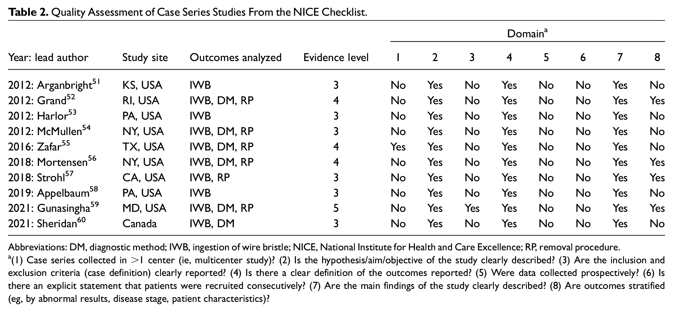
Abbreviations: DM, diagnostic method; IWB, ingestion of wire bristle; NICE, National Institute for Health and Care Excellence; RP, removal procedure.
(1) Case series collected in >1 center (ie, multicenter study)? (2) Is the hypothesis/aim/objective of the study clearly described? (3) Are the inclusion and exclusion criteria (case definition) clearly reported? (4) Is there a clear definition of the outcomes reported? (5) Were data collected prospectively? (6) Is there an explicit statement that patients were recruited consecutively? (7) Are the main findings of the study clearly described? (8) Are outcomes stratified (eg, by abnormal results, disease stage, patient characteristics)?
The literature search revealed in total 91 cases among 47 case reports and 10 case series. The summary of these articles is presented in Supplemental Table S1 (available online). Table 3 breaks down the qualitative data from these articles and separates statistical analysis by initial bristle anatomic location. In pooled analyses of patient and treatment characteristics, patients had an equal sex distribution (50.5% female, 48.4% male, 1.1% unreported, P = .31). Ages ranged from 6 to 92 years (mean, 43.8; SD, 20.7) and were not significantly different with respect to delay (P = .22) or anatomic location (P = .15). No information was indicated on the health or dental status of the patients. The mean bristle length was 2.05 cm (SD, 0.77) and was not different by anatomic location (P = .60) or delayed presentation (P = .08). The specific brand, age, or condition of wire grill brushes was not reported.
Summary of Data.
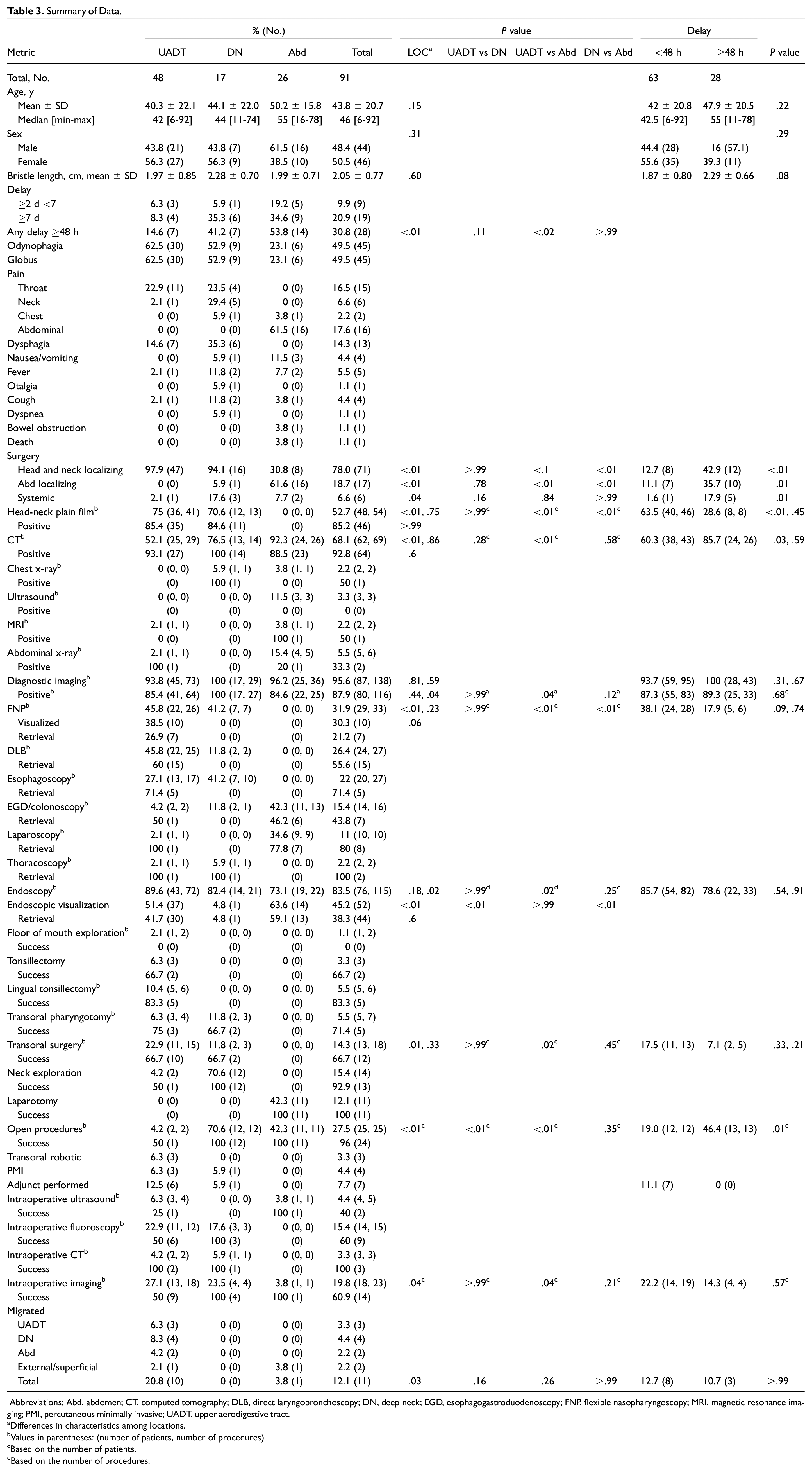
Abbreviations: Abd, abdomen; CT, computed tomography; DLB, direct laryngobronchoscopy; DN, deep neck; EGD, esophagogastroduodenoscopy; FNP, flexible nasopharyngoscopy; MRI, magnetic resonance imaging; PMI, percutaneous minimally invasive; UADT, upper aerodigestive tract.
Differences in characteristics among locations.
Values in parentheses: (number of patients, number of procedures).
Based on the number of patients.
Based on the number of procedures.
Presentation
We identified 48 cases that primarily presented with bristle in the UADT, of which the most common presenting symptoms were odynophagia and globus pharyngeus (both 62.5%) and throat pain (22.9%). In 21% (10/48) of UADT cases, bristle migration was seen after initial presentation.13,14,17,28,39,40,43,50,53,58 Migration locations included posterior oropharyngeal wall, 40 lateral oropharyngeal wall,17,39 submental space, 50 infrahyoid DN, 28 unspecified subsite of the DN, 43 lumen of alimentary tract, 53 intra-abdomen, 58 and superficially toward the skin. 13 Notably, 1 of these migrations was identified when a patient had magnetic resonance imaging (MRI) several months after a known retained bristle and subsequently developed abdominal pain. 58 Additionally, there was 1 bristle penetrating the thyroid and protruding from the pyriform sinus. 5 Although this case was classified as being within the UADT based on our standardized definition of anatomic location, it was complicated by a thyroid abscess necessitating neck exploration in conjunction with removal by direct laryngobronchoscopy. 5 One patient underwent mediastinoscopy in 1952 after a perforated esophagus and migration. 14
Seventeen cases were identified with a grill bristle primarily in the DN, of which the most common presenting symptoms were odynophagia and globus pharyngeus (52.9% for both), generalized neck pain (29.4%), and generalized throat pain (23.5%). There were 4 cases of thyroid involvement.16,19,20,26 In one of these cases, the bristle was left in place and followed with serial imaging for 8 months, with the patient remaining asymptomatic. 26 After initial identification, none of the cases in the DN resulted in further bristle migration. Four complications as a result of bristle ingestion were noted in this group: 1 pierced and thrombosed internal jugular vein, 7 1 esophageal perforation leading to abscess formation, 3 neck abscess after initially missed diagnosis, 2 and a lingual abscess after prolonged missed diagnosis. 4
Twenty-six cases were identified in the abdomen, of which the most common presenting symptoms were generalized abdominal pain (61.5%) and globus pharyngeus (23.1%). Patients with bristles in the abdomen were more likely (61.5%) to have reported abdominal localizing symptoms as compared with UADT (0%, P < .01) or DN (5.9%, P < .01). A bristle lodged within the abdominal cavity was significantly more likely to have presented with a delay ≥48 hours as compared with a bristle lodged in the UADT (53.8% vs 14.6%, P < .02). There was 1 case in this group where the bristle migrated after diagnosis, which involved eventual defecation of the bristle. 58 Complications of inadvertent bristle ingestion in this group were as follows: 1 ureter obstruction causing hematuria, 2 cases suspicious for appendicitis,8,12 1 case of sepsis leading to death in a patient with acute myeloid leukemia, 9 and pancreatic injury, 38 as well as abscesses of the extraluminal duodenum, 6 abdominal wall, 59 and peritoneum. 36
We identified delay ≥48 hours after ingestion in 14.6% (7/48) of UADT cases, 41.2% (7/17) of DN cases, and 53.8% (14/26) of abdominal cases. Patients with a bristle in the abdomen were significantly more likely to have delayed presentation beyond 48 hours as compared with those with bristles in the UADT (P < .02). There was no significance when comparing the frequency of delay in the DN to other locations. Additionally, we identified several cases of missed diagnosis.4,5,18,33,36,38,51,74
No initial anatomic location was significantly more likely to have bristle migration after initial presentation. No anatomic location was significantly more likely to have systemic symptoms upon presentation. However, patients that presented with a delay ≥48 hours were significantly more likely to present with systemic symptoms (17.9% vs 1.6%, P < .01).
Diagnostics Imaging
Overall, diagnostic imaging was used in 95.6% of cases (87/91) in 138 separate procedures. Computed tomography (CT) imaging was performed 29 times among 52.1% (25/48) of UADT cases, 14 times among 76.5% (13/17) of DN cases, and 26 times among 92.3% (24/26) of abdominal cases. The frequency of CT scan usage was significantly higher for patients with a bristle in the abdomen as compared with UADT (P < .01) but not the DN. CT scan was successful in 93.1% (27/29) of the attempts for UADT, 100% (14/14) of those for DN, and 88.5% (23/26) of those for the abdomen.
Although no statistical analysis was possible because of data heterogeneity, plain film imaging was performed less frequently and had a lower success rate overall. Neck plain film imaging was performed 41 times among 75% (35/48) of UADT cases and was successful in identifying the bristle in 85.4% (35/41). Neck plain films were performed 13 times in 70.6% (12/17) of DN cases and was successful in 84.6% (11/12) of these attempts. One DN case (5.6%) included a chest x-ray. 22 This was the modality that initially detected the bristle. Abdominal plain film imaging was performed 5 times among 14.5% (4/26) of abdominal cases. This was successful in 1 of these attempts (20%). 35 Ultrasonography was performed 3 times in 12.5% (3/26) of abdominal cases6,35,38 but was unsuccessful in all of these.
We identified 2 cases of inadvertent MRI usage. One postmenopausal woman had an abdominal CT scan concerning for malignancy, with subsequent MRI during her workup, which clearly identified the bristle. 36 The second case occurred after a bristle was initially identified on abdominal CT scan but was conservatively managed and not removed; subsequent MRI for unrelated shoulder pain resulted in acute abdominal pain and bristle migration to the omentum. 58
Endoscopy
Endoscopy was performed in 89.6% (43/48) of all UADT cases, which resulted in successful retrieval in 41.7% (30/48). An equal number of patients underwent flexible nasopharyngoscopy (FNP) or direct laryngoscopy, with each procedure occurring in 45.8% (22/48) of the UADT cases. However, successful FNP visualization occurred in 38.5% (10/26) of attempts and successful retrieval in 26.9% (7/26).34,53,56 Of these, 6 were with a channeled endoscope53,56 and 1 with a Kelly clamp. 6 Conversely, the bristle was successfully removed in 60% (15/22) of direct laryngoscopy attempts. Esophagogastroduodenoscopy (EGD) was performed in 4.2% (2/48) of primary UADT cases, with 1 successful retrieval. 23 There was 1 primary UADT case where the bristle was removed by laparoscopy after being later identified in the abdomen during unrelated MRI. 58 In another case, the bristle migrated to the superior mediastinum and was thoracoscopically removed after a neck exploration for the approach. 14
We found endoscopy use in 82.4% (14/17) of DN cases, which resulted in 1 successful visualization and subsequent retrieval, which was performed thoracoscopically in a case where it had penetrated the esophagus into the mediastinum. 37 FNP was performed on 41.2% of all DN cases, direct laryngoscopy in 11.8%, and EGD in 11.8%.
Endoscopy was performed in 73.1% (19/26) of abdominal cases and resulted in successful retrieval in 59.1% (13/26). This was most commonly EGD or colonoscopy (42.3%, 11/26) and less commonly laparoscopy (34.6%, 9/26). Laparoscopy was performed 10 times among 10 of 91 patients: 9 bristles primarily presented in the abdomen,8,24,35,52,59 and 1 was inadvertently swallowed during endoscopy. 58 It was successful 80% (8/10) of the time. Both laparoscopic failures were converted to open procedures, which were successful.8,59 Of the EGD attempts, 46.2% (6/11) were successful, and 77.8% (7/9) of laparoscopy attempts proved successful.
Surgical Intervention
Transoral surgery was the least successful procedural class overall, with a successful retrieval occurring in 66.7% (10/15) of attempts among 22.9% (11/48) of UADT cases. Similarly, 3 transoral surgery attempts were made in 2 of 17 (11.8%) DN cases with a successful retrieval occurring in 66.7% (2/3). For medially located bristles rather than laterally located bristles, transoral surgery had a higher rate of success by means of lingual tonsillectomy (83.3% success rate)45,54,57 or pharyngotomy (75% success rate).17,20,40 There was no significant difference in attempts at, or successful retrieval during, transoral surgery between UADT and DN cases.
Open surgery (neck exploration) was attempted 2 times in 4.2% (2/48) of primary UADT cases, with a successful retrieval occurring in 50% (1/2). Neck exploration was attempted 12 times among 70.6% (12/17) of DN cases, with a successful retrieval occurring in 100% (12/12). Open surgery (laparotomy) was attempted 11 times among 42.3% (11/26) of abdominal cases, with a successful retrieval occurring in 100% (11/11). UADT cases were significantly less likely to undergo open surgery as compared with the DN or abdomen cases (P < .01).
Other Adjuncts
Intraoperative imaging was performed 23 times among 19.8% (18/91) of cases. Intraoperative imaging was much more likely to be used for UADT cases than abdominal cases (27.1% vs 3.8%, P = .04). Intraoperative ultrasound was attempted 4 times among 6.3% (3/48) of UADT cases39,43,50 and was successful in 25% (1/4). 39 Both UADT cases of unsuccessful intraoperative ultrasound assistance progressed to intraoperative CT (2/48, 4.2%), which proved successful in both attempts.43,50 Twelve attempts of fluoroscopy occurred among 11 of 48 cases in the UADT.17,20,27,28,40,45,46,50,54,56 It was successful in 50% (6/12).17,20,27,28,46,56 One failed attempt resulted in subsequent CT for more precise localization, followed by a successful fluoroscopy-guided second attempt in conjunction with direct laryngoscopy. 46 Three unsuccessful cases progressed to successful retrieval by lingual tonsillectomy.45,54 One case had a failed fluoroscopy in conjunction with direct laryngoscopy, with postoperative CT misinterpreting residual bristle as calcification of the internal carotid artery. 40 This patient returned for follow-up 1 month later with persistent pain; the bristle had migrated to the retropharyngeal space and was removed by transoral pharyngotomy. The remaining failed fluoroscopy attempt was in conjunction with oral exploration of the tongue and floor of mouth, and this bristle was removed transcutaneously under CT guidance. 50 Interestingly, there was 1 UADT case where transoral surgery was attempted without intraoperative imaging or other adjunct and was successful. 52
Intraoperative fluoroscopy was attempted in 17.6% (3/17) of DN cases and was 100% successful.3,15,44 In 1 case, the initial attempt was a pharyngotomy with the assistance of a metal detector, 15 but a second pharyngotomy was successful with fluoroscopic guidance. One patient had an esophageal perforation with abscess and had a successful fluoroscopically guided neck exploration. 3 In another patient, a technique termed per-oral endoscopic myotomy was used, in which a point of incision was selected with fluoroscopic guidance and a transverse mucosal incision was made endoscopically in close proximity to the object and dissected for bristle retrieval. 44 There were no attempts at intraoperative ultrasound or CT for cases that primarily presented in the DN, although bristles in 2 primary UADT cases had migrated to the DN prior to successful attempts at CT-guided removal after an ultrasound attempt.43,50 Another primary UADT case included successful fluoroscopy-guided neck exploration after migration to the DN. 28
Among the abdominal cases, there was 1 attempt of intraoperative ultrasound (3.8%, 1/26). 6 In this case, endoscopic ultrasound was used in conjunction with EGD to place a stent to drain an abscess in the bowel wall and retrieve the bristle. There were no attempts at fluoroscopy or CT guidance within the abdomen.
There were 3 reports of transoral robotic lingual tonsillectomy. Strohl et al cited 2 successful removals of bristles by lingual tonsillectomy,43,57 while Karatayli et al indicated 1 such case after failed retrieval with direct laryngoscopy and intraoperative fluoroscopy. 45
There were 4 cases of minimally invasive surgical procedures. Three cases primarily presented with bristle in the UADT.27,43,50 One of these migrated to the DN, and the bristle was percutaneously removed under intraoperative CT guidance with a Chiba needle and vascular sheath. 43 The other case, from our institution, 50 migrated to the submental space on follow-up imaging after 2 failed floor of mouth explorations. Neuroradiology was consulted for percutaneous removal. Ultrasound-assisted removal was initially attempted, which moved the bristle more superficially; however, given the artifact from repeated attempts, the decision was made to finish removal with CT localization, which was successful. In 1 case, up-biting endoscopic scissors were used to remove a bristle from the posterior oropharyngeal wall during direct laryngoscopy. 27 The last minimally invasive case involved the aforementioned per-oral endoscopic myotomy described by Andrawes et al to remove a bristle from the DN. 44
Discussion
This systematic review identified over a century of cases of grill brush bristle injuries, with the first case published in 1928 by Graham in reference to a patient who presented in 1914. 12 Since that time, multiple innovative techniques were developed and published: Ballenger et al first reported successful thoracoscopy in 1952. 14 Arnold and Som attempted intraoperative utilization of a metal detector and described the first use of fluoroscopy. 15 Naunheim et al cited the first minimally invasive procedure with fluoroscopy in conjunction with suspension microlaryngoscopy. 27 Strohl et al first detailed transoral robotic lingual tonsillectomy for bristle retrieval. 57 Volders and Heran first reported transcutaneous removal with CT guidance. 43 Ricardo et al used endoscopic abdominal ultrasound and stent placement to retrieve a bristle. 6
Overwhelmingly, the most common presenting symptoms for wire bristle ingestion are localized to the neck or UADT and include odynophagia and/or globus, as reported in 49.5% (45/91) of cases. Additional symptoms, such as neck pain (29.4% of DN cases) and abdominal pain (61.5% of abdominal cases), can help to increase suspicion for a bristle lodged in the DN or abdomen. Nevertheless, as these symptoms overlap with many other possible pathologies and as patients presented ≥48 hours after the initial wire bristle ingestion in 30.8% (28/91) of all cases, a high index of suspicion is needed by the clinician. The tendency of grill bristles to penetrate tissues and migrate through structures adds to the complexity and constellation of symptoms that may be experienced and lends itself to missed diagnosis.4,5,18,33,36,38,51,74
Our proposed approach to the suspected wire bristle ingestion is summarized in Figure 2 . In symptomatic patients with a history of barbequed food ingestion, clinicians should have a high index of suspicion during history and physical examination. If the bristle is readily accessible on bedside examination, clinicians should remove it if possible. 60 If the bristle is not readily identified, they should perform CT imaging of the suspected area, which had a 92.8% success rate overall but was performed 68.1% of the time. Given the risk of serious complications and the ability for 3-dimensional localization of wire bristles and other foreign bodies, we believe that this is an essential component of the initial workup. Patients presenting with gastrointestinal symptoms, such as abdominal pain, nausea, or vomiting, should undergo abdominopelvic CT. Providers should use clinical judgment if other areas should be imaged, such as the chest.14,37 Occasionally, multiple anatomic CT scans need to be obtained because of the ingestion of multiple bristles lodged in different areas. 75 Combined with a high index of suspicion from the patient’s history, a correct diagnosis and anatomic localization can be made in most cases. MRI should not be considered in the routine workup as it can cause bristle migration.36,58
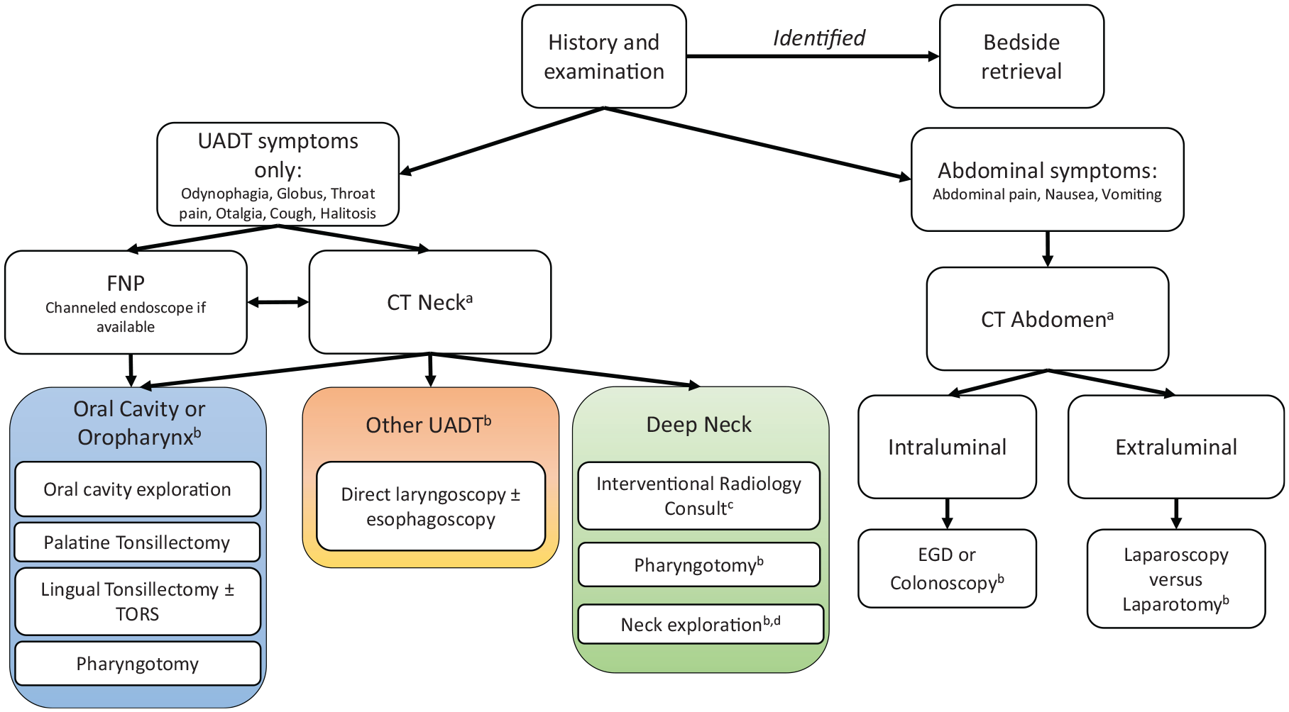
Proposed algorithm for management of bristle ingestions. aConsider additional imaging and consultations as clinically appropriate. bConsider intraoperative imaging. cif feasible. dTarget approach based on imaging. CT, computed tomography; EGD, esophagogastroduodenoscopy; FNP, flexible nasopharyngoscopy; TORS, transoral robotic surgery; UADT, upper aerodigestive tract.
Depending on the anatomic location where the bristle is lodged, a treatment plan can be developed. FNP can be performed at bedside for suspected UADT bristles without sedation and has minimal risk. FNP can be diagnostic and potentially therapeutic if a channeled scope is available53,56 or if the operator is able to use an instrument while operating the scope. 6 Channeled endoscopes have been described for removing fishbones in similar situations. 77 Given the low rate of success of transoral surgery (66.7%) and the potential for morbidity with open or transoral surgery, we feel that FNP should be attempted prior to more invasive modalities. If the bristle is clearly visualized but unable to be retrieved, the patient should be brought to the operating room for laryngoscopy, possibly in conjunction with esophagoscopy, as this also has minimal risk and morbidity and was successful in 68.2% (15/22) of attempts. Depending on the location of the bristle within the UADT, transoral surgery can be attempted. Of the transoral surgical procedures, lingual tonsillectomy and pharyngotomy had relatively high rates of success for medially located bristles (83.3% and 75%, respectively). Nevertheless, given transoral surgery’s overall lower likelihood of success, adjunctive modalities should be considered, as bristles are easily displaced during retrieval in the oral cavity.17,50
Intraoperative imaging can be considered with fluoroscopy or intraoperative CT.17,20,27,40,45,46,54,56 Fluoroscopy was the most frequently used intraoperative imaging modality (in 22.9% of UADT cases), but intraoperative CT may allow for more precise 3-dimensional localization, although in the 2 cases that intraoperative CT was employed, the bristles had migrated to the DN,43,50 but this was successful both times. Of bristles that presented in the UADT, 52.1% (25/48) were removed from the UADT without imaging, although 22 of these were endoscopically removed.31,34,41,48,53,55,56 The remaining 2 patients who underwent transoral surgery without interoperative imaging had robotic removal. 57 Robotic surgery can be a useful adjunct for bristles located in the lingual tonsil or vallecula due to excellent 3-dimensional visualization and bimanual dexterity. 57 Challenges of robotic surgery include the availability of both a surgical robot and surgeon with robotic surgical skills, as well as the robot being large and cumbersome to maneuver which makes intraoperative imaging difficult.
For bristles presenting within the DN or migrating to the DN,14,28,43,50 successful removal was achieved by transoral surgery via pharyngotomy,15,44 transcervical open surgery,2-4,7,16,19,21,22,28,54,56,60 or minimally invasive surgery.43,44,50 Minimally invasive surgery with intraoperative imaging should be considered an option given the potential for less morbidity.27,43,44,50 All minimally invasive reports were successful,27,43,44,50 but this may be confounded by reporting bias. If minimally invasive surgery is not an option, transoral surgery is an option, but this has a lower success rate of 66.7% overall. Most DN bristles in fact required transcervical open surgery, which had a 92.9% success rate. Intraoperative imaging with fluoroscopy or intraoperative CT should be considered for open surgery as well, as successfully used in the 3 cases for which it was employed.3,16,28
If a bristle is identified intraluminally within the abdomen, then a consultation should be conducted for EGD ± colonoscopy, with surgery as a contingency. Although EGD ± colonoscopy was successful in 42.3% of such attempts, the potential benefit of the procedure outweighs the minimal risk. Unsuccessful intra- or extraluminal bristles can be removed endoscopically by laparoscopy or a laparotomy. Laparoscopy had an 80% success rate with 2 failures that required laparotomy,8,59 while laparotomy had a 100% success rate.
Limitations
This systematic review includes case reports and low-quality case series, hampering the quality and quantity of data available for analysis. Given the relative rarity of the diagnosis, higher-quality studies are unlikely to be obtained, however. In addition, due to the possibility of publication bias toward complex cases, this systematic review could overestimate the complications associated with wire bristles.
Conclusion
Wire grill brush bristles are hazardous foreign bodies when ingested that can readily penetrate tissues and cause various injuries. Patients with suspected wire bristle ingestions should undergo early CT imaging for precise localization and subsequently have their removal approach be guided by specific location. Proximal locations in the UADT had a lower success rate at initial retrieval, and providers should consider the use of adjunctive modalities, such as intraoperative imaging, possibly with percutaneous approaches to optimize their chance.
Supplemental Material
sj-docx-1-oto-10.1177_01945998211062156 – Supplemental material for Hazards and Management of Wire Bristle ingestions: A Systematic Review
Supplemental material, sj-docx-1-oto-10.1177_01945998211062156 for Hazards and Management of Wire Bristle ingestions: A Systematic Review by Nathaniel Miller, Michael Noller, Matthew Leon, Yonatan Moreh, Nora L. Watson, Justin Costello and Steven Hong in Otolaryngology–Head and Neck Surgery
Footnotes
The views expressed in this manuscript are those of the authors and do not reflect the official policy or position of the Department of the Army, Department of Defense, or the US government.
Author Contributions
![]() ;
;
Disclosures
Supplemental Material
Additional supporting information is available in the online version of the article.
References
Supplementary Material
Please find the following supplemental material available below.
For Open Access articles published under a Creative Commons License, all supplemental material carries the same license as the article it is associated with.
For non-Open Access articles published, all supplemental material carries a non-exclusive license, and permission requests for re-use of supplemental material or any part of supplemental material shall be sent directly to the copyright owner as specified in the copyright notice associated with the article.