
Abstract
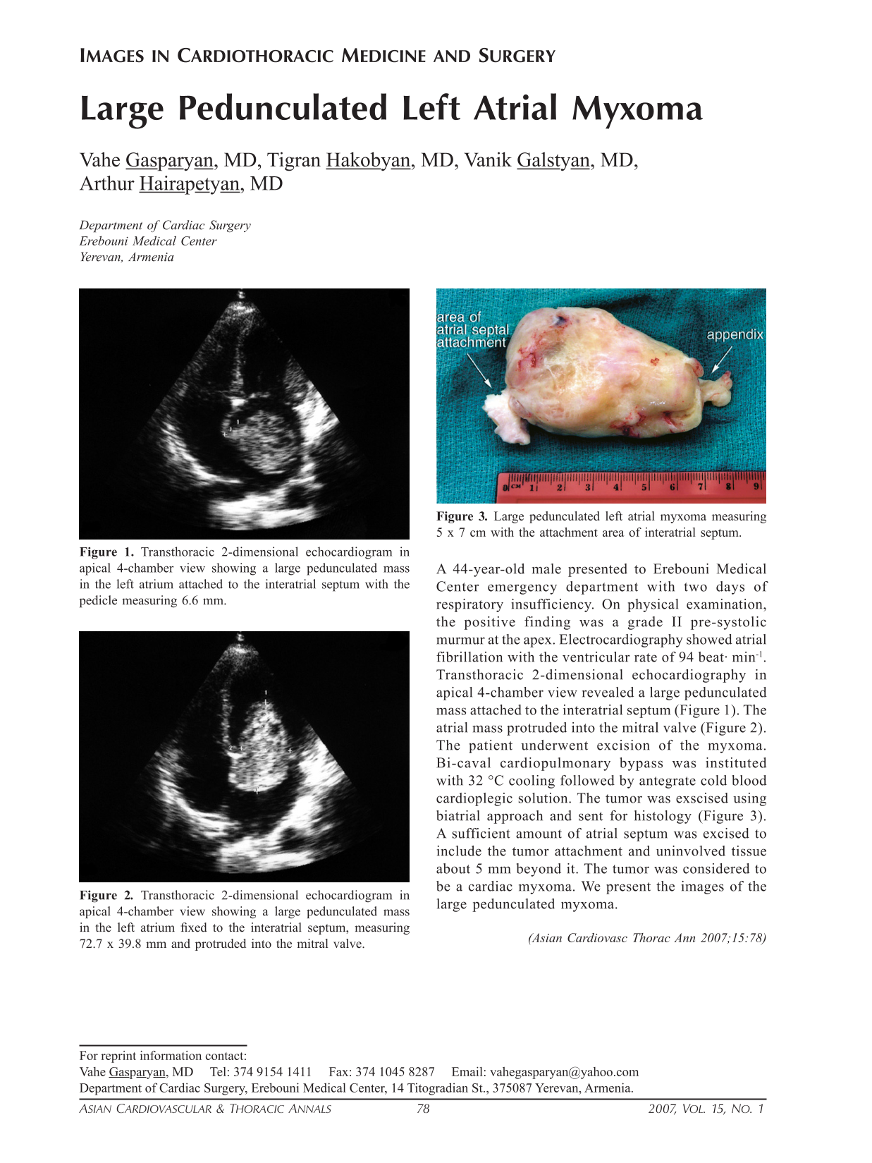
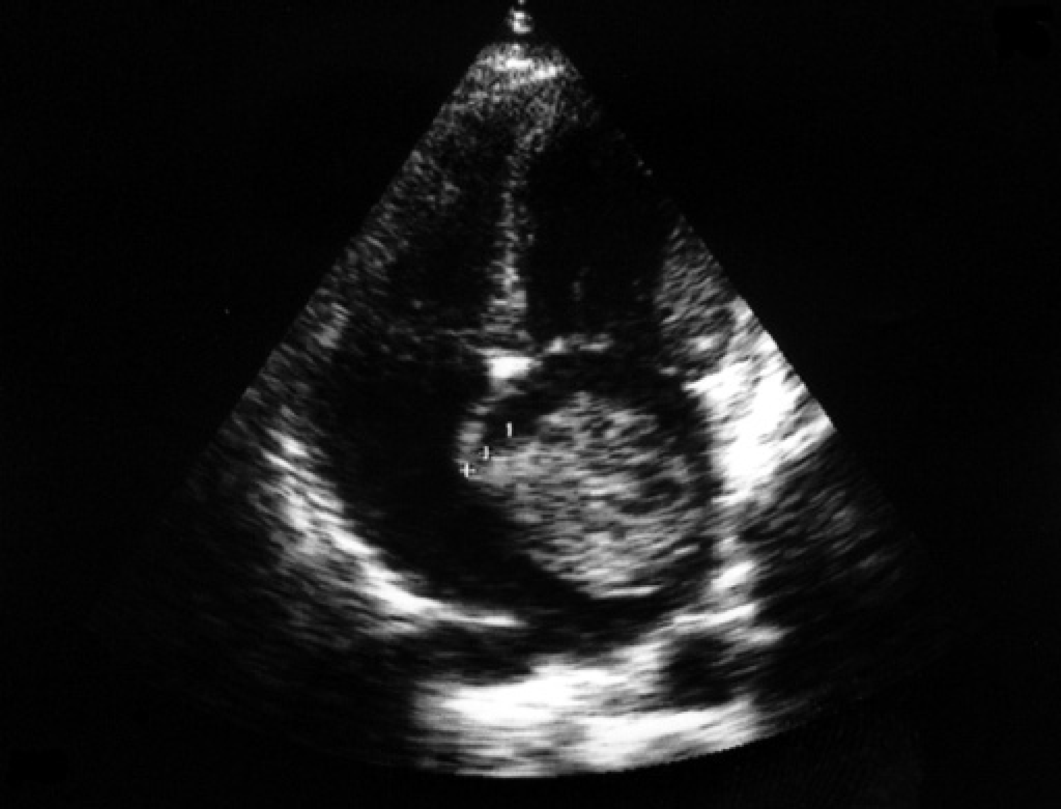
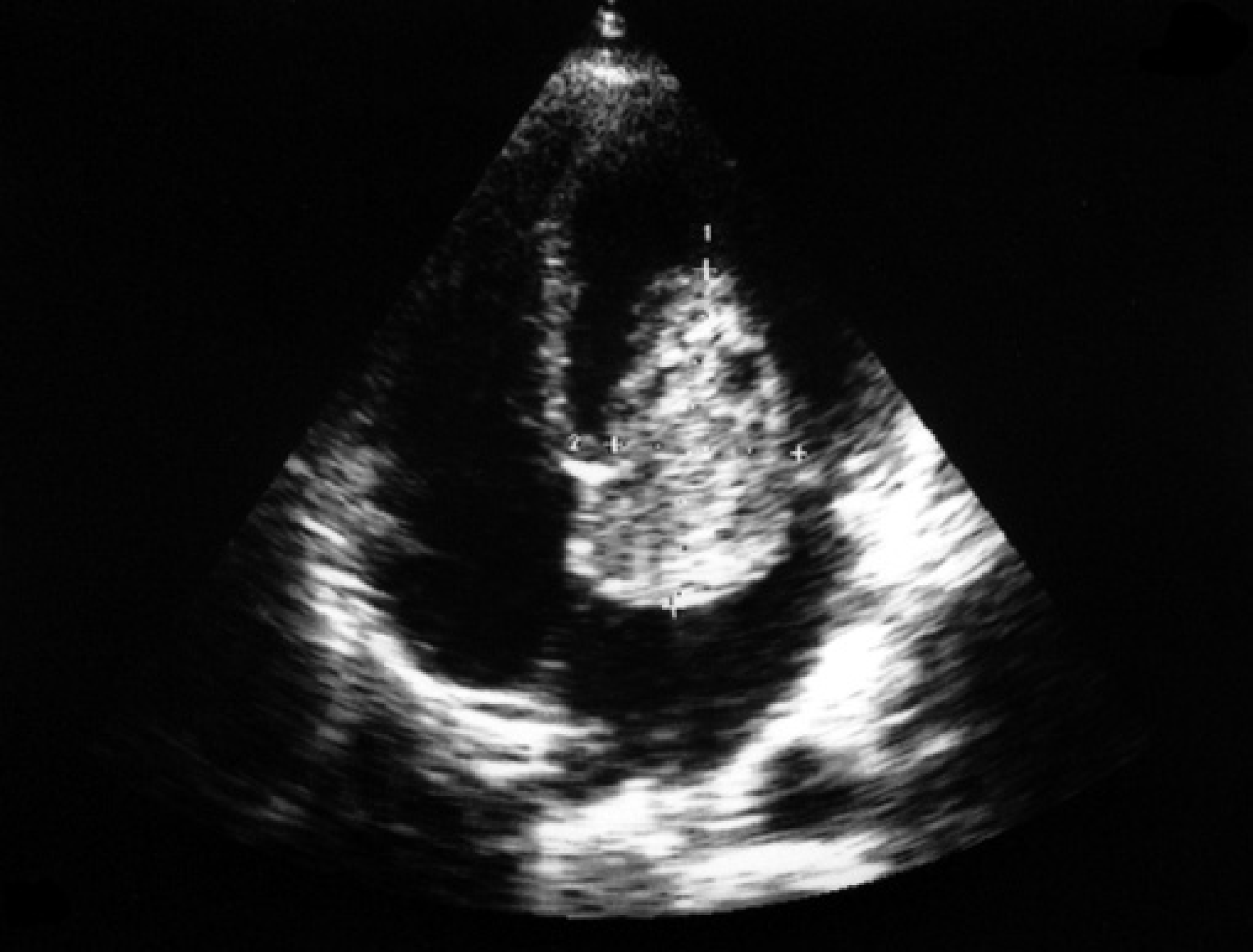
A 44-year-old male presented to Erebouni Medical Center emergency department with two days of respiratory insufficiency. On physical examination, the positive finding was a grade II pre-systolic murmur at the apex. Electrocardiography showed atrial fibrillation with the ventricular rate of 94 beat· min−1. Transthoracic 2-dimensional echocardiography in apical 4-chamber view revealed a large pedunculated mass attached to the interatrial septum (Figure 1). The atrial mass protruded into the mitral valve (Figure 2). The patient underwent excision of the myxoma. Bi-caval cardiopulmonary bypass was instituted with 32 °C cooling followed by antegrate cold blood cardioplegic solution. The tumor was exscised using biatrial approach and sent for histology (Figure 3). A sufficient amount of atrial septum was excised to include the tumor attachment and uninvolved tissue about 5 mm beyond it. The tumor was considered to be a cardiac myxoma. We present the images of the large pedunculated myxoma.
Transthoracic 2-dimensional echocardiogram in apical 4-chamber view showing a large pedunculated mass in the left atrium attached to the interatrial septum with the pedicle measuring 6.6 mm.
Transthoracic 2-dimensional echocardiogram in apical 4-chamber view showing a large pedunculated mass in the left atrium fixed to the interatrial septum, measuring 72.7 × 39.8 mm and protruded into the mitral valve.
Large pedunculated left atrial myxoma measuring 5 × 7 cm with the attachment area of interatrial septum.