
Abstract
Radiofrequency ablation of pulmonary vein ostia does not provide complete and long-term elimination of atrial fibrillation. Combining this procedure with local radiofrequency application on sites with strong vagal reflexes results in partial parasympathetic denervation and increases the antiarrhythmic effect. A novel catheter-ablation technique to modify ganglionated plexi in the left atrium was assessed in 58 patients (mean age, 52.1 ± 1.9 years, 67% male) with drug-refractory atrial fibrillation, which was chronic in 21 (36%; mean duration, 14.3 ± 2.9 months; range, 5–39 months). The mean left atrial volume was 93.1 ± 6.1 mL. The patients underwent ablation of 4 areas of ganglionated plexi in the left atrium, with no circumferential ablation of the pulmonary veins; atrial fibrillation ceased immediately in 94.1% of them. Transient vagal bradycardia was seen in 93% of patients. For 7.2 ± 0.4 months after the procedure, 86.2% of them were free from arrhythmias, and no antiarrhythmic drugs were administered. Ganglionated plexi ablation is an efficient treatment for atrial fibrillation.
Introduction
Atrial fibrillation (AF) is the most common type of arrhythmia and a serious problem with risks of stroke, thromboembolic complications and heart failure. Pulmonary vein (PV) radiofrequency (RF) ablation is currently the standard intervention for AF in patients without cardiac pathology. 1 Nevertheless, its efficacy is suboptimal. 2 Studies have shown the role of sympathetic-vagal misbalance as an inducer of AF. 3 –5 Pappone and colleagues 6 identified the sites of nerve fibers using vagal reflexes during RF ablation, and treated these areas in addition to PV ablation, giving a better overall outcome. They proposed a maximum extension of vagal denervation that would abolish all vagal reflexes in the left atrium, which is critical in achieving the best possible result. However, the use of ganglionated plexi ablation alone resulted in a high long-term rate of AF relapse, due to regeneration of nerve fibers. 7,8 High-frequency stimulation with registration of vagal reaction was used to define ganglionated plexi concentrations in these studies. We used an alternative approach of extended RF ablation based on anatomical data of ganglionated plexi development in atrial myocardium.
Patients and Methods
The study included 58 patients with AF who underwent catheter ablation by the proposed method from December 2005. Their ages ranged from 41 to 67 years (mean age, 52.1 ± 1.9 years), and 39 (67%) were men. Concomitant cardiac pathology was excluded, based on echocardiography and angiography. Serum thyroid hormones were measured to exclude hypothyroidism. Arterial hypertension was documented in 11 (19%) patients. The paroxysmal type of AF was recorded in 15 patients (26%; paroxysm frequency, 9.2 ± 1.1 per year), the persistent type in 22 (38%; mean episode duration, 21.3 ± 5.7 days), and the chronic type in 21 (36%; mean duration, 14.3 ± 2.9 months; range, 5–39 months). The overall mean duration of arrhythmia was 6.9 ± 0.7 years (range, 1–14 years). Four (7%) patients with chronic AF developed acute cerebral ischemic attacks, one had an ischemic stroke 2 years earlier. Clinical symptoms of heart failure were observed in 43 (74%) patients, 27 (47%) were in New York Heart Association functional class I, 13 (22%) in class II, and 3 (5%) in class III. Left atrial enlargement (56.9 ± 3.9 mm) was diagnosed in 41 (72%) patients. The left atrial volume (measured by computed tomography) was 53–141 mL (mean, 93.1 ± 6.1 mL), and it exceeded 100 mL in 19 (33%) cases. Preventive antiarrhythmic therapy (class I or III drugs) was ineffective in all patients. They were on oral anticoagulation at least 1 month before ablation therapy, and transthoracic and transesophageal echocardiography was performed to exclude left atrial thrombi. The study was approved by the hospital ethics committee, and written informed consent was obtained from all subjects.
Three-dimensional maps were constructed for the left and right atria and the coronary sinus using a Carto XP electroanatomic mapping system (Biosense Webster, Diamond Bar, CA, USA). After transseptal catheterization, systemic anticoagulation was achieved with intravenous heparin to maintain an activated clotting time of 250–350 sec. Bipolar electrograms were recorded between a bandpass filter of 30–500 Hz, using a GE Prucka CardioLab (GE Medical Systems, Milwaukee, WI, USA) recording system. Surface electrocardiograms from leads I, II, and V1 were recorded continually throughout the study. During the procedure, no selective search of ganglionated plexi concentration was performed, and high-frequency stimulation was not applied. The ganglionated plexi locations were defined on the basis of anatomical data. Armour and colleagues 9 described the sites of all important ganglionated plexi in the human heart. To standardize the procedure of catheter ablation, 4 areas were defined (as projected from inside the left atrium) for RF energy delivery (Figure 1): area no. 1 (left superior) was adjacent to the left superior PV ostium at 8–1 o'clock on the vein ostium circumference, area no. 2 (left inferior) was adjacent to the left inferior PV ostium at 5–10 o'clock, area no. 3 (right superior) was adjacent to the right superior PV ostium at 7–1 o'clock, area no. 4 (right inferior) was adjacent to the right inferior PV ostium at 2–8 o'clock. The sequence of treatment was chosen randomly. In most cases (83%), the numbered areas were treated in ascending order. Radiofrequency ablation was performed with an irrigated catheter (3.5-mm electrode tip, 2 bipolar electrode pairs, with a distance of 2 mm between electrodes; (NaviStar Termocool, Biosense Webster, Diamond Bar, CA, USA) with power settings of 35 to 40 W, an irrigation rate of 17 mL·min−1, and application duration of 45–60 sec. The endpoint of the procedure was complete elimination of the atrial potential in the numbered areas, with registration of an isoelectric line and absence of vagal reflexes during the final RF applications. Pulmonary vein electrical activity was not investigated, and its disappearance was not the aim of the intervention. The cavotricuspid isthmus was ablated in 28 (48%) patients; the indications were: history of documented independent paroxysms of typical atrial flutter in 5, episodes of large-wave fibrillation with a surface electrocardiogram identical to typical atrial flutter in 5, transformation of AF into typical atrial flutter during RF ablation in 3, and induction of typical atrial flutter following RF ablation in the left atrium in 15. Warfarin was prescribed for all patients for 1 month after RF ablation, to prevent thromboembolic complications. Those who had a recurrence of AF within 4 weeks were treated with a class I or III antiarrhythmic drug for 4–6 weeks.
Schematic representation of ganglionated plexi ablation; the endocardial surface of the left atrium is shown (from inside, after removal of anterior wall); nos. 1–4 indicate areas of radiofrequency ablation.
Patients were examined at 1, 3 and 6 months after the procedure with 24-hour Holter monitoring and echocardiography. The primary endpoint of the study was freedom from atrial tachyarrhythmias (> 30-sec duration), including AF and atypical atrial flutter. Because early recurrences of atrial tachyarrhythmias within the first month after ablation may be a transient phenomenon, this time interval was excluded from analysis. Heart rate variability as an indicator of autonomic nervous system activity was evaluated before and after RF ablation (9.1 ± 2.1 days) in 17 patients with paroxysmal AF given no antiarrhythmic drugs during the study period.
Statistical analysis of 24-hour monitoring data was performed using the standard deviation of normal beat-to-normal-beat intervals (SDNN) over the whole period analyzed, and the root mean squared of successive differences (rMSSD) between adjacent RR interval pairs. Spectral analysis of short 5-min portions of the electrocardiogram was also carried out. The absolute values (msec) of the power of the low and high frequencies were investigated as well as their relative characteristics. The balance of vegetative influences was assessed by the spectral low to high-frequency ratio (LF/HF). The mean sinus rate and time-domain (standard deviation of RR intervals and root mean square of differences of adjacent RR intervals), and the frequency domain (low and high frequency, and LF/HF analyses of heart rate variability were obtained by 24-hour Holter monitoring before and after ablation.
All data are given as mean ± standard deviation. Student's t test was used to estimate the significance of differences between means of normally distributed variables, with p = 0.05 (significance for medians, 95%). The chi-squared test was employed for discrete variables. To assess statistical differences of changes in basic intra and intergroup characteristics, the nonparametric Wilcoxon-Mann-Whitney U test was used.
Results
During ablation, 75–135 RF applications were made (21.9 ± 3.1 in each of the 4 areas; Figure 2). There were no complications. The fluoroscopy time was 5–28 min (mean, 9.1 ± 1.3 min), and the total procedure duration was 110–195 min (mean, 135.4 ± 12.1 min). Changes in AF dynamics during ablation were analyzed. At the time of the procedure, AF was observed in 34 (58.6%) patients; it was preexisting in 28, and developed at catheter introduction or the first RF application in 6. In 32 of these 34 cases, AF terminated during ablation; sinus rhythm was restored in the other 2 by transthoracic electrical cardioversion after the procedure. Before sinus rhythm had stabilized, 14 patients developed 1–4 short episodes (several seconds) of sinus rhythm restoration between AF recurrences (“stop and restart”; Figure 3À). Atrial fibrillation initiation (Figure 3B) at the time of ablation was recorded in 6 patients when the initial 2 areas were treated. Atrial fibrillation ceased in the final stages of RF application, regardless of the sequence of ablation of vegetative ganglia. We believe this chronological sequence of events indicates that the AF sustaining mechanisms were consecutively eliminated as ganglionated plexi were “turned off”. Pacing performed after completion of the ablation procedure did not induce any sustained paroxysm of AF. Unsustainable paroxysms that faded away spontaneously in 3–120 sec (mean, 11.9 ± 5.1 sec) were observed in 8 (13.8%) isoproterenol-free subjects, and later in 11 (19%) who had isoproterenol. Thus, even aggressive pacing coupled with pharmacological agents did not induce even short-term AF in 47 (81%) patients.
3D-imaging of the left atrium during ganglionated plexi ablation in the posterior and anterior projections in 4 patients; (A) Isolated ganglionated plexi ablation, (B) Ablation combined with CTI block; pink markers denote pulmonary vein ostia, red markers show radiofrequency applications. CTI = cavotricuspid isthmus.
Rhythm transformation effects during ganglionated plexi ablation; (À) “Stop and restart”, (B) Initiation of atrial fibrillation. Speed = 25 mm·sec−1.
Chronotropic reactions (heart rate decrease of 8–15 beats per minute) were registered almost throughout 67.3% ± 9.2% of all RF applications. In addition, 252 episodes of pronounced bradyarrhythmia were documented in 93% of patients; 1–12 (mean, 4.3 ± 0.9) per procedure. These periods (Figure 4) presented as: sinus bradycardia with heart rate < 40 beats per minute (68 episodes; 27%); sinus node arrest with pauses > 2 sec (43 episodes; 17.1%); atrioventricular block grade I, II, or III (29 episodes; 11.5%); decreasing heart rate or pause for > 3 sec in patients with AF (112 episodes; 44.4%). These events were transient and faded spontaneously after the manipulation was terminated. Nevertheless, a progressively decreasing heart rate (or even asystole) required short-term electrical pacing in 13 (5.1%) cases. In 7 patients, ablation was performed with intravenous isoproterenol infusion to prevent further episodes of critical bradycardia.
Intraoperative vagal bradycardias; (À) Sinus bradycardia (32 beats per minute) + complete atrioventricular block, asystole for 5.4 sec (speed, 10 mm·sec−1); (B) Asystolic pause for 8.8 sec during atrial fibrillation (speed, 10 mm·sec−1); (C) Sinus rhythm reinstatement through asystole for 4.9 sec (speed, 25 mm·sec−1).
The patients were followed up for 6–8 months (mean, 7.2 ± 0.4 months). All were in sinus rhythm during that period. Six (10%) patients are currently taking antiarrhythmic drugs. During follow-up, 14 paroxysms were documented in 8 (14%) patients; 10 stopped spontaneously and 4 required pharmacological cardioversion. Repeat ablation was performed in 3 patients at 7, 7 and 9 days after the initial procedure. Ablation of the cavotricuspid isthmus for typical atrial flutter was undertaken in 2 patients, and segmental isolation of the right inferior PV ostium for frequent blocked extrasystoles originating from the vein's sleeve was carried out in one. Analysis of post-ablation parameters (Table 1) showed lower values of heart rate variability: SDNN was reduced by 30.2% (p < 0.001), and rMSSD decreased by 47.4% (p < 0.001). Spectral analysis showed that activity of the low-frequency spectrum increased by 16.7% (p < 0.01; sympathetic tone prevailed) while high-frequency spectrum activity decreased by 15.1% (p < 0.001; vagal block was observed). As a result of these effects, LF/HF rose by 37.2% due to a prevailing sympathetic nervous system. Ganglionated plexi ablation therefore weakened the heart's parasympathetic innervation and enhanced sympathetic innervation. Comparison of heart rate variability criteria in patients free from AF and those with AF recurrence after ablation revealed significant differences. Patients with recurrent AF had higher SDNN values (97.5 ± 22.3 vs 79.3 ± 27.8 msec; p < 0.001), higher rMSSD values (30.8 ± 6.9 vs 20.7 ± 7.1 msec; p < 0.001), and lower LF/HF (1.29 ± 0.08 vs 1.53 ± 0.07; p < 0.001), indicating less pronounced parasympathetic denervation compared to patients free from AF as a result of effective RF ablation.
Dynamics of Heart Rate and Heart Rate Variability before and after Plexi Ablation
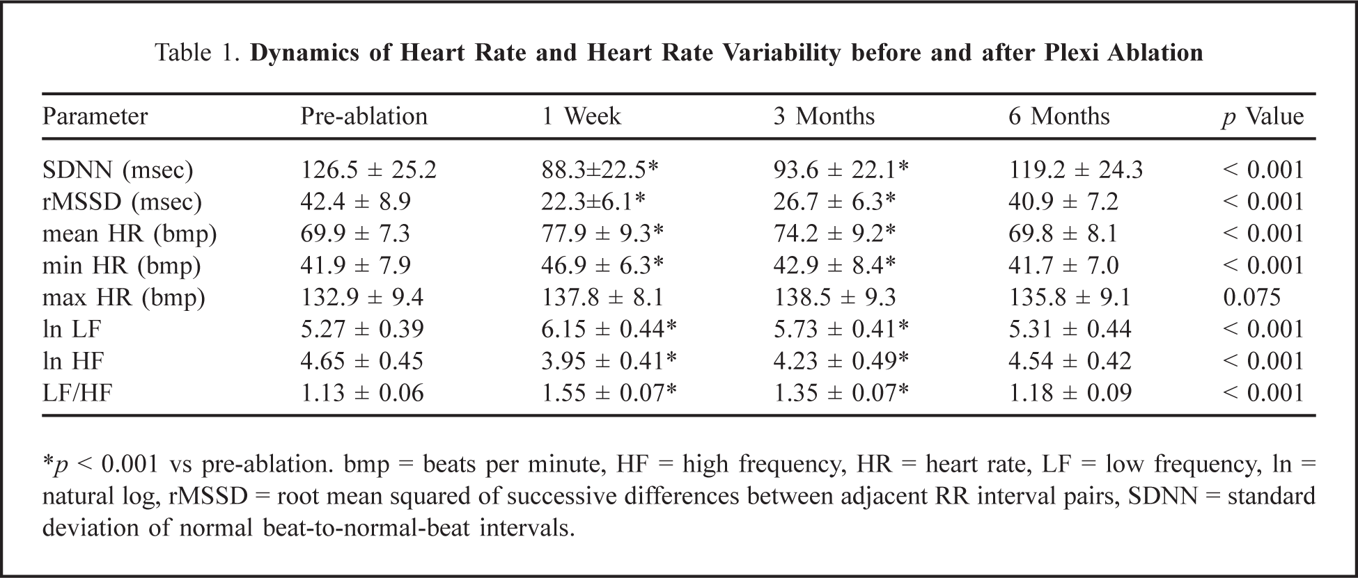
p < 0.001 vs pre-ablation. bmp = beats per minute, HF = high frequency, HR = heart rate, LF = low frequency, ln = natural log, rMSSD = root mean squared of successive differences between adjacent RR interval pairs, SDNN = standard deviation of normal beat-to-normal-beat intervals.
Discussion
This study showed that isolated ablation, aimed at left atrial ganglionated plexi only, is as effective as PV isolation. Thus anatomical substrates of the autonomic nervous system play the main role in AF development. As more than a third of our patients were in chronic AF before treatment, the fact that 86.2% were free from AF as a result of this approach is very encouraging. These ganglionated plexi have well-studied and well-described anatomy. 9,10 Tan and colleagues 11 demonstrated that nervous tissue has maximum density 5–10 mm from atriovenous anastomosis, the central site where RF energy is applied. Normally, sites of ganglionated plexi have no electrophysiologic voltage markers, so they were approached empirically, based on anatomical data. Advantages of this approach include no need for activation mapping or any preliminary electrophysiological study, no obstacles to sinus excitation spreading in the left atrium, no ablation lines on the posterior wall of the left atrium (avoiding esophageal damage), no risk of PV stenosis and no arrhythmogenic effects (no left atrial tachycardia). Thus, we have developed a safe method with a complication risk comparable to that of Lasso-technology, and clinical effects surpassing those of circumferential isolation of PV ostia.
All patients maintained the same PV electrical activity as before ablation. Thus the ablation effect can only be explained by vegetative denervation of the heart, which was confirmed by strong intraoperative vagal reflexes and denervation criteria after ablation. As the distribution of vegetative ganglia conforms to the principle of individual variability, extended ablation was used in each area. This could be avoided if we had in-vivo visualization and exact localization of ganglionated plexi in each patient. Schauerte and colleagues 3 recently demonstrated that catheter RF modification of plexi adjoining the right pulmonary artery and caval veins in dogs prevents induction of AF mediated through the vagus. These findings provided prerequisites for our study. Recent experiments at the University of Oklahoma Health Sciences Center indicate that irritation of plexi at the base of PV ostia leads to critical bradycardia and triggers AF from the plexus area (effects analogous to those intraoperatively). 4,12,13 Cholinomimetics (carbocholine), 20-Hz stimulation pulses, and multiple extrastimuli during the refractory period of the atrial myocardium were used as specific plexi activators to ensure selective excitation of nervous tissue. 5,12 “Neutralization” of plexi structures by local anesthesia or RF ablation prevented these effects. 5,13 This group was the first to apply experimental approaches in clinical practice, restoring sinus rhythm in 84% of patients by targeting plexi in the left atrial region. Thereafter, they showed that plexi ablation improved the outcome of concomitant isolation of PV ostia by 28%. Prior to their studies, targeted RF ablation of ganglionated plexi was used only to treat syncope caused by parasympathetic influences. 14 Later studies found lower efficacy of ganglionated plexi ablation. 7,8 Scanavacca and colleagues 8 reported 4 of 7 treated patients needed secondary classical isolation of PV ostia. The substantially higher efficacy of ganglionated plexi ablation in our patients may be due to our novel approach.
Hsieh and colleagues 15 first used vegetative profile changes of heart rate and demonstrated an effect of parasympathetic denervation in RF ablation, defining it as “autonomic dysfunction”. A similar effect was reported using focal ablation, isolation of PV ostia, and linear applications in the left atrium or even the maze-III procedure. 6,16,17 Pappone and colleagues 6 showed that RF vegetative denervation based on detection of vagal reflexes decreased AF recurrence after PV isolation over 12-months. This study shifted emphasis towards vegetative denervation, which was no longer considered a complication and became the aim of ablation and an imperative for its success. Vagal bradycardias are known to result from thermal RF stimulation of parasympathetic fibers. 18 Vagal reflexes are seen in 5% of cases during ablation of supraventricular tachycardias in the septal region, and in no more than 34.3% of cases during ablation of the PV and posterior wall of the left atrium. 6,19 At the same time, the number and intensity of vagal reactions in our study significantly exceeded all published data, and comprised 93%, indicating that ablation was performed in areas of more highly concentrated nervous tissue compared to other techniques. It appears that the core of this technique is elimination of a critical amount of plexi sites. The chronological pattern of rhythm transformation supports this assumption; AF induction at the start of ablation, short-term termination followed by a restart in the middle of the procedure, and sinus rhythm restoration at the end. This means that vagal reflexes triggered by manipulations in the first 2 areas indicate how accurately the autonomic nervous system was targeted, while the final terminating effect shows how deep the impact was. These findings do not exclude reinnervation during follow-up, and reports show that autonomic system misbalance is restored within 6 months after catheter ablation, thus domination of the sympathetic link of the autonomic nervous system decreases. 6 This is probably the reason for AF relapses after selective ganglionated plexi ablation. 8 After anatomical plexi ablation, we also observed the restoration of autonomic tonus by 6 months; however, this did not influence the efficacy of the procedure. This might be explained by the fact that anatomical plexi ablation stimulates the growth of new healthy tissue rather than restoration of existing sick nervous tissue, because of the degree of damage.
This study opens new perspectives in the treatment of AF but poses a number of questions, such as, who are the best candidates for this procedure? A number of studies confirmed that patients with AF and no structural heart changes have no alterations of vagal activity before they develop AF, which might be a limitation of this approach. 20 It should be remembered that the baseline autonomic misbalance has its individual specifics and varying spectral pattern, therefore, not every AF patient can benefit from targeting the peripheral parasympathetic nervous system. Another question is the duration of the effect of plexi ablation. Our follow-up was 7.2 ± 0.4 months, so long-term effects are unknown. Three studies of heart rate variability after PV RF ablation reported signs of denervation persisting for only 3–6 months, which provoked AF recurrence in some patients. 6,16,17 Longer follow-up is important to determine the long-term safety and efficacy of this ablation strategy. Finally, during follow-up, AF recurrence was limited by patient symptoms and ambulatory recordings, and we cannot exclude asymptomatic episodes of AF after the procedure. Nevertheless, it was concluded that RF catheter ablation of atrial sites of all important ganglionated plexi may prevent AF recurrence in patients with paroxysmal and chronic AF. Our findings should stimulate new studies aimed at defining the role of mapping and ablation of ganglionated plexi.