
Abstract
A 2-year-old boy with cyanosis was found to have normal situs and looping with anomalous drainage of a right-sided superior vena cava to the left atrium, and intact interatrial septum in association with anomalous drainage of the left pulmonary veins to the right superior vena cava. He underwent successful surgical repair of this rare congenital malformation.
Introduction
Anomalies of systemic venous return are extremely heterogeneous congenital malformations with characteristics ranging from completely normal physiology to severe forms of right-to-left shunting requiring surgical treatment. Anomalous drainage of a right-sided superior vena cava (SVC) to the left atrium (LA) is one of the rarest variants of systemic venous return anomalies, characterized by right-to-left shunt physiology and cyanosis. So far, less than 20 such cases have been reported. 1 –4 This anomaly is usually diagnosed early in life, but it is occasionally detected in older patients with cyanosis, dyspnea, cardiomegaly, or cerebral embolus or abscess.
Case Report
A 2-year-old boy, otherwise asymptomatic, had been noted to have cyanosis at 4 months of age. His growth and milestones of development were normal. There was no history of recurrent respiratory infections or features suggestive of heart failure. On physical examination, he was found to be cyanotic with finger clubbing, and his resting arterial oxygen saturation was 80%. His pulse, respiratory rate, and blood pressure were normal. A cardiovascular system examination revealed normal heart sounds and no murmur. Chest radiography showed a normal cardiothoracic ratio and normal vascular pattern. An electrocardiogram was within normal limits for age. Echocardiography indicated normal situs and looping with isolated drainage of a right-sided SVC to the LA. The interatrial septum was intact, and the drainage of the inferior vena cava and coronary sinus were normal.
An agitated-saline contrast echocardiogram demonstrated drainage of a right-sided SVC to the LA. Cardiac magnetic resonance imaging confirmed drainage of a right SVC to the LA and absence of a defect in the interatrial septum (Figure 1). Cardiac catheterization showed systemic oxygen saturation of 82% and a stepdown of saturation in the LA. A pulmonary angiogram in levo phase revealed abnormal drainage of the left pulmonary veins to the SVC (Figure 2). A right internal jugular venogram ascertained drainage of a right SVC to the LA. Surgical correction was performed under standard cardiopulmonary bypass using bicaval and aortic cannulation. The preoperative findings were confirmed at operation, and the pulmonary veins on the left side joined to form a common pulmonary vein that drained into the right SVC just above the cavoatrial junction. Myocardial protection was achieved with cold blood cardioplegia administered antegradely after aortic cross clamping under moderate hypothermia. Via a right atriotomy, the upper part of the interatrial septum and septum secundum were excised. A glutaraldehyde-treated autologous pericardial patch was used to reroute the right SVC into the right atrium (RA) through an atrial septal defect, and the left pulmonary veins to the LA by suturing the patch to the right SVC wall just above the entry point of the left pulmonary veins, which was brought down along the adjacent left atrial wall to the lower margin of the fossa ovalis. This ensured free drainage of the right SVC into the RA while diverting left pulmonary venous blood to the LA. The right atriotomy was closed, the aortic cross clamp was removed, and de-airing maneuvers were carried out. The child was weaned off cardiopulmonary bypass, and the sternum was closed. He was extubated after 6 hours of mechanical ventilation, and discharged home on the 7th postoperative day. Cardiac catheterization and angiography before discharge showed normal right SVC flow to the RA (Figure 3).
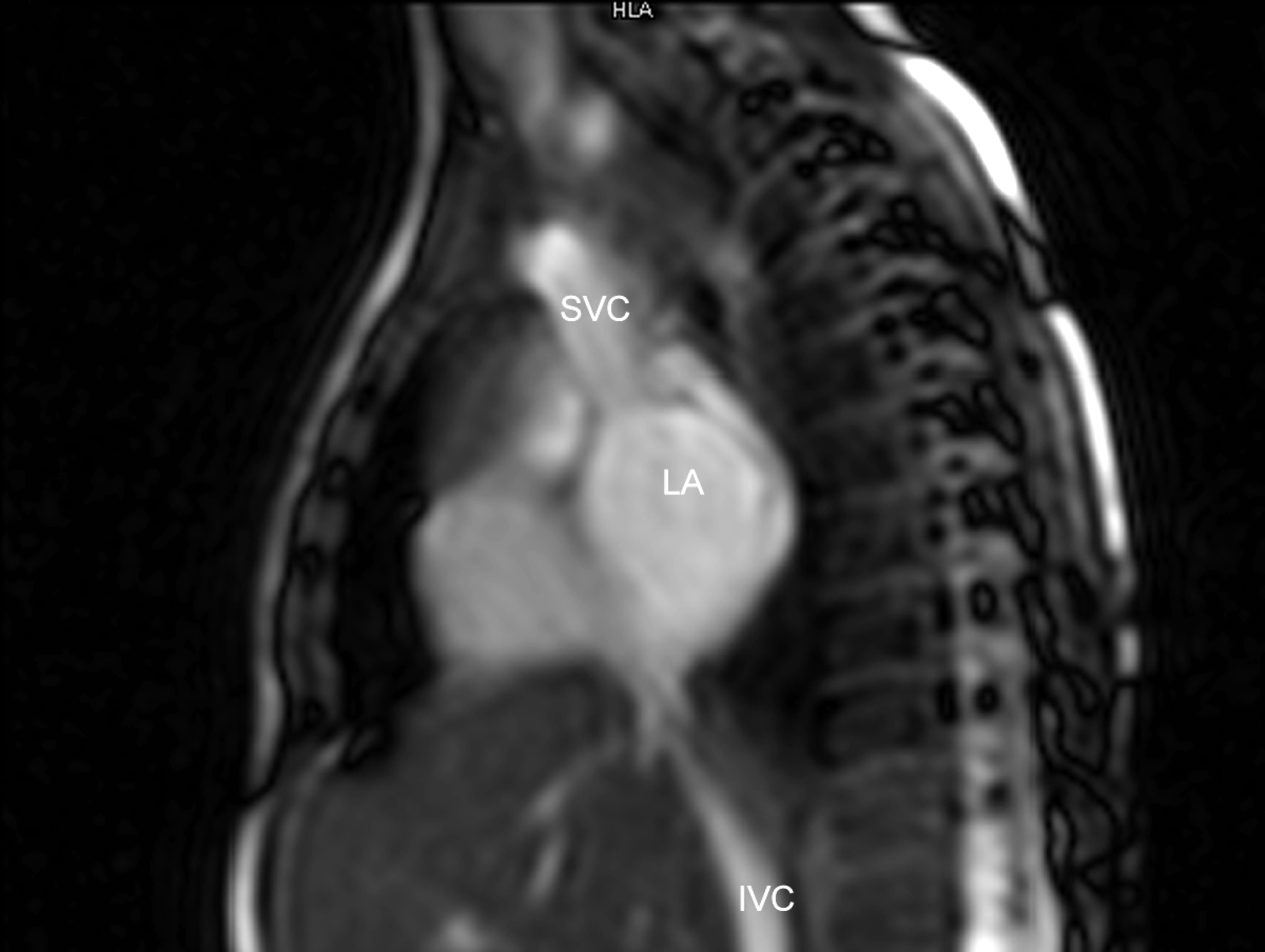
Magnetic resonance image of sagittal slice of the thorax, showing drainage of the right superior vena cava (SVC) to the left atrium (LA). IVC = inferior vena cava.
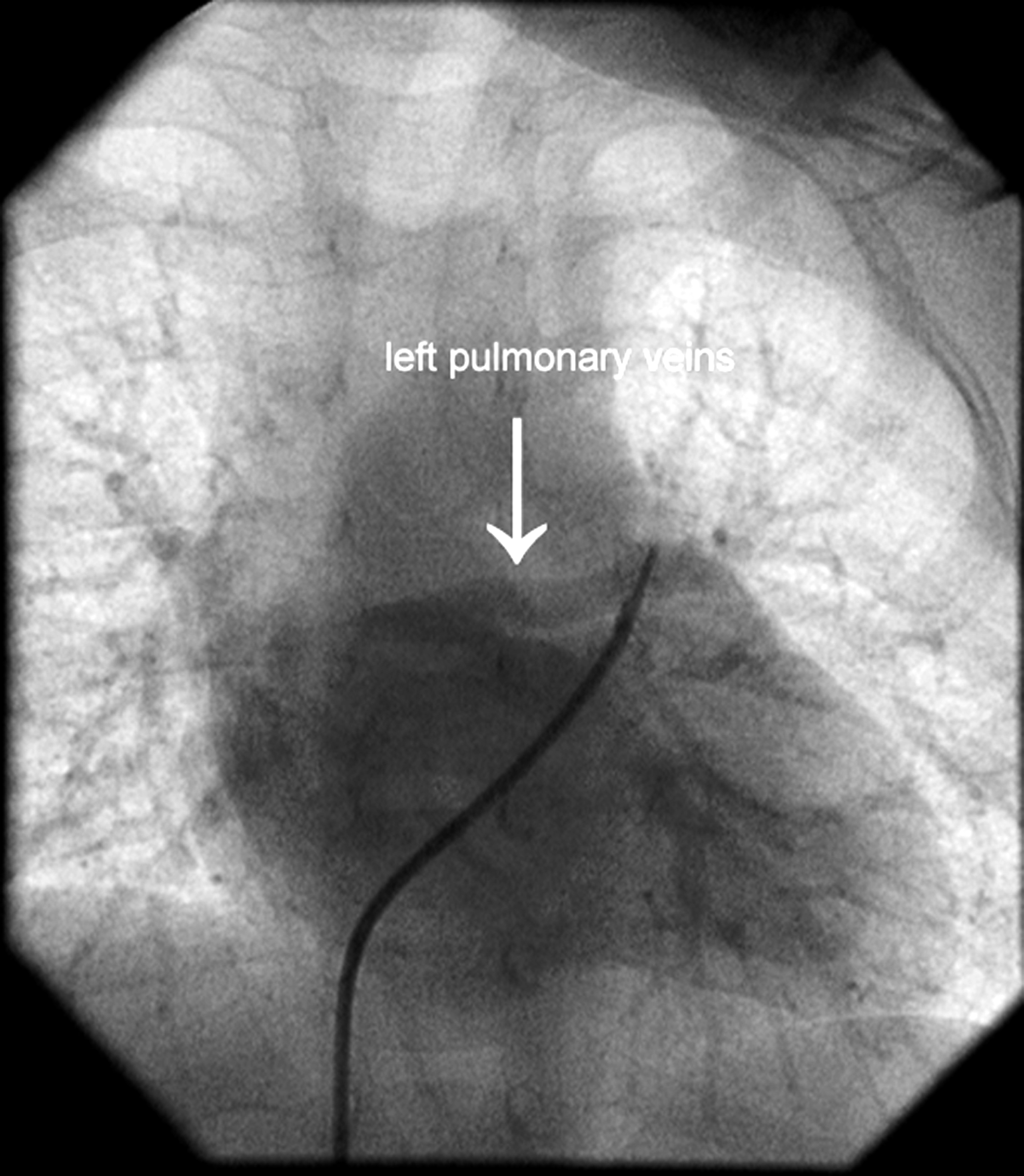
Pulmonary angiography in levo phase, demonstrating anomalous drainage of the left pulmonary veins to the right superior vena cava.
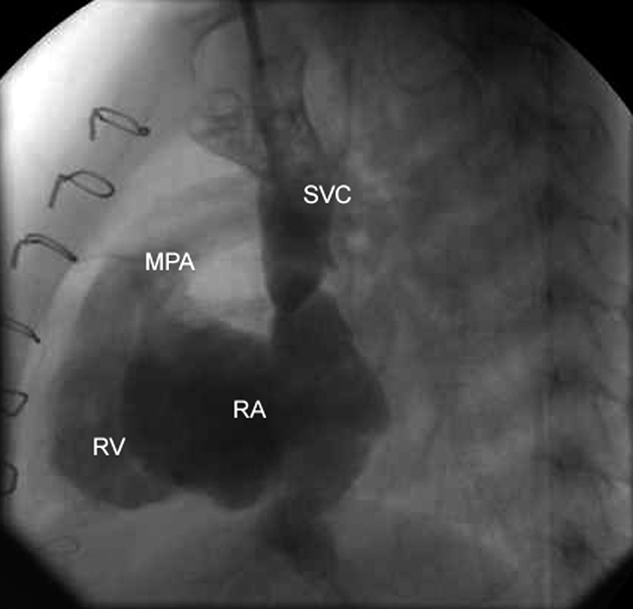
Postoperative internal jugular vein angiogram showing unobstructed drainage of the right superior vena cava (SVC) to the right atrium (RA). MPA = main pulmonary artery, RV = right ventricle.
Discussion
Anomalous drainage of a right SVC to the LA is a cyanotic congenital cardiac malformation that occurs mostly in association with partial anomalous drainage of the right pulmonary veins to the right SVC, with or without a sinus venosus defect. Our case was rare in that this anomaly was associated with anomalous drainage of the left pulmonary veins to the right SVC with an intact interatrial septum. The embryological basis for this anomaly is not well understood. However, it has been postulated that malposition of the right horn of the sinus venosus in a left and cephalad direction results in this anomaly. 5 Van Praagh and colleagues 2 attributed this anomaly to a condition similar to sinus venosus atrial septal defect in association with atresia of the SVC orifice. The pressure difference between the LA and RA determines preferential flow, and if the SVC flow to the LA predominates during fetal life, the proximal segment of the SVC involutes, with atresia or stenosis of the caval orifice to the RA. Echocardiography using agitated normal saline as bubble contrast gives important information to diagnose this anomaly. 3 Magnetic resonance imaging of the heart and/or a jugular venous angiogram are very useful prior to surgical correction.
Several techniques have been used for surgical correction of this anomaly. The first technique employed was clamping of the SVC and anastomosis to the RA. 1 Repair was subsequently accomplished utilizing cardiopulmonary bypass to baffle the SVC to the RA. Kothari and colleagues 4 used a bidirectional Glenn shunt to repair this anomaly associated hypoplastic right ventricle. Although anomalous drainage of the left pulmonary veins to the RA has been reported, its association in a patient with anomalous drainage of a right SVC to the LA has not been described. 6 An appropriate technique has to be chosen based on the anatomical features and associated intracardiac anomalies. Anomalous drainage of a right SVC to the LA should be kept in mind when dealing with a cyanotic child with an apparently normal cardiovascular system on physical examination. Surgical correction is indicated to prevent complications of cyanosis and the risk of systemic embolization.
Presented at the Breakfast Sessions of the 42 nd Annual Meeting of the Society of Thoracic Surgeons, Chicago, IL, USA, January 30–February 1, 2006.