
Abstract
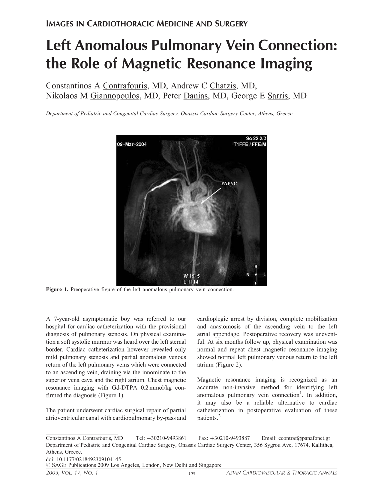
A 7-year-old asymptomatic boy was referred to our hospital for cardiac catheterization with the provisional diagnosis of pulmonary stenosis. On physical examination a soft systolic murmur was heard over the left sternal border. Cardiac catheterization however revealed only mild pulmonary stenosis and partial anomalous venous return of the left pulmonary veins which were connected to an ascending vein, draining via the innominate to the superior vena cava and the right atrium. Chest magnetic resonance imaging with Gd-DTPA 0.2 mmol/kg confirmed the diagnosis (Figure 1).
Preoperative figure of the left anomalous pulmonary vein connection.
The patient underwent cardiac surgical repair of partial atrioventricular canal with cardiopulmonary by-pass and cardioplegic arrest by division, complete mobilization and anastomosis of the ascending vein to the left atrial appendage. Postoperative recovery was uneventful. At six months follow up, physical examination was normal and repeat chest magnetic resonance imaging showed normal left pulmonary venous return to the left atrium (Figure 2).
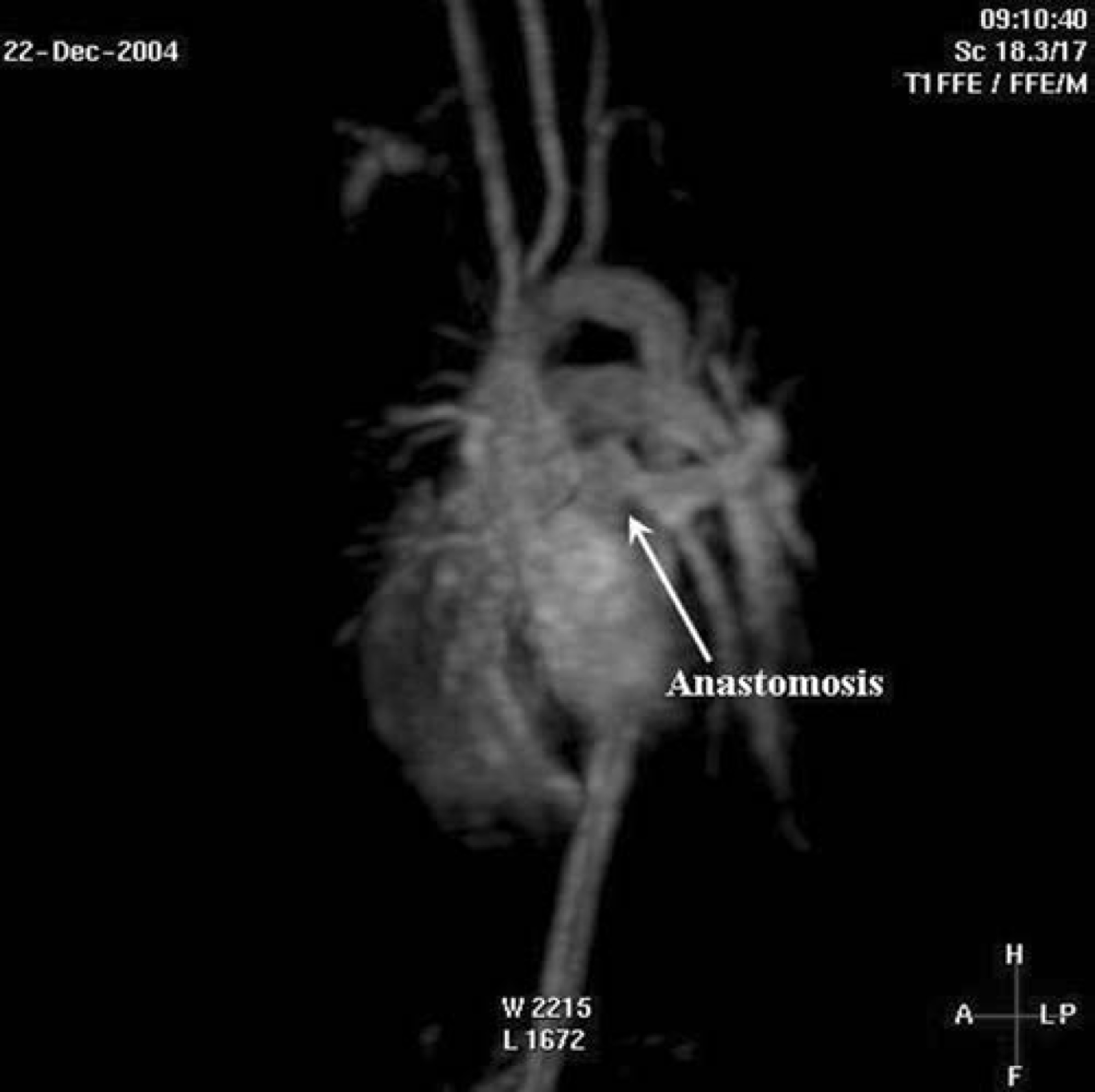
Six months after successful surgical correction. Clear depiction of the LA-ascending vein anastomosis.
Magnetic resonance imaging is recognized as an accurate non-invasive method for identifying left anomalous pulmonary vein connection 1 . In addition, it may also be a reliable alternative to cardiac catheterization in postoperative evaluation of these patients. 2