
Abstract
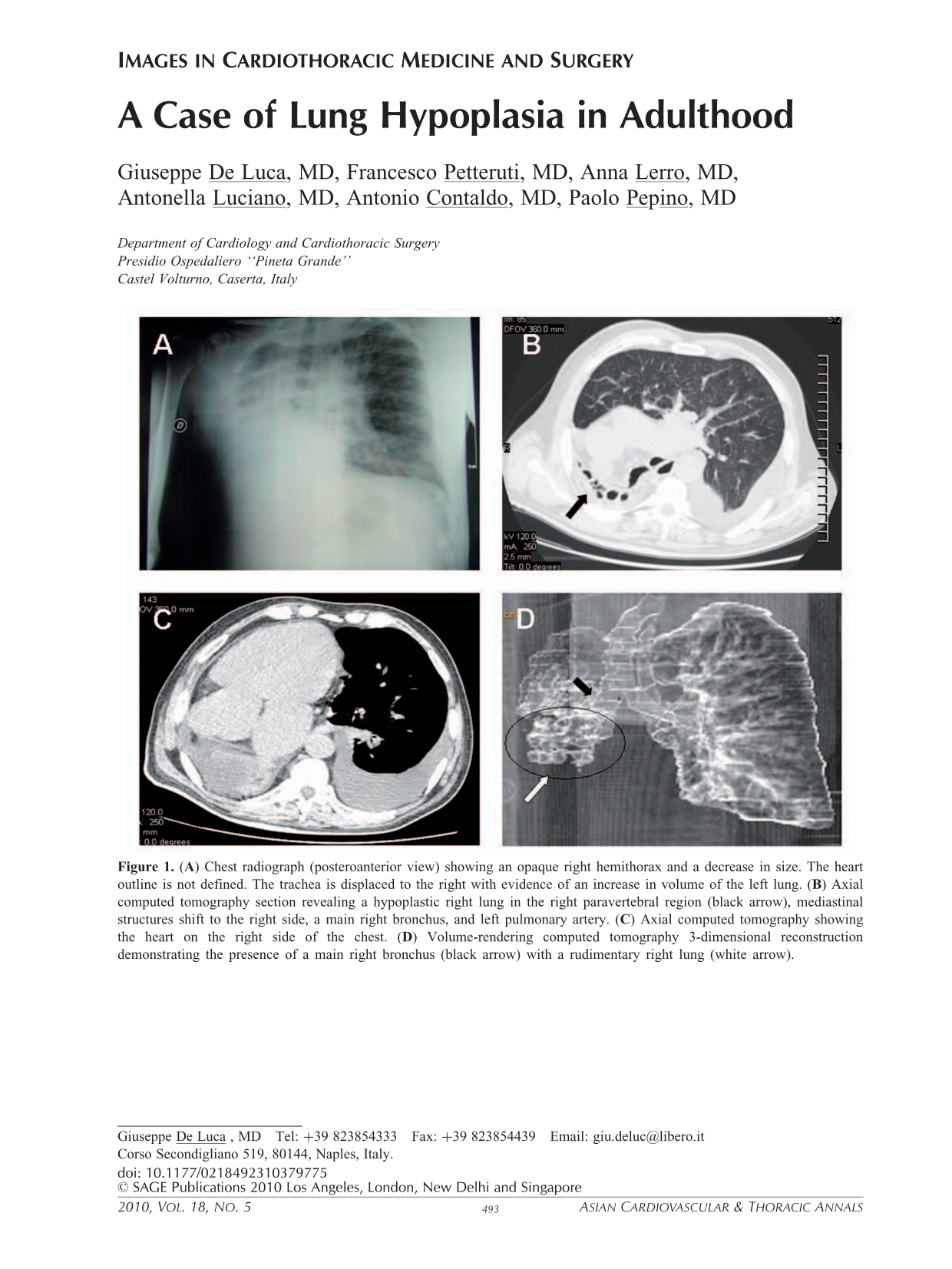
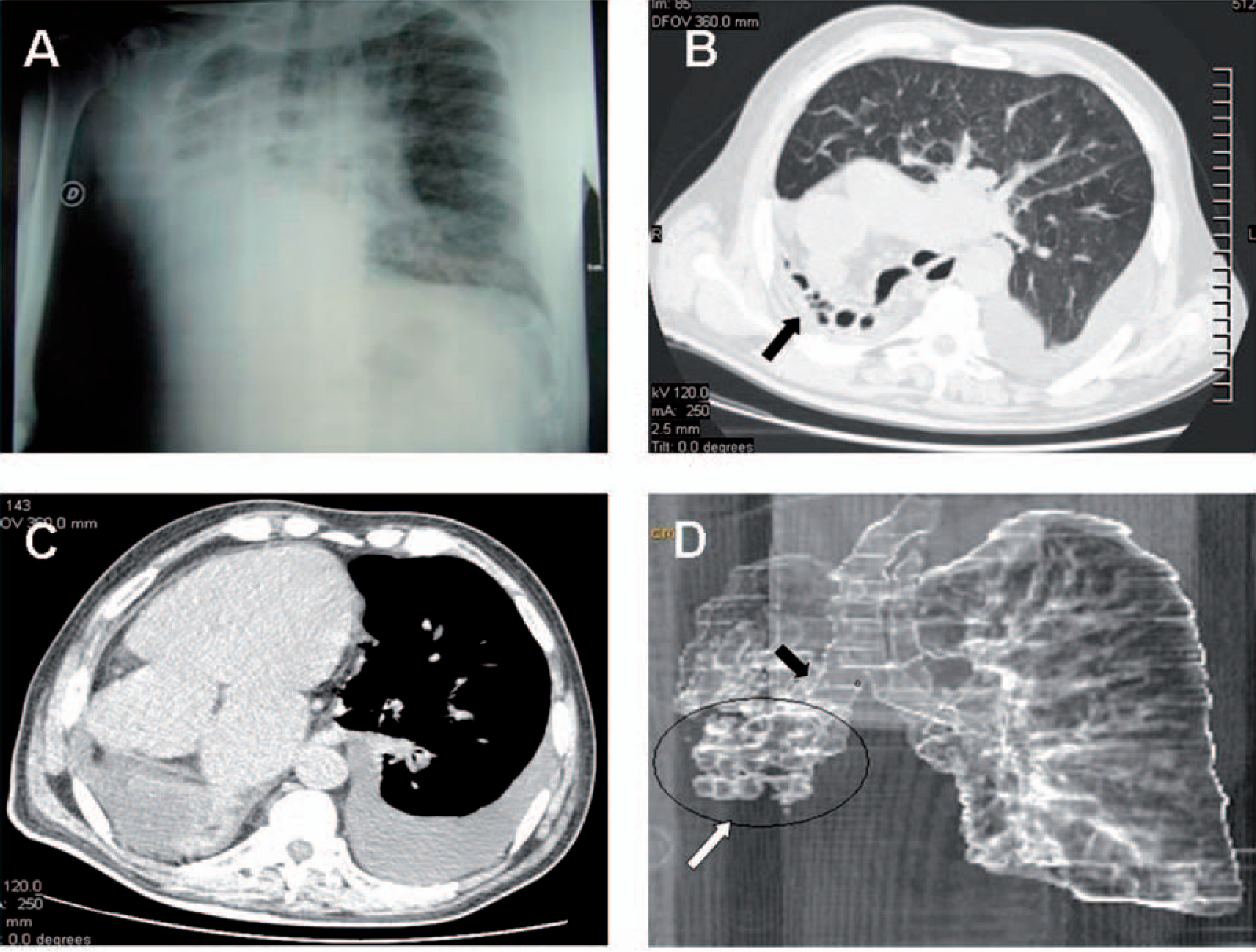
A 64-year-old man was admitted with right thoracic pain. His medical history was unremarkable. Chest examination showed diminished movement of the thorax on the right side. Auscultation revealed no breath sound over the middle lower right hemithorax. Heart sounds were heard on the right side. Electrocardiography and cardiac enzymes were consistent with acute anterior myocardial infarction. Chest radiography showed extensive opacification of the right hemithorax, and the heart outline was not defined (Figure 1A). Echocardiography demonstrated dextroposition of the heart with severe hypokinesy of the interventricular septum and apex. The left ventricular ejection fraction was 25%. Computed tomography (Figure 1B) detected hypoplastic lung tissue with cystic changes in the right paravertebral area, hyperinflation of the left lung with herniation to the contralateral side and displacement of the mediastinum to the right side, and absent right pulmonary artery. The heart was on the right side of the chest (Figure 1C). Volume-rendering computed tomography reconstruction (Figure 1D) showed a main right bronchus and a decreased number of alveoli and alveolar air space. Angiography confirmed critical coronary stenosis, and a stent was inserted. At bronchoscopy, the trachea was seen to deviate to the right side, the carina was visualized, the main right bronchus was present and it showed chronic inflammation. Lung hypoplasia is vary rare in adulthood; in fact the diagnosis is usually established in the prenatal or neonatal periods.
(