
Abstract

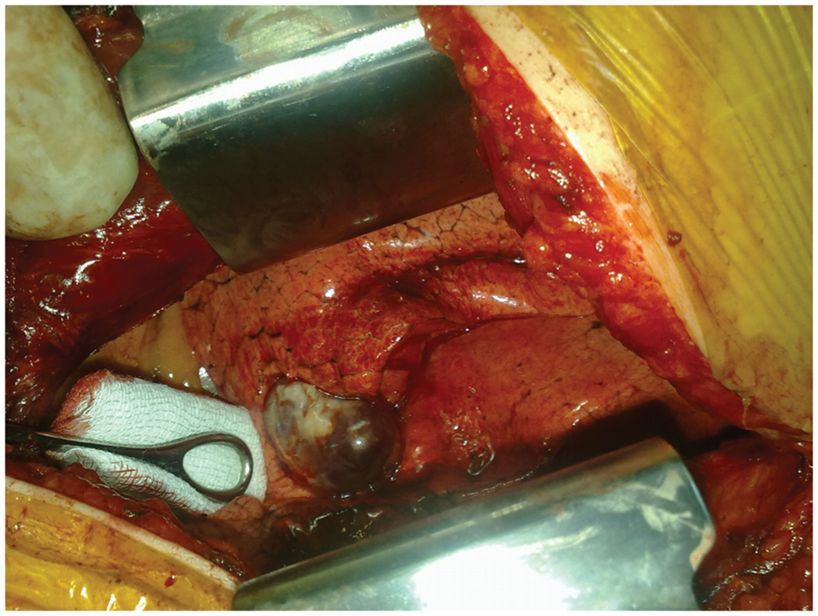
A 55-year-old woman presented with palpitation. A pulmonary mass was detected on chest radiography. Non-contrast computed tomography of the thorax revealed both the feeding artery and evacuator vein, which suggested a pulmonary arteriovenous malformation (Figure 1). Under general anesthesia with a double-lumen endotracheal tube, the patient underwent a left lateral thoracotomy. The pulmonary arteriovenous malformation was located in the lingula segment of the left upper lobe (Figure 2). Lingulectomy was performed without any complication.
(A) The 2 vessels are seen on the left side originating from the hilum of the lung. (B) Computed tomography scan revealing vessels on the 34th cross-section. (C) A later section (38th) clearly demonstrating the course of the vessels to the malformation. (D) Contrast injection demonstrated enhancement of the vessels in mediastinal view. Operative view of the pulmonary arteriovenous malformation, from the side of the surgeon, which is located in the lingula segment of the left upper lobe.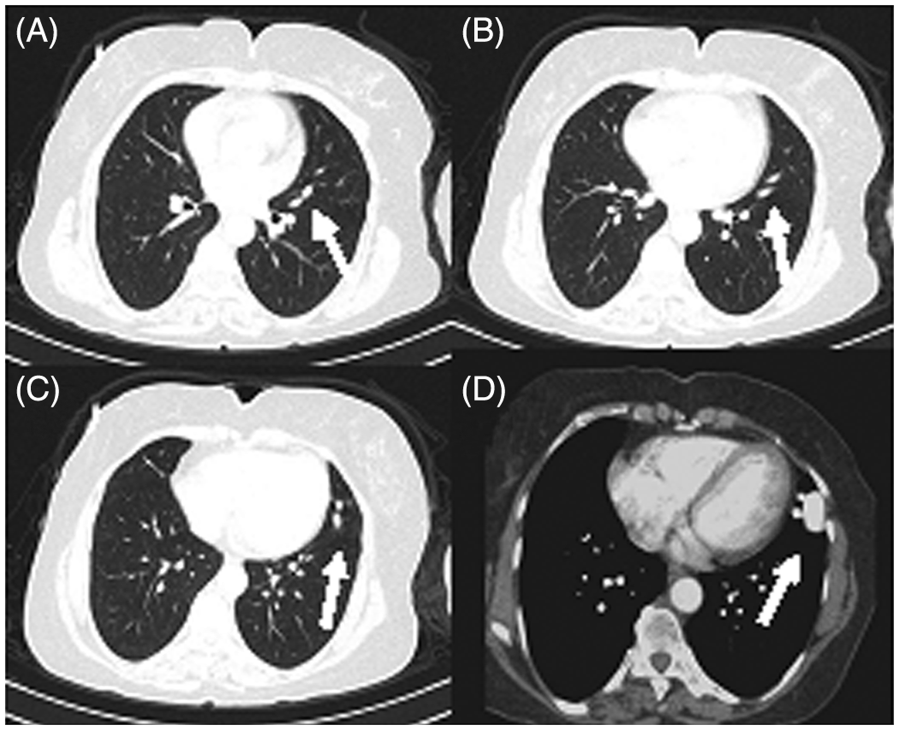
Footnotes
Funding
This research received no specific grant from any funding agency in the public, commercial, or not-for-profit sectors.
Conflicts of interest statement
None declared.