
Abstract
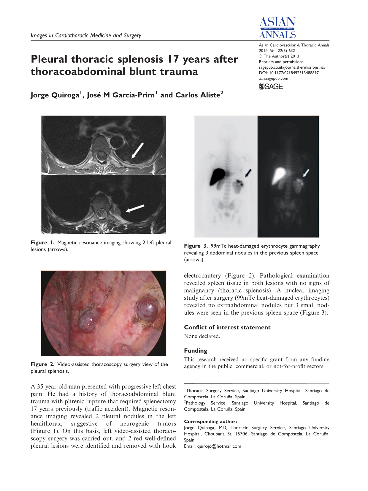
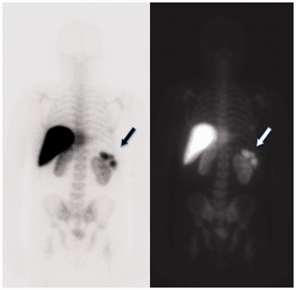
A 35-year-old man presented with progressive left chest pain. He had a history of thoracoabdominal blunt trauma with phrenic rupture that required splenectomy 17 years previously (traffic accident). Magnetic resonance imaging revealed 2 pleural nodules in the left hemithorax, suggestive of neurogenic tumors (Figure 1). On this basis, left video-assisted thoracoscopy surgery was carried out, and 2 red well-defined pleural lesions were identified and removed with hook electrocautery (Figure 2). Pathological examination revealed spleen tissue in both lesions with no signs of malignancy (thoracic splenosis). A nuclear imaging study after surgery (99mTc heat-damaged erythrocytes) revealed no extraabdominal nodules but 3 small nodules were seen in the previous spleen space (Figure 3).
Magnetic resonance imaging showing 2 left pleural lesions (arrows). Video-assisted thoracoscopy surgery view of the pleural splenosis. 99mTc heat-damaged erythrocyte gammagraphy revealing 3 abdominal nodules in the previous spleen space (arrows).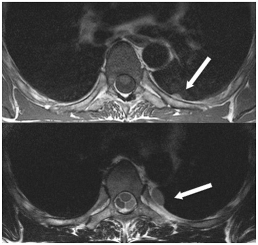
Footnotes
Conflict of interest statement
None declared.
Funding
This research received no specific grant from any funding agency in the public, commercial, or not-for-profit sectors.