
Abstract

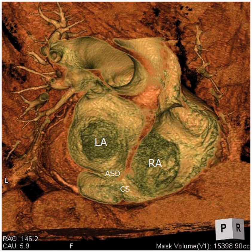
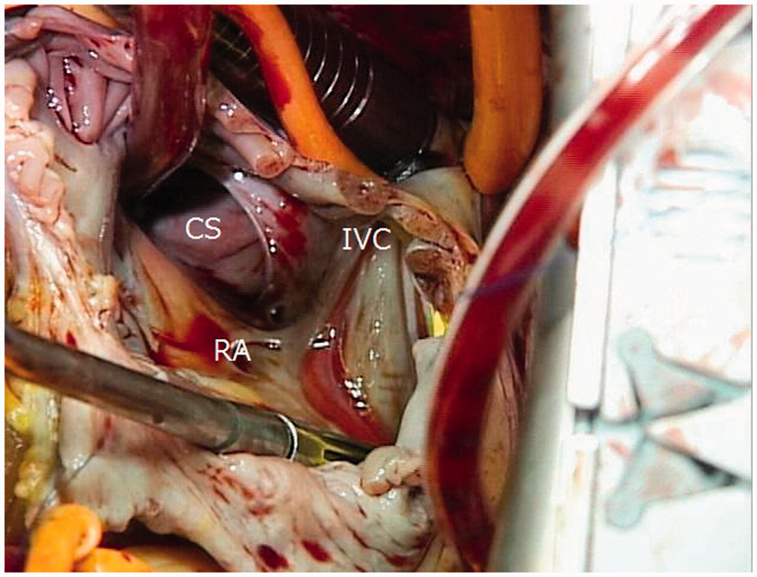
Physical examination a 73-year-old woman with dyspnea revealed a systolic murmur at the 2nd intercostal space along the left sternal border. Chest radiography revealed right heart enlargement. Electrocardiography indicated normal sinus rhythm. Transesophageal echocardiography showed a dilated coronary sinus located at both atria. The pulmonary-to-systemic blood flow ratio was 2.46. Chest computed tomography revealed an unroofed coronary sinus atrial septal defect (Figures 1 and 2). She was diagnosed with unroofed coronary sinus atrial septal defect without persistent left superior vena cava. At surgery, an ostium opening into the right atrium was found (Figure 3). We performed autologous pericardial patch closure of the ostium, leaving the coronary vein draining into the left atrium. The patient was discharged 11 days postoperatively in a satisfactory condition without any critical complications.
Two-dimensional chest computed tomography revealing an unroofed coronary sinus atrial septal defect. ASD: atrial septal defect; CS: coronary sinus; IVC: inferior vena cava; LA: left atrium; RA: right atrium. Three-dimensional chest computed tomography revealing an unroofed coronary sinus atrial septal defect. ASD: atrial septal defect; CS: coronary sinus; IVC: inferior vena cava; LA: left atrium; RA: right atrium. Operative photograph showing the ostium opening into the right atrium. CS: coronary sinus; IVC: inferior vena cava; LA: left atrium; RA: right atrium.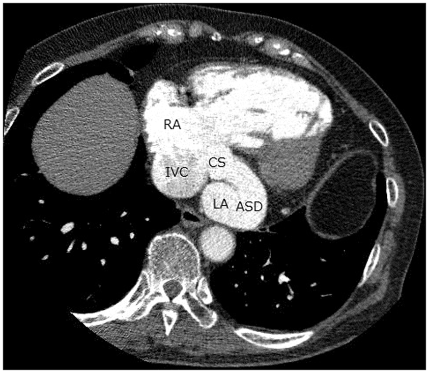
Footnotes
Funding
This research received no specific grant from any funding agency in the public, commercial, or not-for-profit sectors.
Conflicts of interest statement
None declared.