
Abstract
Superior vena cava obstruction can be a serious complication after heart transplantation. A 58-year-old man with ischemic cardiomyopathy underwent orthotopic bicaval heart transplantation. On the 12th postoperative day, one hour after removing the central venous line, he developed sudden onset of facial edema, cyanosis, and tachycardia. Emergency transesophageal echocardiography revealed superior vena caval thrombosis at the site of anastomosis. Considering the risks of surgical reexploration, the superior vena cava was recanalized by stent deployment. All of the patient’s symptoms were relieved a few hours after stent placement.
Keywords
Introduction
Heart transplantation is the treatment of choice for patients with end-stage heart failure who do not respond to medical, interventional, or other surgical treatments. 1 In the early 1960s, the classic technique for heart transplantation was developed, but another technique (bicaval anastomosis) was developed at nearly the same time. 2 Currently, bicaval anastomosis is frequently used because of its hemodynamic benefits due to the fact that sinus node function seems to be better preserved. 3 However, it can have some complications such as vena caval anastomotic stenosis or strictures, which may lead to superior vena cava (SVC) syndrome. If a central venous line (CVL) remains for a prolonged time, it may be one of the causes of secondary thrombosis. We describe the case of a patient with SVC obstruction 2 weeks after heart transplantation, who was treated successfully by endovascular stent placement.
Case report
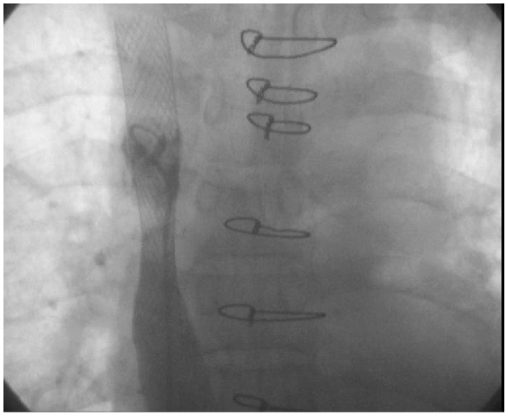
Our patient was a 58-year-old man who had suffered from ischemic cardiomyopathy in the terminal phase with functional class IV and myocardial volume oxygen consumption of 13 cc·kg−1·min−1. He had a history of hypertension, hyperlipidemia, and diabetes mellitus which was controlled with oral hypoglycemic agents. He underwent orthotopic bicaval heart transplantation. Anastomosis of the left atrial cuff, aorta, pulmonary artery and inferior vena cava were performed with 4/0 Prolene sutures, while the SVC anastomosis was performed with 5/0 Prolene sutures. The technique for SVC anastomosis, like the other anastomoses, was running sutures. As the diameter of the anastomosis was small, the traction applied to the sutures was not very great, so that the pursestring effect and tightening of the anastomosis were minimal. The patient was weaned off cardiopulmonary bypass with adrenaline 0.1 µg·kg·−1·min−1 for inotropic support. Intraoperative transesophageal echocardiography (IOTEE) showed no gradient across the anastomotic sites including the SVC anastomosis. As the IOTEE did not show any gradient across the SVC anastomois, and as the anastomosis seemed to be perfect, so the gradient across the SVC anastomosis did not measured directly. During the first 24 postoperative hours, the patient was extubated, and the inotrope dose was tapered and discontinued on the 5th postoperative day. Transesophageal echocardiography on the first postoperative day revealed a normal ejection fraction with mild to moderate right ventricular dysfunction and no evidence of any torsion or gradient in the SVC, inferior vena cava, aorta, or pulmonary artery anastomotic sites. On the 12th postoperative day, prior to discharge and one hour after removing the CVP line, the patient developed sudden onset of facial edema, cyanosis, and tachycardia. Emergency transesophageal echocardiography revealed SVC thrombosis at the site of anastomosis. Intravenous heparin 800 U·h−1 was started. Computed tomographic angiography revealed a thrombus in the SVC extending to the left jugular vein and left subclavian vein. Venography showed complete obstruction of the proximal SVC with an enlarged patent azygos vein (Figure 1). Considering the diagnosis of SVC syndrome and the risks of surgical reexploration, we decided to perform stent placement. As the face and neck of the patient were very edematous and he was restless, any manipulation of that area was avoided, and a wire was passed via the right femoral vein through the inferior vena cava and right atrium to the site of obstruction, and a stent was deployed successfully (Figure 2). All of the patient’s symptoms were relieved a few hours after stent placement. Warfarin 5 mg daily was started, and the international normalized ratio was kept between 2 and 2.5 for 3 months. The patient was discharged home a few days after the endovascular procedure, and during 4 years of follow-up, he has been asymptomatic with no complaint.
Venography showing obstruction of the proximal superior vena cava (lower arrow) with an enlarged patent azygos vein (upper arrow). The superior vena cava became patent after deployment of a stent.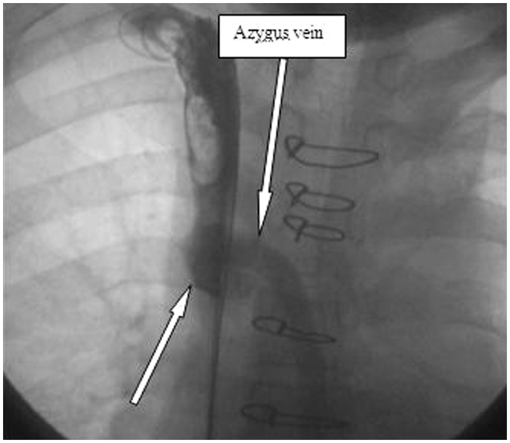
Discussion
The SVC is the second largest vein in the human body. Factors contributing to SVC obstruction can be malignant or benign in nature. Most cases arise from malignant and neoplastic conditions. SVC obstruction in benign conditions is rare and it can be caused by histoplasmosis infection, thrombophlebitis, tuberculosis, central venous catheters, pacemaker catheters, defibrillators and resynchronizations, mediastinal fibrosis because of granulomatous disease or radiation, intrathoracic goiter, ventriculoatrial shunts, and aortic aneurysms. Rarely, vena caval stenosis is a complication after heart transplantation using the bicaval anastomosis technique.3,4 The incidence of this complication seems to be greater in the pediatric heart transplant group. 5 Mismatch of the SVC diameters of the recipient and donor may cause late stenosis of the SVC due to tightening of the suture line or intimal hyperplasia. 6 Because there was no gradient in the SVC in the early postoperative course of this patient and he was asymptomatic, a clot was the most likely cause of his subsequent symptoms.
It is recommended that the duration of CVL placement should be based on the clinical condition of the patient, 7 so it is not standard practice to leave a CVL in a patient until the time of discharge. The tip of the CVL may cause trauma to the endothelium, or it may be a primary nidus for thrombosis. Because the symptoms in our patient started after removing the CVL, the CVL catheter might have allowed minimal SVC patency for the passage of the blood. There was no evidence of any abnormality in venous drainage in the upper part of the body before removing the CVL. Therefore, the CVL might have played a role not only in SVC obstruction but also in delayed presentation of SVC syndrome. Considering that our patient was taking corticosteroids and immunosuppressive medications, surgical reexploration would have involved a higher risk than an endovascular procedure. The endovascular procedure avoided a second operation in this critically immunosuppressed patient. Furthermore, the follow-up results after stent placement in our patient confirmed the safety of this procedure, even early after transplantation. This case suggests that SVC obstruction following bicaval heart transplantation can be safely treated by endovascular procedures, and the CVL should be withdrawn as soon as the patient’s condition is suitable.
Footnotes
Acknowledgement
The authors gratefully thank Dr. Negar Salehi, Cardiologist and Interventionalist, Rajaie Heart Center, for assistance in deployment of the stent, and also Ms. Laila Salimi, RN, the coordinator of heart tansplant team for follow up of the patient and collection of data.
Funding
This research received no specific grant from any funding agency in the public, commercial, or not-for-profit sectors.
Conflict of interest statement
None declared.