
Abstract

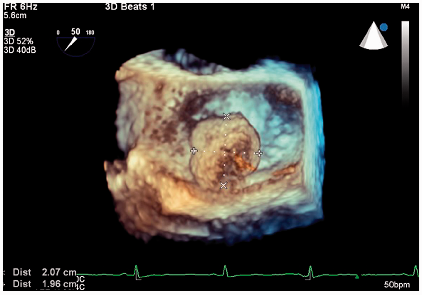
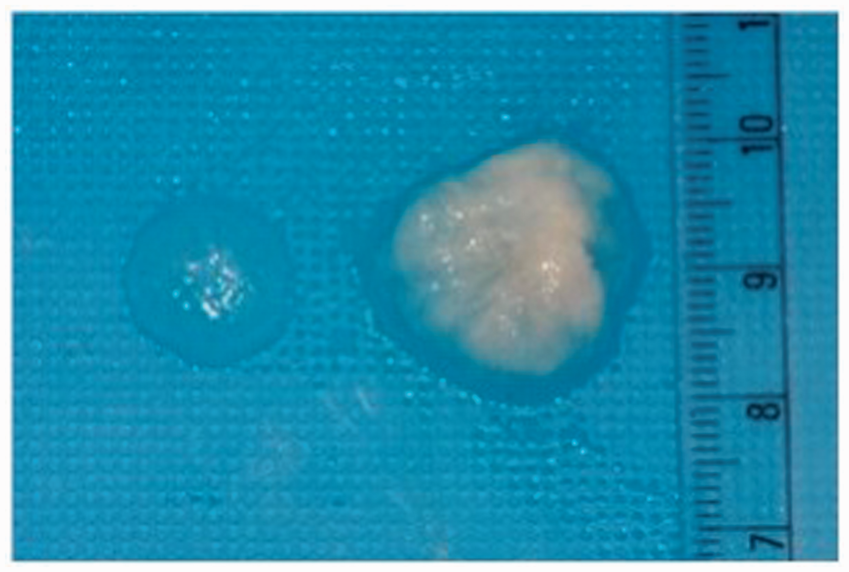
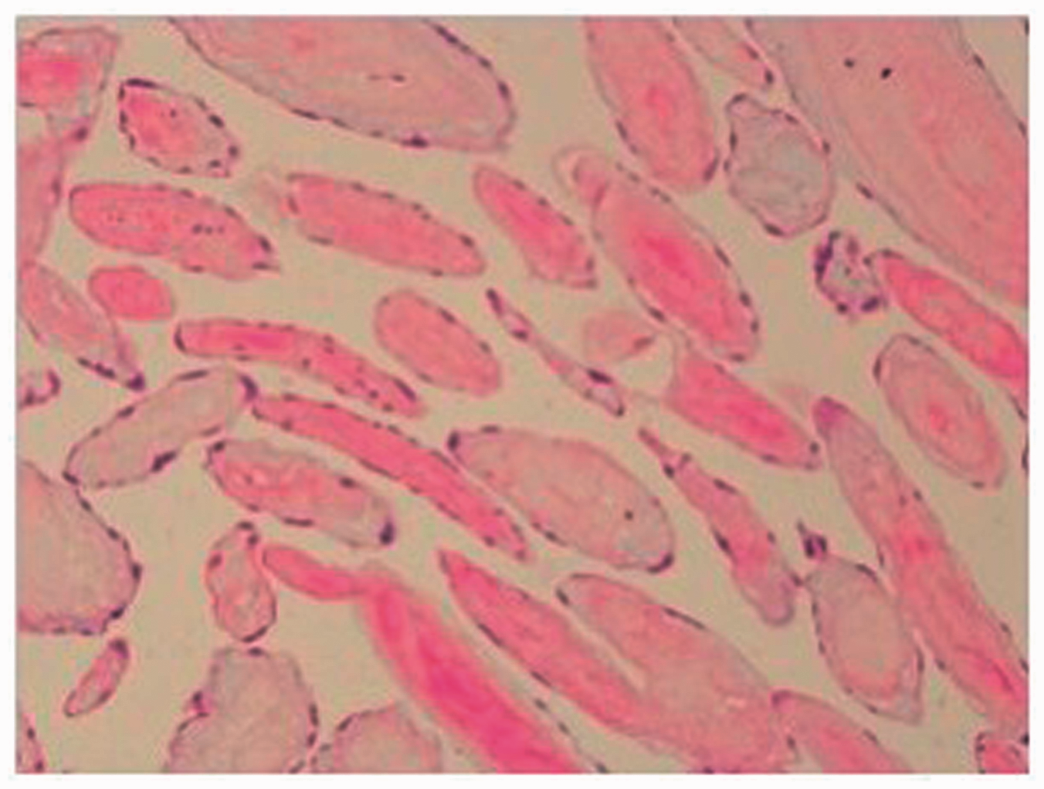
A 52-year-old asymptomatic man, on follow-up after a primary percutaneous coronary intervention to the left anterior descending artery, was diagnosed with a freely floating soft tissue mass measuring 23 × 25 mm with equal echogenicity with a stalk attached to the posterior mitral leaflet, on transthoracic echocardiography. Preoperative 2-dimensional (Figure 1) and 3-dimensional (Figure 2) transesophageal echocardiography showed the characteristic stippled appearance with shimmer of the peripheral edge and echolucency within the tumor, suggestive of cardiac papillary fibroelastoma. Intraoperatively, a 20 × 25 × 25-mm soft pedunculated polypoid mass was found attached to the left atrial wall 3 mm from the mitral valve P1 region. Postoperative transesophageal echocardiography following excision showed no mitral regurgitation. Histopathology showed a papillary tumor composed of filiform processes with a central acellular collagen core surrounded by myxomatous matrix. The peripheral rim and core showed coarse elastin fibrils lined by endothelial cells, confirming the diagnosis of cardiac papillary fibroelastoma (Figures 3 and 4).
Two-dimensional transesophageal echocardiography showing a 1.65 × 1.85-cm pedunculated freely mobile cardiac papillary fibroelastoma in the left atrium with a stippled appearance, shimmer of the peripheral edge, and echolucency within. Three-dimensional transesophageal echocardiography showing the cardiac papillary fibroelastoma arising from the left atrium very near the P1 segment of the mitral valve. Gross histopathological specimen of the cardiac papillary fibroelastoma. Histology of the cardiac papillary fibroelastoma. Hematoxylin and eosin stain, original magnification ×100.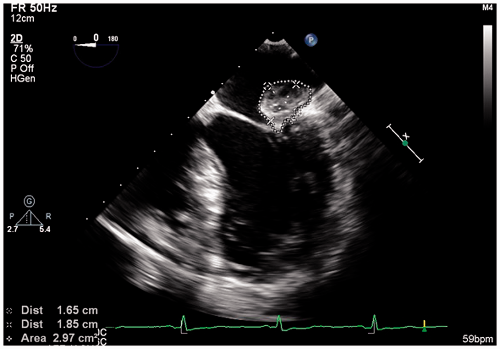
Footnotes
Declaration of conflicting interests
The author(s) declared no potential conflicts of interest with respect to the research, authorship, and/or publication of this article.
Funding
The author(s) received no financial support for the research, authorship, and/or publication of this article.