
Abstract
Background
Transcatheter aortic valve implantation has been established as a safe and effective treatment option for patients at high or prohibitive surgical risk. However, some patients may not be suitable for the transfemoral approach due to severe iliofemoral disease or aneurysmal disease of the thoracoabdominal aorta. The aim of this case series was to evaluate the feasibility and clinical outcomes of the transaortic approach.
Methods
From May 2015 to June 2016, 5 patients (mean age 78.4 ± 3.9 years) with severe symptomatic aortic stenosis underwent transaortic transcatheter aortic valve implantation after a heart team discussion. They were considered to be at high surgical risk and ineligible for the transfemoral approach due to iliofemoral or thoracoabdominal aortic disease.
Results
A CoreValve Evolut R was successfully deployed in all 5 patients. We performed 4 right mini-parasternal incisions and one J-incision partial sternotomy. None of the patients required permanent pacemaker implantation, one required reopening of the mini-parasternal incision for postoperative bleeding. Follow-up echocardiography one month after the procedure showed improvement in the mean aortic gradient (from 63.2 to 8.3 mm Hg) and aortic valve area (from 0.62 to 2.2 cm2). None of the patients had more than mild paravalvular leakage. There was no intraoperative or 30-day mortality.
Conclusion
Transaortic transcatheter aortic valve implantation is a safe and feasible option for patients with severe aortic stenosis who are considered unsuitable for transfemoral aortic valve implantation.
Keywords
Introduction
Severe aortic stenosis (AS) is a common valvular heart disease with an increasing incidence in the elderly population. Transcatheter aortic valve implantation (TAVI) was developed to provide an alternative for patients considered to be at high or prohibitive surgical risk. TAVI is most commonly performed by a transfemoral approach, and its safety and efficacy have been established in many studies.1,2 Other approaches include transapical, 3 transaortic, 4 and trans-subclavian. 5 The transfemoral approach requires suitable iliofemoral and thoracoabdominal aortic anatomy, and carries a risk of access-site complications in patients with small tortuous atherosclerotic vessels. Concomitant thoracoabdominal aneurysmal disease also increases the risk in transfemoral TAVI. Transapical TAVI was introduced as an alternative for patients who are not suitable for transfemoral TAVI, but the requirement of a thoracotomy and direct left ventricular (LV) apical puncture has been associated with risks of LV rupture, LV dyskinesia and aneurysms as well as mitral and aortic injuries. 6 The transaortic approach has been described in case series worldwide, and is a safe and feasible option for TAVI in severe AS patients. 7 In our case series, we describe the procedural and short-term results of our initial experience of transaortic TAVI with CoreValve Evolut R (Medtronic Inc., Minneapolis, MN, USA) implantation via right anterior mini-parasternal and ministernotomy approaches in patients with severe AS.
Patients and methods
Baseline characteristics of 5 patients undergoing transcatheter aortic valve implantation.
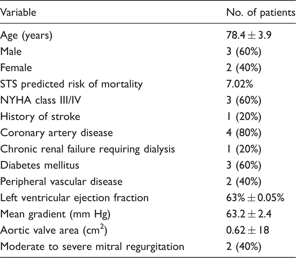
NYHA: New York Heart Association; STS: Society of Thoracic Surgeons.
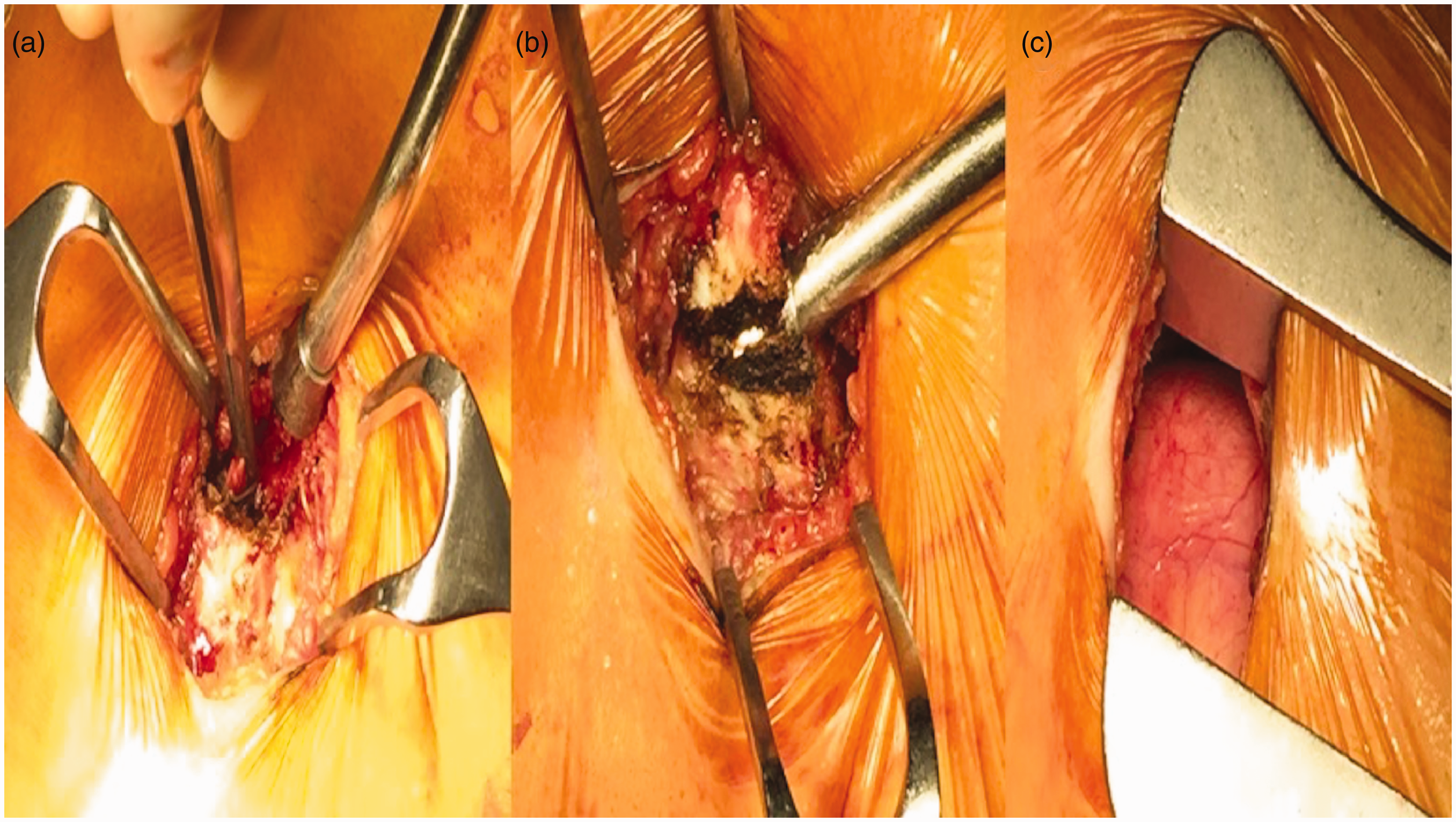
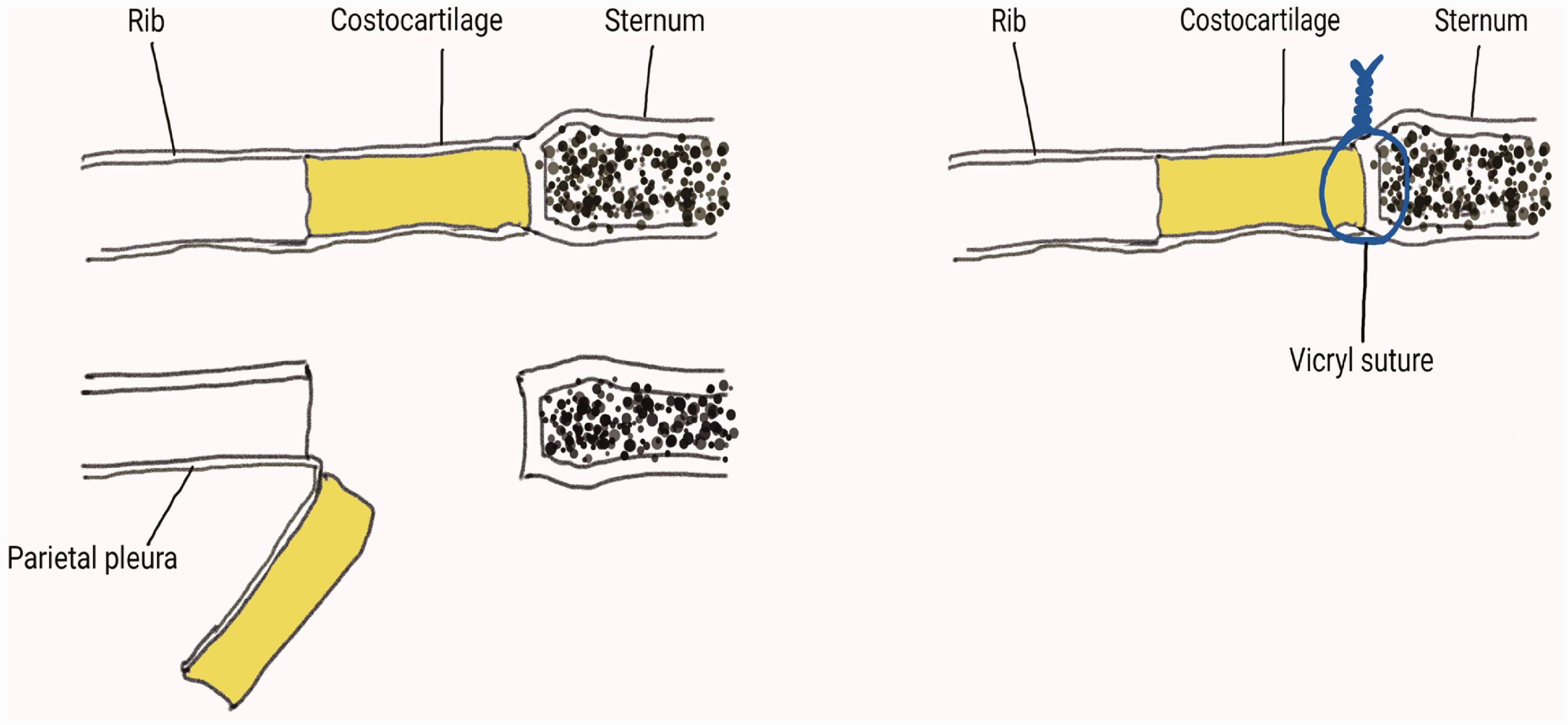
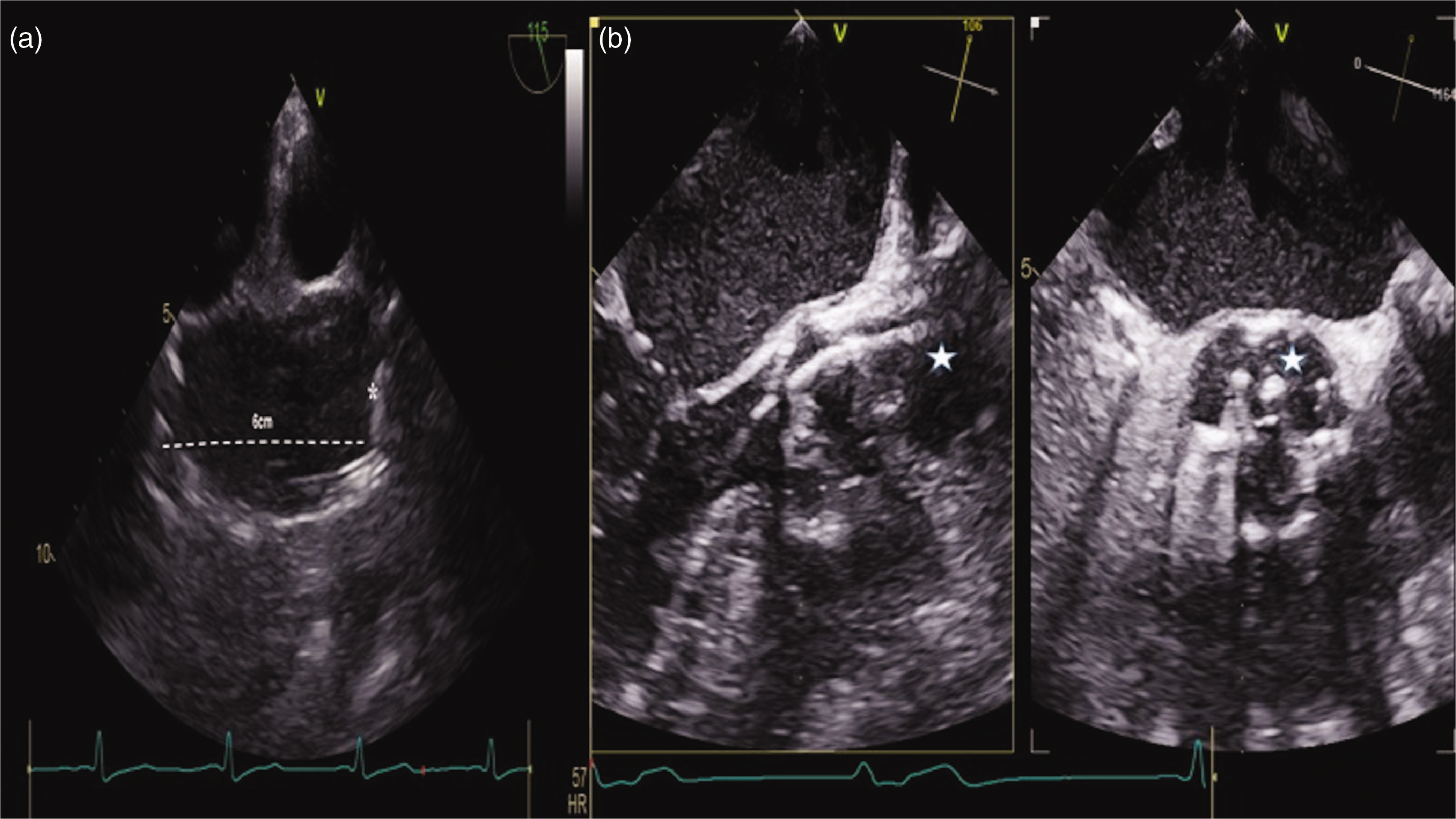
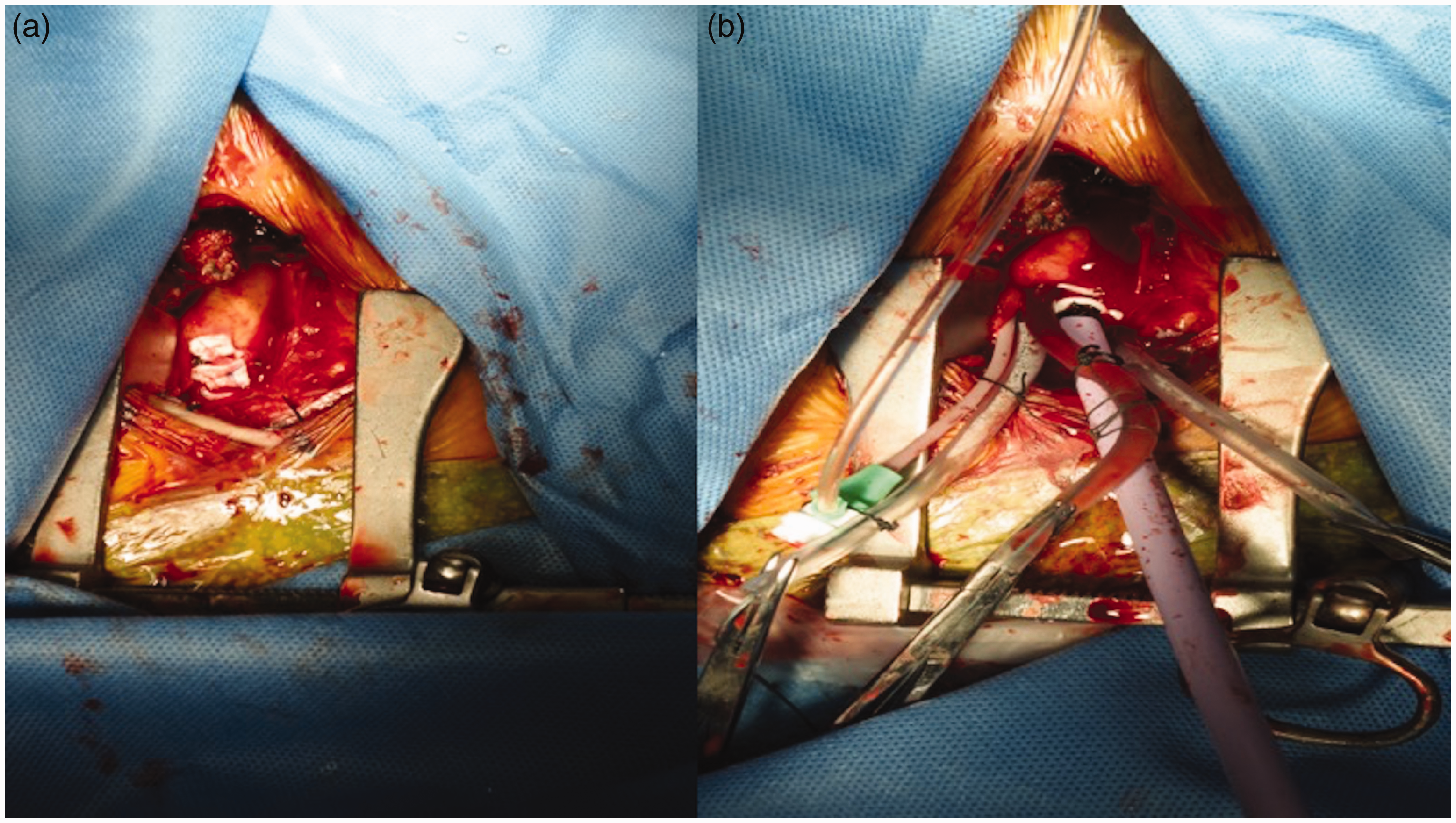
All transaortic TAVI procedures were performed by the heart team in a hybrid theatre equipped with DynaCT (Siemens Artis Zeego). Four patients had a right mini-parasternal incision and one had a J-incision partial sternotomy. For procedures carried out via a parasternal incision, double-lumen intubation was required. A 4-cm parasternal vertical incision was made over the 3rd intercostal space (Figure 1). The right internal mammary artery was ligated. The junction of the costal cartilage and the sternum was disarticulated and retracted inferolaterally below the 3rd rib, away from the wound (Figure 2, Figure 3). A temporary pacing wire was inserted into the right ventricle via the internal jugular or femoral vein. Ascending aortic angiography via a 6 F pigtail catheter inserted through the femoral artery was undertaken to mark the aortic root. A minimum of 6 cm between the aortic annulus and the aortic entry site is needed for safe deployment of the CoreValve because it is at least 5.5 cm in height. Two concentric pursestring sutures were applied around the entry site, and the ascending aorta was cannulated by the Seldinger technique. Under transesophageal echocardiographic guidance, the aortic valve was crossed with a straight-tip guidewire with a selective catheter (Figure 4). After confirming the guidewire and catheter position to be at an appropriate angle, an 18 F Cook sheath was prepared (Figure 5) and inserted via one of the pursestring sutures, and the CoreValve Evolut R bioprosthesis was introduced under angiographic and fluoroscopic guidance over a preshaped Confida guidewire (Medtronic Inc., Minneapolis, MN, USA) under rapid ventricular pacing at 120 beats per minute (Figure 6). An aortogram was obtained after deployment to confirm the valve location and absence paravalvular leakage, and to assess the patency of the coronary arteries (Figure 7). The 18F Cook sheath was removed from the aortic root with the pursestring sutures tied. The costal cartilage was opposed to the sternum with one Vicryl suture, and the wound was closed in layers.
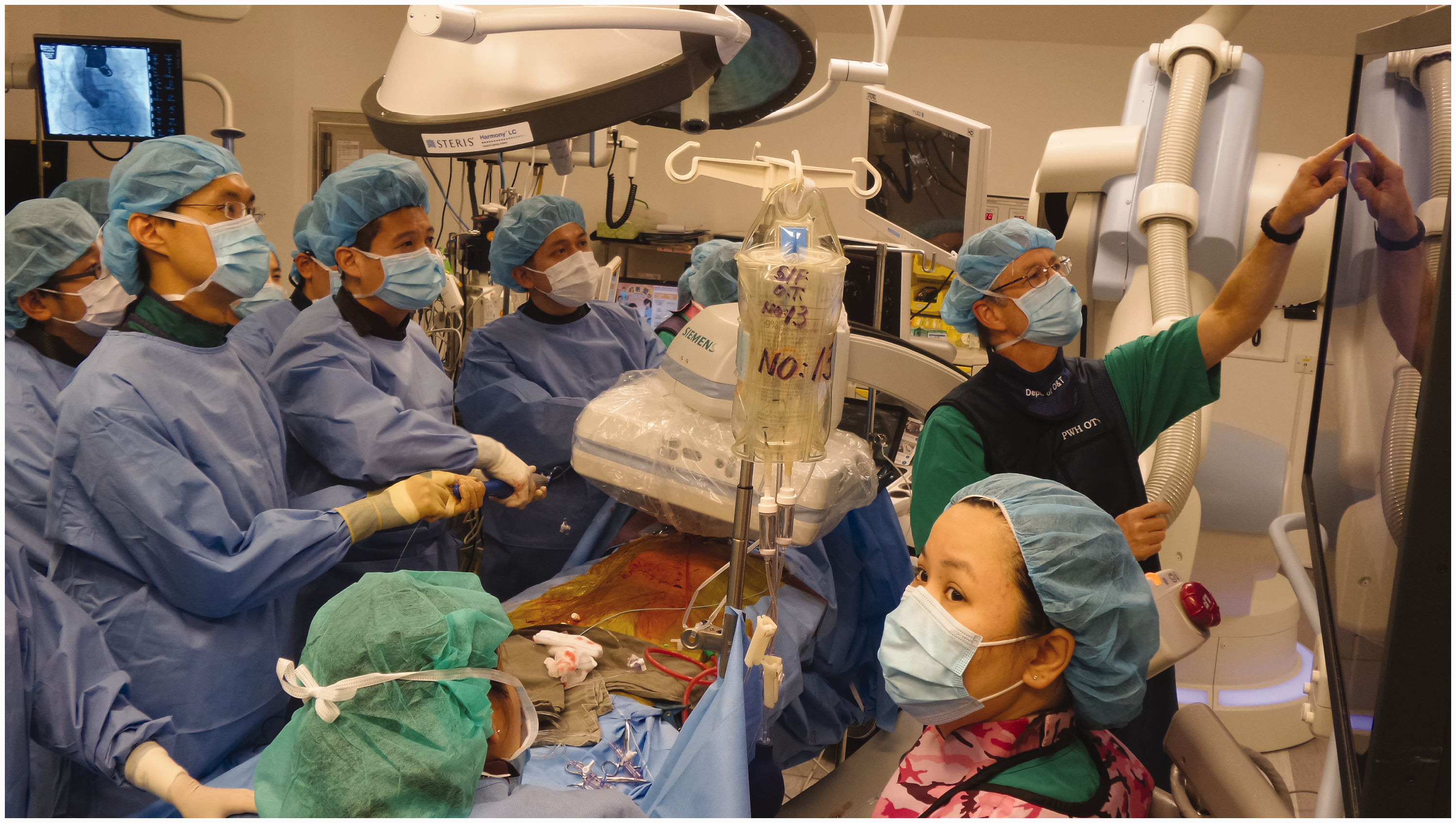
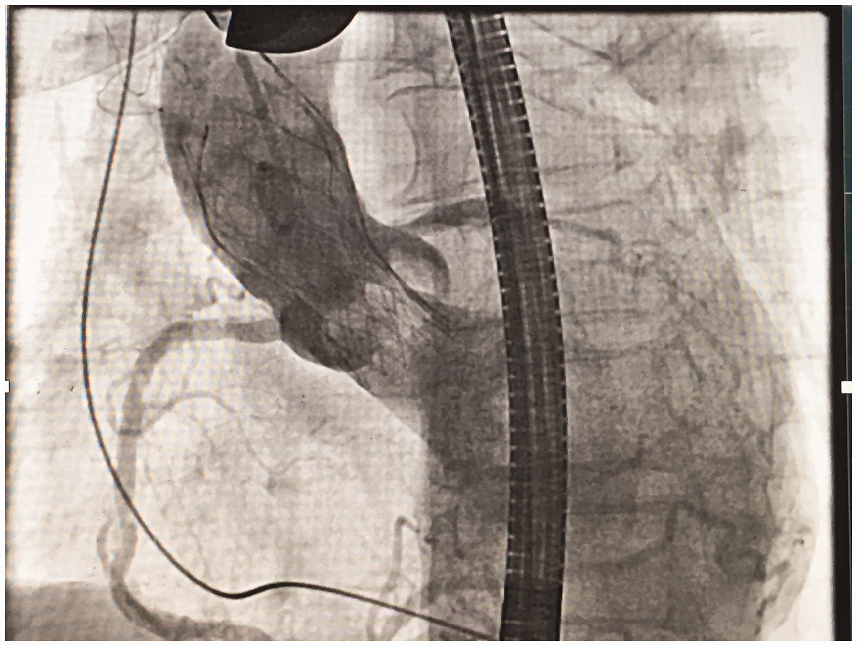
The 4-cm mini-parasternal incision. (a) The right internal mammary artery is ligated. (b) The 3rd costal cartilage is disarticulated at its junction with the sternum. (c) The aorta and planned entry site are exposed after inferolateral retraction of the costal cartilage. Diagram showing disarticulation of the costal cartilage from the sternum. (a) The asterisk (*) represents the surgeon’s finger indenting the aortic wall 6 cm above the aortic valve. (b) transesophageal echocardiographic guidance with the guidewire (white star) in the ascending aorta, and crossing the aortic valve coaxially. (a) The 2 concentric pursestring sutures applied to the ascending aorta in preparation for aortic cannulation. (b) The 18 F sheath inserted into the ascending aorta, secured and tied with a snugger. Operating setup during deployment of the aortic valve prosthesis, with operators standing on the same side of the patient. Final aortogram after valve implantation, showing a satisfactory valve position and patent coronary arteries.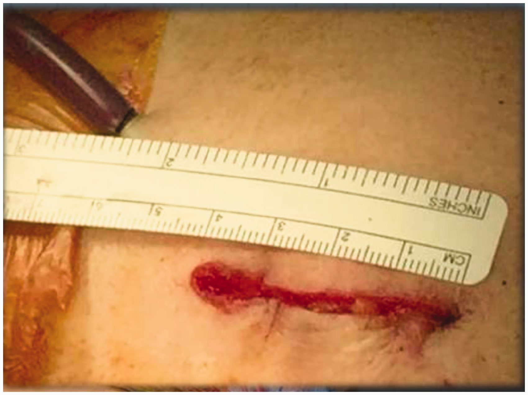
According to the Valve Academic Research Consortium consensus, 8 successful valvular deployment is defined as successful vascular access and deployment of the device in the correct position with normal performance of valve, a mean gradient <20 mm Hg or peak velocity <3 m s−1, and aortic valve area/indexed effective orifice area >1.2 cm2 with no moderate to severe paravalvular leak. Operative outcome was defined as any event occurring during the procedure or within 24 h after the procedure. Adverse events within 30 days were considered procedure-related.
Results
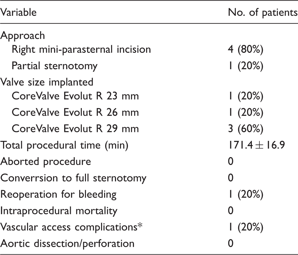
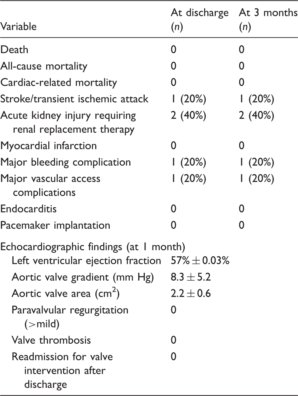
Transaortic TAVI was performed successfully in all 5 patients. Three patients were excluded from transfemoral TAVI due to narrow iliofemoral vessels (<5 mm); one had tortuous iliofemoral vessels bilaterally, and one had a thoracoabdominal aneurysm (Figure 8). After implantation, the on-table LV systolic to aortic pressure gradient was 0.4 mm Hg. The other procedural and clinical outcomes are shown in Tables 2 and 3. There was no procedural death and no conversion to a full sternotomy. Mean total procedural time was 171.4 ± 16.9 min. One patient had an ischemic cerebrovascular accident, 2 had acute kidney injury requiring short-term renal replacement therapy, and one had significant bleeding from the aortotomy site requiring reopening of the parasternal incision for hemostasis. No patient had clinical instability of the sternochondral joint, and none required implantation of a permanent pacemaker. All patients were discharged with satisfactory prosthesis function. Echocardiography one month after TAVI showed improvement in the mean aortic valve gradient (from 63.2 ± 12.4 to 8.3 ± 5.2 mm Hg) and aortic valve area (from 0.62 ± 0.18 to 2.2 ± 0.6 cm2). None of the patients had more than mild paravalvular leakage. At the 3-month follow-up, there was no mortality; mean follow-up was 3.6 months.
(a) Small and narrow iliofemoral vessels (<5 mm). (b) A thoracoabdominal aortic aneurysm. Procedural information on transcatheter aortic valve implantation in 5 patients. Aortic sheath insertion-site bleeding leading to reoperation for hemostasis. Clinical and echocardiographic outcomes.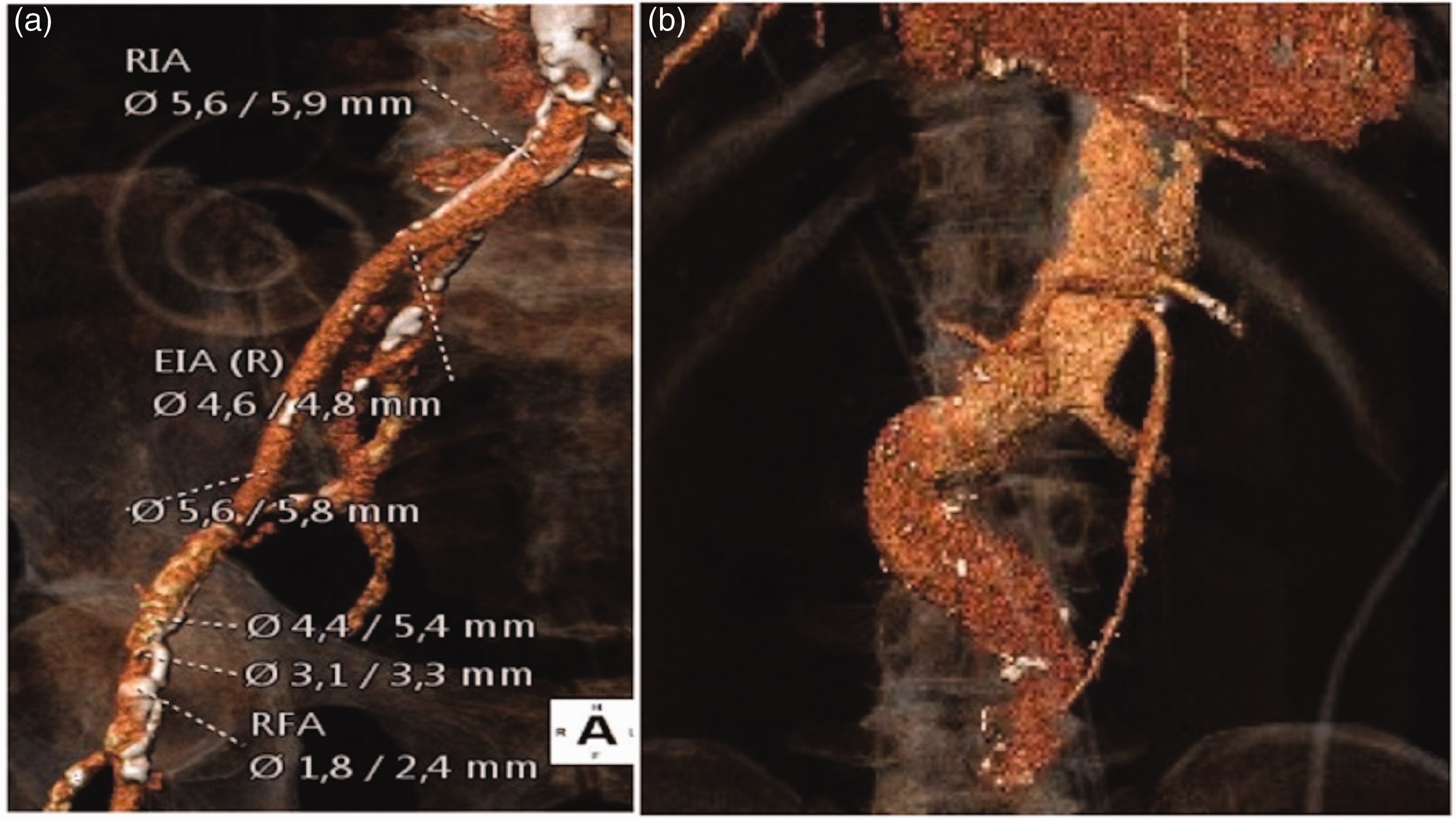
Discussion
SAVR has become the standard treatment for severe AS, with reasonable rates of mortality and morbidity. However, the risk associated with SAVR increases with multiple comorbidities and advanced age. From a large retrospective analysis of the Society of Thoracic Surgeons (STS) database, the 30-day mortality in patients with an STS predicted mortality score >8% was 12.9%, in contrast to 1.7% in patients with STS scores <4%. 9 Randomized controlled trials have shown that TAVI is superior to balloon aortic valvuloplasty and medical treatment in patients at prohibitive surgical risk, and equivalent to SAVR in high-risk surgical patients.10,11 The recent PARTNER 2 trial further substantiated the claim of parity between TAVI and SAVR in intermediate surgical risk patients. 12 Technological advancements and improvements in procedural management have resulted in increased TAVI success rates worldwide. The two most commonly deployed bioprostheses for TAVI are the balloon-expandable Edwards-Sapien prosthesis (Edward LifeSciences, Irvine, CA, USA) and the self-expanding CoreValve Revolving prosthesis. Since the first successful percutaneous implantation of an aortic valve in 2002, several approaches for TAVI have been developed for effective implantation of the aortic prosthesis. 13 With device modifications, implantation success rates have increased, and the incidence of major vascular complications has declined. Nonetheless, vascular complications are still reported in up to 32% of percutaneous transfemoral TAVI cases in large trials and registries.14,15 Major vascular complications are associated with higher 30-day and long-term mortality in TAVI patients. Commonly, patients with severe AS also have concomitant peripheral vascular disease and atherosclerosis, due to overlapping etiologies and risk factors. Excessively calcified and tortuous iliofemoral vessels often preclude the transfemoral approach.
A transapical TAVI approach via delivery systems through the LV apex has been reported. 16 This approach offers an alternative for patients considered unsuitable for transfemoral TAVI, but it has significant complications including apical puncture site bleeding or rupture, myocardial tears, damage to the mitral valve, coronary vessel injury during puncture repair, and aortic trauma. In the long-term, this approach is associated with LV apical false aneurysms, LV apex hypokinesia, and arrhythmias. Data from analysis of the Transapical Delivery of Cribier-Edwards Aortic Bioprosthesis Clinical feasibility study, and series such as the PARTNER trial, showed worse clinical outcomes with the transapical approach.11,16 The transaortic approach has emerged as another option for patients considered unsuitable for transfemoral TAVI. This is commonly performed via a right mini-parasternal incision or partial sternotomy, and both approaches are familiar to cardiac surgeons. Technically, due to the close proximity between the entry site and the aortic annulus, a stable platform is created for coaxial delivery of the valve with better angulation towards the annulus, enabling more precise device delivery. Overall, the transaortic approach offers better control of the delivery system. Theoretically, by avoiding manipulation of the aortic arch, the risk of thromboembolic stroke should be minimized. Contraindications to transaortic TAVI include porcelain aorta and hostile mediastinum. However, the right mini-parasternal approach does not require reentry via the sternum, and is considered feasible in patients with previous history of cardiac surgery.
The use of intraprocedural transesophageal echocardiography can further improve implantation success by providing real-time information on anatomical landmarks such as the aortic valve, aortic annulus, and proximal segments of the coronary arteries. Newer generations of echocardiography devices can now provide 3-dimensional reconstructions for reference. Real-time visualization of the aortic valve during guidewire crossing and valve deployment ensures optimal coaxial positioning of the wire and valve with the aortic root, enhancing deployment accuracy and preventing suboptimal outcomes. After deployment, echocardiography can assess the seating of the prosthesis and document the site and severity of paravalvular leaks. In our case series, we have demonstrated that transaortic TAVI is safe and feasible in patients with severe AS and concomitant peripheral vasculopathy. The best TAVI approach should ensure minimal procedural risk whilst offering the best outcome and recovery. Transaortic TAVI is a viable alternative in patients with small tortuous iliofemoral vessels, thoracoabdominal aneurysms, and LV dysfunction.
Footnotes
Declaration of conflicting interests
The author(s) declared no potential conflicts of interest with respect to the research, authorship, and/or publication of this article.
Funding
The author(s) received no financial support for the research, authorship, and/or publication of this article.