






Abstract
Aim
We aimed to report the experience of aortic valve reconstruction with autologous pericardium using Ozaki’s procedure in Vietnam.
Methods
The study included consecutive patients with isolated aortic valve disease who underwent Ozaki’s procedure in our hospital between June 2017 and August 2019. Aortic valve leaflets were reconstructed with autologous pericardium using Ozaki’s procedure.
Results
Sixty-one patients were included (mean age 55.8 years; 41 were male): 24 with aortic stenosis, 17 with aortic regurgitation, and 20 with both. Of the 61 patients, 16 had a bicuspid aortic valve, and 5 had infective endocarditis. The preoperative peak and mean gradient pressure gradients were 91.7 ± 16.1 mm Hg and 55.3 ± 10.3 mm Hg, respectively. Surgery was performed via a full or partial sternotomy. The procedure was successful in 59 cases. Two patients were converted to prosthetic valve replacement. The aortic crossclamp time was 110.9 ± 20.5 minutes. Intraoperative transesophageal echocardiography showed a mean pressure gradient of 8 ± 2 mm Hg and an aortic valve area of 3.04 ± 0.44 cm2. The mean follow-up period was 18.5 ± 5.7 months. One patient died in hospital due to cardiac tamponade. One patient underwent reoperation due to infective endocarditis 6 months after surgery. Another died at 8 months after surgery due to a mediastinal abscess. The surviving patients had no aortic regurgitation or mild aortic regurgitation at the last follow-up visits.
Conclusions
Aortic valve reconstruction with autologous pericardium provided good outcomes in our study.
Keywords
Introduction
Aortic valve disease is a common valvular disorder with a prevalence of more than 2% in elderly patients.1,2 In developed countries, the most common causes of aortic valve disease are degeneration, calcification, and congenital defects,3,4 whereas post-rheumatic valvular diseases and infection are the leading causes of this disease in developing countries. 5 Since aortic valve replacement surgery was first performed in 1960, it has remained the primary therapeutic choice in many cardiovascular surgery centers worldwide. 6 However, aortic valve replacement surgery has several limitations. For example, mechanical prosthetic valves require life-long anticoagulant treatment, which is associated with the risk of major bleeding or prosthetic valve thrombosis.7,8 Bioprosthetic valves are also associated with risks, such as valve degeneration, calcification, or immune reactions, especially in young patients.9,10 Furthermore, the hemodynamics of prosthetic valves are inferior to those of the native valves due to their smaller effective valvular areas. 11 Specifically in developing countries, where there are many difficulties in terms of postoperative treatment and follow-up, valvuloplasty surgery is increasingly focusing on methods of preserving the aortic valve, including valvular commissurotomy and extending the leaflets. 12
Ozaki’s procedure is a promising method to independently reconstruct the three leaflets of the aortic valve using autologous pericardium treated with 0.6% glutaraldehyde. The first surgery was performed in Japan in 2007. 13 In a report on midterm results of this technique, Ozaki and colleagues 14 reported several advantages including no need for postoperative anticoagulant, a low pressure gradient, a high effective valvular orifice area, and preservation of aortic root physiology. In Vietnam, surgical aortic valve replacement has been implemented routinely and there are also concerns about anticoagulation-related bleeding or infective prosthetic valve endocarditis, especially in patients lacking regular monitoring. Accordingly, we started implementing aortic valve reconstruction surgery by Ozaki’s procedure from June 2017 after being trained and certified by Professor Ozaki. This study aimed at examining the clinical characteristics of patients with aortic valve disease and early outcomes of aortic valve reconstruction surgery by Ozaki’s method in our center.
Patients and methods
The study was approved by the research committee of E Hospital and Hanoi Medical University (number NCS 06/HMUIRB). Between June 2017 and February 2019, Ozaki’s procedure was performed in all patients diagnosed with isolated aortic valve disease who had indications for surgery based on American Heart Association guidelines 2017, 15 had indications for bioprosthetic valve replacement, contraindications for anticoagulants, were not able to have regular monitoring of coagulant parameters, preferred not using anticoagulants, or had a small aortic annulus diameter that could lead to prosthesis-patient mismatch. 16 The procedure was contraindicated in patients who were undergoing reoperation. All surgeries in this study were performed by two surgeons who were both trained directly by Professor Ozaki. Data were collected from medical records at hospital discharge and clinic visits at 1, 3, and 6 months, and 1 year after surgery. All patients agreed to participate in this study.
Between June 2017 and August 2019, 61 patients met the inclusion criteria and underwent Ozaki’s procedure in our center. The preoperative baseline characteristics of our study cohort are summarized in Table 1. Dyspnea was the most common presenting symptom, followed by chest pain. Syncope was uncommon. Most of the patients were in sinus rhythm at the time of hospital admission, with atrial fibrillation recorded in only one (1.6%) patient. On echocardiography, 20 patients had isolated aortic stenosis with a maximal pressure gradient of 91.7 ± 16.1 mm Hg and a mean pressure gradient of 55.3 ± 10.3 mm Hg. Twenty patients had a combination of aortic stenosis and regurgitation. Five patients were diagnosed with endocarditis. Impaired left ventricular ejection function occurred in 11 (18%) patients.
Preoperative baseline characteristics of 61 patients undergoing aortic valve reconstruction.
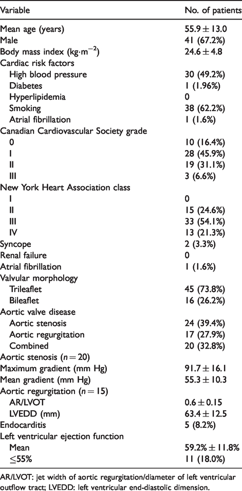
AR/LVOT: jet width of aortic regurgitation/diameter of left ventricular outflow tract; LVEDD: left ventricular end-diastolic dimension.
All patients underwent surgery under general anesthesia. A central venous line was established, and invasive blood pressure monitoring was performed. Other preoperative procedures were similar to those used in open heart surgery. All patients underwent transesophageal echocardiography in operating room before and after surgery. The aortic valve was reconstructed according to the techniques described by Ozaki and colleagues. 13 Sternotomy or a minimally invasive J- or L-shape sternotomy (upper half) was performed. The pericardium was dissected and harvested using a Harmonic knife (8 × 8 cm). The diaphragmatic face was preferred as it is thicker than other parts of the pericardium. The harvested pericardium was treated with 0.625% glutaraldehyde for 10 min and washed with normal saline for 6 min. This step was repeated three times (18 min in total; Figure 1). 13 After processing the pericardium, cardiopulmonary bypass was instituted with one arterial and one venous cannula. The left heart suction line ran from the right upper pulmonary vein through the mitral valve to the left ventricle. Cardioplegia was delivered through the aortic root or directly through the coronary orifice, and body temperature was reduced to 32°C. The aorta was opened along the circumference 1.5 cm above the right coronary root. All leaflets of the aortic valve were removed. The size of the aortic valve annulus was measured using a prosthetic valve measuring tool, and the distance between the commissures were measured using an AVNeo kit. The pericardium was cut to form three leaflets using Ozaki’s block based on the measured size, prioritizing the largest leaflet at the diaphragm side. The leaflets were sutured to the annulus using 4/0 suture. The 3 respective commissures were reconstructed (Figure 2). In patients with a bileaflet valve, we divided the annular circumference to create three leaflets. In those with endocarditis affecting one leaflet, reconstruction was performed on the affected leaflet only while preserving the other two.

Autologous pericardium harvested and treated with 0.6% glutaraldehyde solution.

Aortic valve reconstruction with pericardium under direct vision and on transesophageal echocardiography.
After surgery, the patients were monitored in the intensive care unit. An antiplatelet agent was prescribed for all patients who did not have indications for vitamin K antagonist anticoagulants.
Preoperative demographic, clinical, laboratory, echocardiographic, and electrographic data were collected. Operative parameters, including operative time, extracorporeal circulation time, aortic crossclamp time, size of the aortic valve annulus, leaflets, and number of reconstructed leaflets, were also collected. After the surgery, the duration of mechanical ventilation, length of stay in the intensive care unit, and surgical complications, such as hemorrhages, reoperation, and renal failure, were recorded. Transthoracic echocardiography was performed 1 week after surgery, and all echocardiographic parameters were recorded.
Categorical data are presented as frequencies and percentages while continuous variables are presented as mean ± standard deviation if normally distributed, or median and interquartile range otherwise. All analyses were performed using software, and a two-tailed p value < 0.05 was considered statistically significant.
Results
Table 2 summarizes the intraoperative parameters of the 61 patients. Fifty-six (91.8%) patients underwent surgery via sternotomy, and the surgery was performed via minimally invasive sternotomy (J- or L-shaped) in 5 patients. Two patients were converted to valve replacement surgery after attempts to perform Ozaki’s procedure had failed. Intraoperative transesophageal echocardiography showed a mean pressure gradient of 8 ± 2 mm Hg and an aortic valve area of 3.04 ± 0.44 cm2. The postoperative details are summarized in Table 3. One patient required reoperation and died on postoperative day 1 due to cardiac tamponade. The echocardiography findings 1-week after surgery are listed in Table 4. Echocardiography was performed 1-week after surgery in 58 (95%) patients and showed mild or no aortic regurgitation. The mean orifice area and pressure gradient were 3.04 ± 0.44 cm2 and 6.8 ± 1.4 mm Hg, respectively. The left ventricular ejection fraction was preserved. Fifty-eight patients were followed-up for a mean of 18.5 ± 5.7 months and their characteristics are summarized in Table 5. One patient presented severe aortic regurgitation due to endocarditis at 6 months after surgery, who was not the one diagnosed with endocarditis before surgery, and benefited from a reoperation with bioprosthetic valve replacement. Another patient died from an abscess on the mediastinum and aortic root at 8 months postoperatively. The patient with mediastinitis was readmitted at 4 months postoperatively due to surgical wound infection and a sternal fistula. She was reoperated on to remove the infected tissue and the surgical wound was taken care of for 4 months, but she eventually died because of a mediastinitis-induced aortic perforation. The risk factors in this patient were female sex, advanced age, and a very thin sternum. Of the 56 patients who had 1-year follow-up data, 55 had no or mild aortic regurgitation, and one had mild to moderate aortic regurgitation. The aortic valve orifice area was 2.9 cm2, and the mean pressure gradient was 6.95 mm Hg.
Intraoperative parameters of 61 patients undergoing aortic valve reconstruction.

Postoperative details of 61 patients undergoing aortic valve reconstruction.

Echocardiography 1-week after surgery in 58 patients (excluding two conversions and one death).

Follow-up results 1-year postoperatively in 58 patients (excluding two conversions and one death on day 1).

Discussion
Aortic valve replacement surgery using a prosthetic valve is still standard practice in many cardiovascular surgical centers worldwide, including Vietnam. 6 Ozaki’s procedure has been performed since 2007 at Toho University Hospital. Ozaki and colleagues11,13,14 have reported positive short- and midterm outcomes of the procedure, with many advantages, such as no need for postoperative vitamin K antagonists, a low pressure gradient, and a good valvular orifice area. We have been implementing Ozaki’s procedure in our hospital since June 2017 after being trained by Professor Ozaki. In this study, we evaluated short-term outcomes of the procedure in 61 patients with isolated aortic valve disease.
Two patients in our study were converted to aortic valve replacement surgery. This rate was higher than that reported by Ozaki and colleagues 14 and Reuthebuch and colleagues, 17 in which no conversion was required. In these two patients, after Ozaki’s procedure, when the cardiopulmonary bypass ended, their left ventricular function was poor. Transesophageal echocardiography raised the suspicion that the left coronary cusp was obstructing the coronary ostium and causing myocardial ischemia. Therefore, we decided to convert to valve replacement surgery, which resulted in a normally functioning heart. In Ozaki’s procedure, the reconstructed cusps are at the level of the sinotubular junction, which is higher than that of natural cusps.13,14 We believe that a large left coronary leaflet can potentially obscure the left coronary ostium because the rate of postoperative coronary artery stenosis or occlusion after aortic valve replacement surgery ranges from 0.5–3%. 18 One patient in our study died on day 1 post-surgery due to cardiac tamponade. The 30-day mortality was 1.6% in our study (Table 2), which is comparable to the 1.88% rate in the study by Ozaki and colleagues, 14 and lower than the 3.33% rate in the study by Reuthebuch and colleagues (Table 4). 17 The 30-day mortality rate of aortic valve replacement surgery using prosthetic valves is reported to be 2%–3% post-surgery. 18
In our patients, intensive care unit stay was 4.7 days. Because as this is a new technique, we kept the patients in the intensive care unit for longer than usual. Nevertheless, the mean ventilation time in our study was 12.6 hours, which is comparable with conventional aortic valve replacement surgery. From a technical point of view, we performed Ozaki’s procedure via minimally invasive sternotomy (J- or L-shape) in 5 patients and achieved successful results, suggesting that this less invasive technique can be applied in Ozaki’s procedure. The aortic crossclamp time and duration of extracorporeal circulation in our study were 111 ± 20 min and 139 ± 29 min, respectively. In terms of these parameters, Ozaki and colleagues 13 reported these times as 98.5 ± 7.3 min and 142.5 ± 3.4 min, respectively, in 88 patients. In the present study, the effective valve orifice area (3.0 cm2) and pressure gradient (6.8 ± 1.4 mm Hg) were comparable to those of Ozaki and colleagues 14 and Reuthebuch and colleagues. 17
We reconstructed only one leaflet in two patients. One of these patients experienced endocarditis of the left coronary cusp, and the other was diagnosed with noncoronary cusp prolapse. In our patients, we followed all the measuring, cutting, and suturing techniques of the Ozaki procedure. Twenty-two (36.1%) patients who had a small aortic annulus (< 21mm) had good outcomes. Ozaki and colleagues 11 previously reported good results in this subgroup of patients, especially when transcatheter aortic valve implantation or a non-scaffold prosthetic valve was not feasible. 17 In a study by Ozaki and colleagues, 14 the midterm results (5-year follow-up) showed that 4.2% of patients required reoperation. In our study, one (1.6%) patient required reoperation due to endocarditis at 8 months post-surgery. In contrast, a previous study reported a 1%–6% rate of endocarditis after prosthetic aortic valve replacement surgery. 19 At 1-year follow-up, 55/56 patients in our study were taking aspirin, and one was taking a vitamin K antagonist for comorbid atrial fibrillation. No thromboembolic events were recorded.
This was a single-center study with a small sample size. The follow-up time was short, which impedes comparisons with outcomes of aortic valve replacement surgery. We will continue to follow-up our patients to evaluate long-term outcomes. However, according to the short-term outcomes reported in the present study and the midterm results reported by Ozaki’s group, aortic valve reconstruction surgery using the Ozaki procedure in Vietnam is safe, effective, and associated with good short-term outcomes. This could be a promising therapeutic option in patients who are unable to have close monitoring or want to avoid using anticoagulants.
Footnotes
Acknowledgements
We would like to thank the patients who trusted us and agreed to undergo the Ozaki procedure and voluntarily participated in this study. We would like to thank Dr. Ngo Thi Hai Linh for assisting with language in the writing process. Thank you to the anesthesiologists, cardiologists, and nurses of the Cardiovascular Center, E Hospital, for participating in the diagnosis, treatment, and surgery of the patients.
Declaration of conflicting interests
The author(s) declared no potential conflicts of interest with respect to the research, authorship, and/or publication of this article.
Funding
The author(s) received no financial support for the research, authorship, and/or publication of this article.
