
Abstract
Background
Gastroesophageal reflux disease (GERD) is associated with epithelial injury and inflammation of the upper gastrointestinal tract. Long-term pharmacological therapies may present limitations, encouraging interest in complementary approaches.
Aim
This study evaluated the in vitro wound healing and anti-inflammatory activity of the multicomponent gel food supplement Reflus Gel® (RG), formulated for gastric acidity management.
Methods
Wound healing was assessed using a scratch assay on fully confluent Caco-2 cells, while anti-inflammatory activity was evaluated by measuring interleukin (IL)-1β, IL-6, and tumour necrosis factor (TNF)-α release in lipopolysaccharide-stimulated THP-1-derived macrophages by enzyme-linked immunosorbent assay.
Results
RG enhanced wound closure at 24 and 48 h compared with untreated control (p < 0.0001) and reduced IL-1β (p < 0.01), IL-6 and TNF-α (p < 0.0001) secretion in stimulated macrophages.
Conclusions
These findings indicate that RG promotes cell migration and modulates inflammatory responses in vitro, suggesting a potential role as a complementary strategy in GERD management.
Keywords
Introduction
Gastroesophageal reflux disease (GERD) is a common chronic disorder affecting approximately 14% of the global adult population (Xie et al., 2025). It is characterised by reflux of gastric contents into the oesophagus, which results in epithelial damage (Fass et al., 2021). Proton pump inhibitors are the mainstay of treatment, but frequently provide incomplete relief or require long-term use with associated adverse effects (Kröner et al., 2021). These limitations have encouraged increasing interest in complementary approaches (Komolafe et al., 2025).
Beyond acid exposure, GERD involves epithelial barrier dysfunction and inflammation (Phillips et al., 2025). Reflux injury induces the release of proinflammatory mediators, such as IL-1β, IL-6 and TNF-α, which inhibit epithelial repair and sustain mucosal damage (Zheng and Tao, 2025). Recent advances in drug delivery systems aim to improve the efficacy of anti-inflammatory compounds across pathological contexts (Bukke et al., 2024; Chandrakala et al., 2025). These developments reflect a growing interest in modulating inflammation and supporting tissue homeostasis (Medhi et al., 2025; Zehravi et al., 2025).
Reflus Gel® (RG) is a multicomponent gel food supplement formulated to counteract gastric acidity and protect the oesophageal mucosa. Its composition includes sodium alginate, potassium citrate, Emblica officinalis extract, methylsulfonylmethane (MSM) and sodium hyaluronate. Some of these components are individually described to exert wound healing and anti-inflammatory activities: Emblica officinalis extract, MSM, and sodium hyaluronate have been reported to contribute to mucosal healing and to modulate inflammatory responses through distinct pathways (Butawan et al., 2017; Yadav et al., 2017; Yue and Shao, 2025).
Despite the documented properties of the individual ingredients, the biological activity of this formulation as a whole has not been investigated. Therefore, the aim of this study was to evaluate the in vitro effects of RG on epithelial wound healing and inflammatory cytokine release in Caco-2 epithelial cells and lipopolysaccharide (LPS)-stimulated THP-1-derived macrophages, respectively.
Materials and methods
Materials and chemicals
RG sticks were kindly provided by Euro-Pharma S.r.l. (Torino, Italy). Bacterial LPS from Escherichia coli (O111:B4) (cat. no. L2630), dimethyl sulfoxide (DMSO) (cat. no. D4540), human IL-1β enzyme-linked immunosorbent assay (ELISA) kit (cat. no. RAB0273), human IL-6 ELISA kit (cat. no. EZIL6), human TNF-α ELISA kit (cat. no. EZHTNFA-150K), β-mercaptoethanol (cat. no. M3148), phorbol 12-myristate 13-acetate (PMA) (cat. no. P1585) and thiazolyl blue tetrazolium bromide (MTT) (cat. no. M2128) were obtained from Merck (Darmstadt, Germany). Dulbecco's Modified Essential Medium (DMEM) 4.5 g/L glucose (cat. no. SIAL-DMEM-HPA), non-essential amino acids solution (NEAA) (cat. no. SIAL-NEAA-B), Fetal Bovine Serum (FBS) (cat. no. yourSIAL-FBS-SA), Penicillin and Streptomycin solution (PenStrep) (cat. no. SIAL-PEN/STREP), phosphate buffer solution (PBS) (cat. no. SIAL-PBS-2A), Roswell Park Memorial Institute (RPMI) 1640 culture medium (cat. no. SIAL-RPMI-HA) and sodium pyruvate solution (cat. no. SIAL-SPS) were purchased from SIAL (Rome, Italy). Acetylsalicylic acid (ASA) (cat. no. 158185000) was acquired by Thermo Fisher Scientific (Waltham, Massachusetts, USA).
Cell culture maintenance
Caco-2 (cat. no. HTB-37) and THP-1 (cat. no. TIB-202) cell lines were obtained from ATCC. Both were grown in a CO2 incubator at 37 °C with 5% CO2 and 95% humidity.
Caco-2 cells were cultured in a 75 cm2 flask in DMEM 4.5 g/L glucose supplemented with 10% heat-inactivated FBS, 1% PenStrep and 1% NEAA. Complete medium was changed every 2 days. Cells were subcultured at 80% confluence with Trypsin-EDTA 0.25% and reseeded at a density of 1 × 10⁴ cells/cm2.
THP-1 cells were grown in 25 cm2 flask in RPMI supplemented with 10% heat-inactivated FBS, 1% PenStrep, 1% sodium pyruvate and 1 mM β-mercaptoethanol. Complete medium was added every 2 days. Cells were subcultured once the density exceeded 8 × 10⁵ cells/mL and reseeded at an initial density of 2 × 10⁵ cells/mL.
Cell viability assay
Prior to treatment, the highest non-toxic concentration of RG was determined in Caco-2 and THP-1 cells performing the MTT assay.
Briefly, Caco-2 cells were seeded in 96-well plates at a density of 1 × 10⁴ cells/well in complete medium and allowed to adhere for 24 h. The following day, the medium was removed and cells were washed with PBS. RG was diluted in serum-free medium (120 mg/mL), sterile-filtered, and applied at 0.012–120 mg/mL for 48 h.
THP-1 cells were seeded in complete medium containing 100 nM PMA at a density of 1 × 10⁵ cells/well to induce differentiation into macrophages. After 24 h, cells were washed twice with PBS and incubated overnight in complete medium without PMA. Subsequently, cells were treated with different concentrations of RG in the range 7.5–120 mg/mL for 1 h, after which LPS (10 ng/mL) was added. Cells were then incubated for additional 23 h, for a total treatment duration of 24 h.
MTT reagent (5 mg/mL) was diluted to 0.5 mg/mL in serum-free medium, and 100 µL were added to each well. After 4 h of incubation at 37 °C, absorbance was measured at 570 nm with a reference wavelength of 630 nm using a SpectraMax® Mini multi-mode microplate reader (Molecular Devices, USA). Statistical analysis was performed by one-way analysis of variance (ANOVA). The highest concentration maintaining at least 95% cell viability compared with untreated control (CTL), and showing no statistically significant reduction, was selected as the non-toxic concentration for subsequent experiments in both cell lines.
Wound healing assay
Caco-2 cells were seeded in 12-well culture plates in complete medium. Upon reaching full confluence, a uniform scratch was created using a sterile 200 µL pipette tip. The cells were then washed twice with PBS to remove detached debris and treated with 0.12 mg/mL RG for 48 h. Images of wound closure were captured immediately after scratching (T0), after 24 h (T24) and 48 h (T48) using a VisiCam P6® digital camera connected to an inverted microscope (VISISCOPE® IT417 PH, VWR, USA). The wound areas of treated and untreated control cells were quantified with PROVIEW software and normalised as the percentage of the corresponding initial wound area T0, which was set at 100%. Wound areas of RG-treated cells were then compared with untreated control cells at the corresponding time points by one-way ANOVA. The percentage reduction in wound area of treated cells was calculated relative to untreated cells according to the following equation (1):
Enzyme-linked immunosorbent assay
The levels of IL-1β, IL-6, and TNF-α were quantified in the supernatants of LPS-stimulated THP-1-derived macrophages as previously described (Carpentieri et al., 2022). Two wells without PMA and LPS were included as negative control for differentiation and inflammation (CTL-), while PMA-differentiated THP-1 cells treated with LPS served as positive control (CTL+). ASA was used at 100 µM as a positive anti-inflammatory control to validate the responsiveness of the experimental model (Liu et al., 2017). This concentration was selected within the non-cytotoxic and effective range reported for THP-1 cells (Garzón-Porras et al., 2019). Briefly, cells were seeded in 12-well plates at a density of 1 × 10⁶ cells/well in complete medium supplemented with 100 nM PMA to induce differentiation. After 24 h, cells were washed twice with PBS and incubated overnight in complete medium without PMA. The following day, macrophages were treated with 15 mg/mL RG, following the same conditions described for the MTT assay. After treatment, supernatants were collected, centrifuged at 3600 × g for 5 min to remove cellular debris, and stored at −80 °C until analysis.
IL-1β, IL-6, and TNF-α were quantified using ELISA kits (Merck). Statistical differences between groups were assessed using one-way ANOVA. The percentage of reduction of each cytokine in RG-treated and ASA-treated cells was calculated over CTL+ according to the following equation (2):
Statistical analysis
All experiments were conducted in three biological replicates, corresponding to independent cell passages performed on different days, each in duplicate. Data are presented as mean ± standard error of the mean (SEM). Statistical analyses were carried out using GraphPad Prism 9 (GraphPad Software, San Diego, CA, USA). Comparisons between groups were performed using t-tests or ANOVA, as appropriate, followed by the relevant post hoc corrections. The statistical tests applied to each dataset are reported in the corresponding figure legends. Statistical significance was set at p < 0.05.
Results
Effect of RG on wound closure in Caco-2 confluent cells
To evaluate the effect on epithelial cell migration, fully confluent Caco-2 cells were scratched and treated with 0.12 mg/mL RG, the previously determined non-toxic concentration (Supplemental Material, Figure S1). Comparison at the same time point between RG-treated cells (RG) and untreated control cells (CTL) showed that RG significantly reduced wound area (Figure 1), resulting in a 28.4% ± 2.5% and 43.4% ± 4.2% increase in wound closure at 24 and 48 h, respectively (p < 0.0001) (Figure 2).
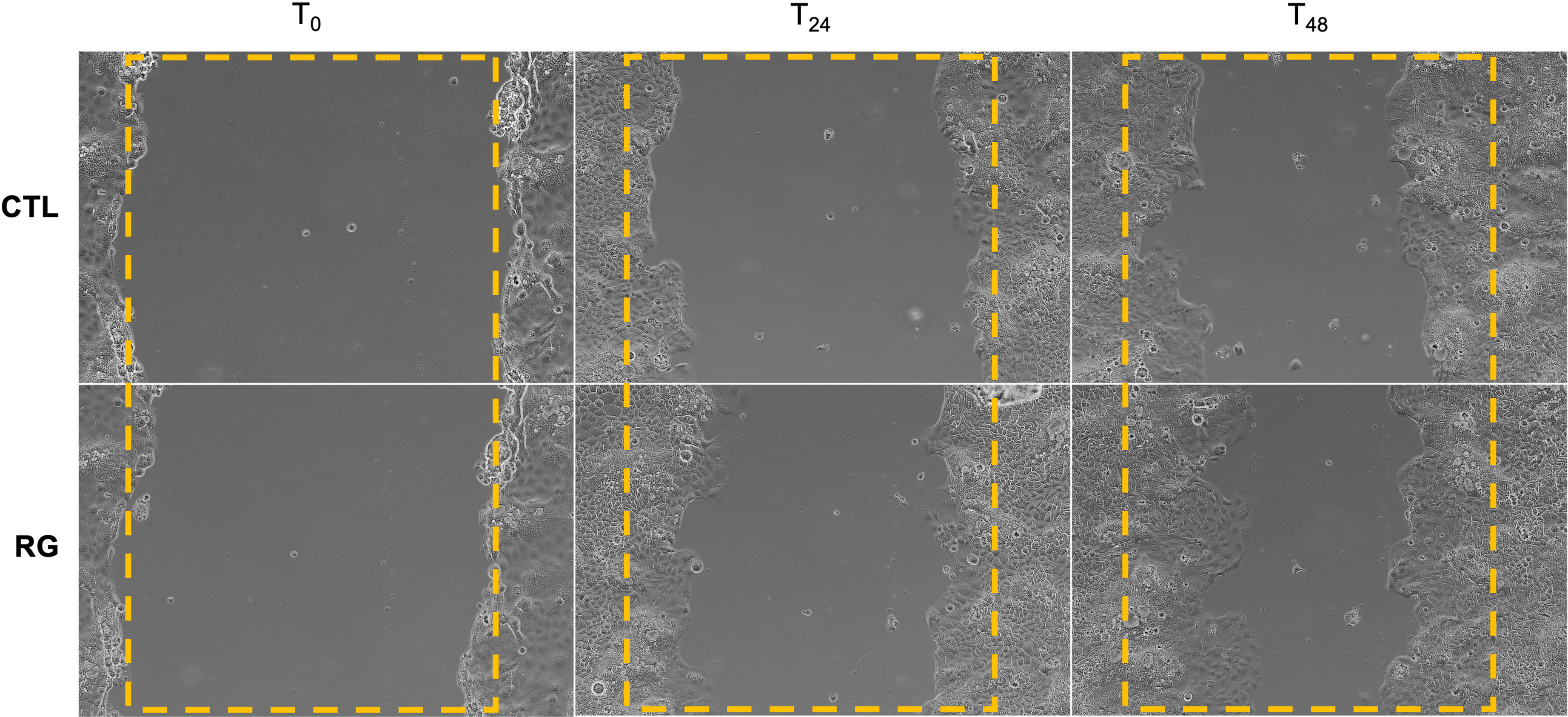
Representative images (Magnification: 10×) of scratched Caco-2 cells at 0 h (T0), 24 h (T24), and 48 h (T48) after treatment with Reflus Gel® (RG) or control medium (CTL).
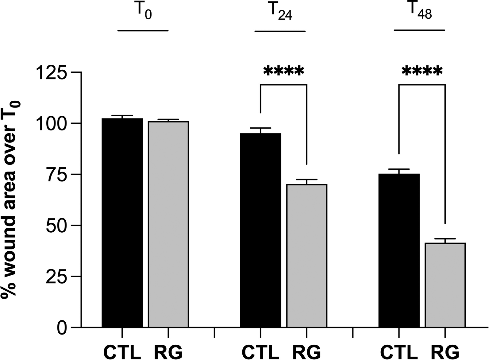
Statistical analysis of wound area differences between untreated control cells (CTL) and RG-treated cells, expressed as a percentage of the initial scratch width at T0. Data are shown as mean ± SEM of three independent experiments, each performed in duplicate. Statistical significance was assessed using one-way ANOVA; ****p < 0.0001. SEM: standard error of the mean; RG: Reflus Gel®; ANOVA: analysis of variance.
Anti-inflammatory effects of RG in LPS-stimulated human macrophages
To assess the anti-inflammatory effect, LPS-stimulated macrophages were treated with 15 mg/mL RG, the previously determined non-toxic concentration (Supplemental Material, Figure S2). The release of IL-1β, IL-6, and TNF-α was evaluated in four experimental groups: undifferentiated THP-1 cells (CTL–, negative control), PMA and LPS-stimulated macrophages (CTL+, positive control), ASA-treated (ASA) and RG-treated macrophages (RG). All conditions were compared with the reference CTL+. CTL– showed significantly lower cytokine levels than CTL+ (p < 0.0001), confirming effective stimulation. ASA reduced IL-1β by 18.4 ± 2.8% (p < 0.05), IL-6 by 26.8 ± 4.1% (p < 0.01), and TNF-α by 23.8 ± 2.5% (p < 0.01) compared with CTL+, confirming the validity of the experimental system. RG significantly reduced the release of IL-1β by 38.7 ± 3.4% (p < 0.01), IL-6 by 44.5 ± 3.9% (p < 0.0001), and TNF-α by 39.0 ± 2.5% (p < 0.0001) compared with CTL+, indicating a reduction of the inflammatory response. Statistical comparisons between groups are shown in Figure 3.
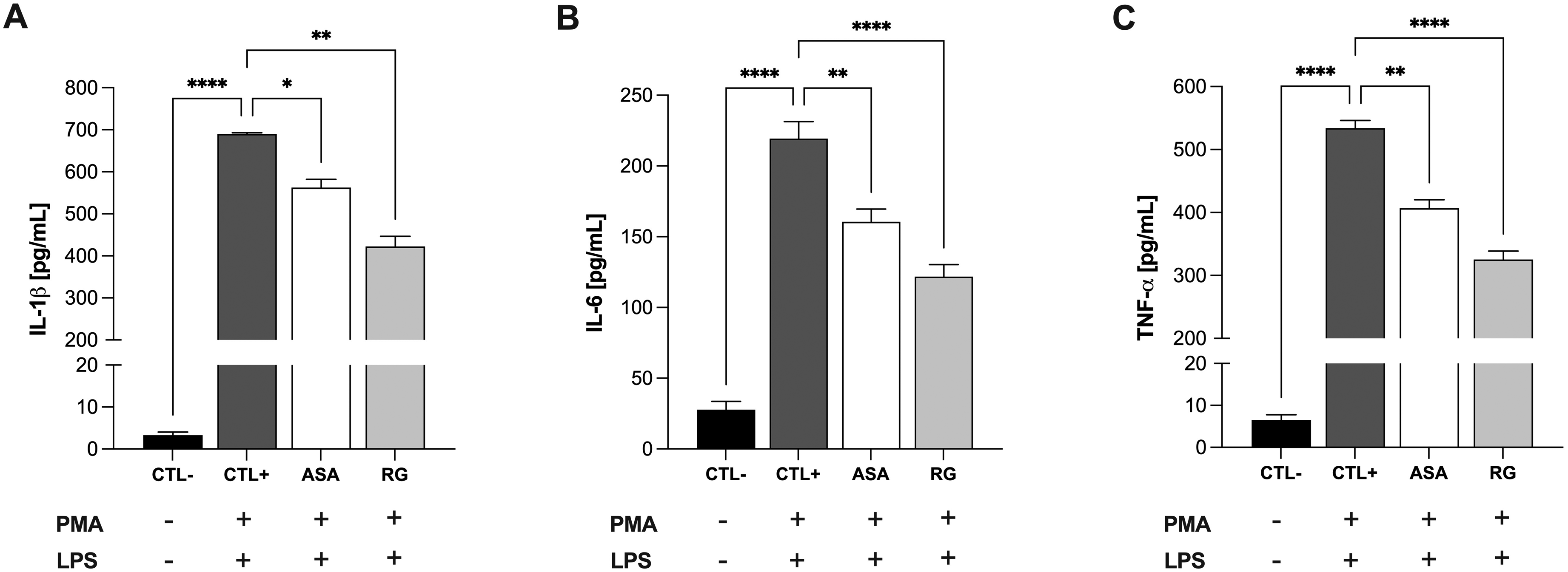
Levels of (A) IL-1β, (B) IL-6, and (C) TNF-α in THP-1 cell culture supernatants, quantified by ELISA. PMA-differentiated and LPS-stimulated THP-1 macrophages served as the positive control (CTL+), while undifferentiated THP-1 cells cultured in the absence of PMA and LPS served as the negative control (CTL−). ASA at 100 μM was used as positive control of anti-inflammatory activity. RG represents LPS-stimulated macrophages treated with the highest non-toxic concentration of Reflus Gel®. The symbols “+” and “–” below the graph indicate the presence or absence of PMA and LPS treatments. Cytokine concentrations in CTL+ samples were compared with those in CTL−, ASA and RG. Data are expressed as mean ± SEM from three independent experiments, each performed in duplicate. Statistical differences were determined by one-way ANOVA followed by Dunnett's post hoc test; *p < 0.05, **p < 0.01, ****p < 0.0001. ELISA: enzyme-linked immunosorbent assay; LPS: lipopolysaccharide; PMA: phorbol 12-myristate 13-acetate; ASA: acetylsalicylic acid; SEM: standard error of the mean; IL: interleukin; TNF: tumour necrosis factor; RG: Reflus Gel®.
Discussion
In this study, we observed that the multicomponent nutraceutical RG was associated with increased epithelial wound closure and reduced inflammatory IL-1β, IL-6 and TNF-α release in vitro.
The enhanced wound closure observed in Caco-2 cells suggests a direct effect of RG on cell migration, a key process in epithelial restitution. In parallel, the significant reduction of macrophage-derived cytokines indicates the anti-inflammatory potential of the formulation. These effects could be interconnected: the anti-inflammatory action may indirectly enhance cell migration by limiting the persistence of inflammatory mediators. This dual activity is particularly relevant in the context of GERD, where inflammation is known to impair mucosal healing (Ustaoglu et al., 2020).
The observed effects may result from the synergistic action of the formulation's ingredients. Emblica officinalis extract, MSM, and sodium hyaluronate have been widely studied for their ability to modulate cell migration and cytokine production through distinct pathways (Ahn et al., 2015; Huang et al., 2024; Marinelli et al., 2022). Given this, their combination may be associated with greater observed effects compared with the individual components. However, some important limitations should be considered. First, the individual contribution of the formulation components was not evaluated; therefore, the possibility of enhanced effects arising from synergistic interactions remains to be clarified. Second, the findings are based on in vitro models, which do not fully reproduce the complexity of the gastrointestinal environment in vivo. Thus, further in vivo and clinical studies are needed to confirm the efficacy of this formulation in the context of GERD. Third, the use of immortalised cell lines may not fully reflect the behaviour of primary human tissues. Fourth, the study did not investigate the specific molecular mechanisms underlying the observed effects, limiting the mechanistic interpretation of the results. Finally, functional assays were performed using a single concentration of RG, preventing the assessment of dose–response relationships.
In conclusion, this work provides preliminary insights into the potential biological effects of the nutraceutical RG, highlighting the need for further investigation in in vivo and clinical settings to assess its suitability and efficacy in more physiologically relevant GERD models.
Supplemental Material
sj-docx-1-nah-10.1177_02601060261452076 - Supplemental material for In vitro wound healing and anti-inflammatory properties of a multicomponent gel food supplement: Preclinical evidence for a complementary approach in gastroesophageal reflux disease management
Supplemental material, sj-docx-1-nah-10.1177_02601060261452076 for In vitro wound healing and anti-inflammatory properties of a multicomponent gel food supplement: Preclinical evidence for a complementary approach in gastroesophageal reflux disease management by Marta Pulcini, Valerio Mori, Maria G Marini and Luca Massaccesi in Nutrition and Health
Footnotes
Acknowledgments
The authors thank Euro-Pharma S.r.l. for providing Reflus Gel® sticks.
Ethical considerations
This article does not contain any studies with human or animal participants.
Consent to participate
Not applicable.
Consent for publication
Not applicable.
Author contributions
Funding
The authors disclosed receipt of the following financial support for the research, authorship, and/or publication of this article: The study received financial support from Euro-Pharma S.r.l.
Declaration of conflicting interest
The authors declared the following potential conflicts of interest with respect to the research, authorship, and/or publication of this article: A patent application (European Patent Application No. EP4059506) related to the formulation described in this study is owned by Euro-Pharma S.r.l and is currently under examination. The sponsor had no role in the design, execution, interpretation, or writing of the study. The presence of this pending patent did not influence the study design, data interpretation, or decision to submit for publication.
Data availability statement
The datasets generated and analysed during the current study are available from the corresponding author upon reasonable request.
Supplemental material
Supplemental material for this article is available online.
References
Supplementary Material
Please find the following supplemental material available below.
For Open Access articles published under a Creative Commons License, all supplemental material carries the same license as the article it is associated with.
For non-Open Access articles published, all supplemental material carries a non-exclusive license, and permission requests for re-use of supplemental material or any part of supplemental material shall be sent directly to the copyright owner as specified in the copyright notice associated with the article.