
Abstract
The preclinical evaluation of novel imaging agents, such as contrast agents for computed tomography (CT), still largely relies on rodent models or simplified in vitro systems, each carrying limitations with regard to ethical burden, physiological relevance or translational value. To address this gap, we propose Galleria mellonella larvae as a scalable, ethically favourable and physiologically relevant in vivo model for use in the early-stage screening of novel CT contrast agents. We established a standardised imaging protocol utilising isoflurane-based sedation and longitudinal micro-CT scanning, and used it to evaluate three commercially available contrast agents: one clinically approved molecular agent and two nanoparticulate formulations that are used for preclinical studies. All agents were systemically distributed, well-tolerated and enabled consistent visualisation of internal anatomy over eight days. While differences in clearance kinetics and route related to particle size or formulation were less pronounced compared to vertebrate models, this larval system reliably reflected biodistribution trends and allowed non-invasive tracking of agent functionality and safety. The use of G. mellonella in this context enables early elimination of less promising candidates and reduces reliance on vertebrates in downstream (pre)clinical development, aligning with the Three Rs principles. Our findings support the use of G. mellonella as a practical intermediate model, bridging in vitro cell-based assays and in vivo rodent studies in the development of next-generation CT and imaging contrast agents.
Keywords
Introduction
Imaging technologies, and X-ray computed tomography (CT) in particular, have revolutionised our ability to investigate both anatomical and physiological processes across clinical and preclinical settings.1,2 CT frequently relies on the use of contrast agents, typically molecules with high atomic numbers (Z), to enhance soft-tissue and vascular visualisation. 3 Clinically, iodinated compounds are most commonly used as intravascular contrast agents, whereas barium sulphate (BaSO4) remains standard for gastrointestinal imaging.4–7 Despite their widespread use and diagnostic value, the rapid renal clearance of iodinated contrast agents may pose risks, including thyroid dysfunction and contrast-induced nephropathy, particularly in individuals with impaired kidney function. 8
Currently, the field of CT is experiencing exciting advancements, with the development of novel imaging techniques such as phase-contrast CT and spectral CT, with the latter including dual-energy and multi-energy photon-counting CT.9,10 These technologies significantly enhance the sensitivity of CT, allowing improved differentiation between soft tissues and contrast agents in vivo. As a result, they enable improved vascular imaging, more accurate tumour characterisation and delineation, and the simultaneous tracking of multiple contrast agents within a single scan.11–13
Beyond expanding diagnostic capabilities, these technologies also stimulate the development of new contrast agents aimed at improving visualisation and tissue discrimination.1,14 In spectral CT, high-Z materials such as lanthanides, tungsten, bismuth and gold are being explored, because their energy-dependent attenuation properties allow more versatile imaging applications beyond conventional iodine-based agents.3,15 In addition, many of these novel agents are designed to retain rapid excretion profiles, helping to reduce systemic retention and potential toxicity. 13
In parallel, high-Z element-containing nanoparticles are being developed as next-generation contrast agents for preclinical micro-CT imaging. These formulations incorporate high-Z material cores to enhance contrast, and are engineered with surface modifications that prolong circulation half-life compared to conventional small molecule contrast agents. 16 As a result, they provide increased vascular contrast and an extended imaging window, making them well-suited for dynamic and longitudinal small-animal studies. 17 Examples include barium-based nanoparticles, (iodine) nano-emulsions and gold(-coated) nanoparticles. 18 However, their toxicity profiles remain poorly understood and difficult to predict. Due to their prolonged in vivo half-life, these nanoparticles have increased potential for interaction with endogenous tissue, and tend to accumulate in various organs and host cells, raising concerns about possible long-term biological effects beyond their imaging purpose. 18 Hence, comprehensive in vivo toxicity and biocompatibility studies remain essential. Despite ongoing progress, the development of non-toxic, high-performing and cost-effective next-generation contrast agents continues to be a significant challenge. 3
Currently, screenings toward safety, biocompatibility and functionality of clinical and preclinical CT contrast agents are mainly performed in in vitro cell culture systems and in vivo rodent models.19–21 While in vitro assays are useful for early evaluation, they do not adequately reflect physiological complexity and are often insufficient for assessing biodistribution, clearance and systemic toxicity, 22 leading to discrepancies with animal studies. 23 On the other hand, although rodent models provide valuable translational insight, their use raises ethical and regulatory concerns and should be minimised wherever possible. 24 Therefore, we propose the invertebrate model, Galleria mellonella, as an intermediate in vivo screening platform to bridge the gap between cell-based assays and mammalian testing, enabling the scalable and ethically favourable evaluation of novel imaging contrast agents.
G. mellonella, commonly known as the greater wax moth, is an invertebrate organism, and therefore a moth-based screening platform can be regarded as a new approach methodology (NAM).25,26 Like other insects, they possess an open circulatory system composed of a haemocoel (body cavity) in which haemolymph (body fluid) is pumped by the hearts of a cylindrical dorsal vessel. This allows for the distribution of nutrients, hormones and immune cells throughout the larva. 27 Metabolic or infection-related waste present in the haemolymph are primarily removed via the Malpighian tubules, the main excretory organs of insects, which actively transport solutes and water from the haemolymph into the gut lumen. The resulting primary urine passes into the hindgut, where further reabsorption occurs before final elimination through the faeces.28,29
The larvae possess other organ systems, including a structurally organised and immunologically active gastrointestinal tract, a tracheal respiratory system and a fat body that is analogous to both the mammalian liver and adipose tissue, reflecting several aspects of mammalian physiological complexity.25,30–32 The innate immune system of G. mellonella is functionally similar to that of vertebrates, and the larvae can survive and function at physiologically relevant temperatures, thereby making them particularly compatible with human-relevant studies.30,33,34 As a result, G. mellonella has become an established in vivo model for evaluating systemic toxicity responses, as well for studying microbial infections and host–pathogen interactions, including implant-associated biofilms, with recent advances in imaging-supported readouts.35–37 These biologically relevant advantages have led to its extensive use in toxicity and efficacy studies of various chemical and pharmaceutical compounds.30,33,34,37–43 Compared to in vitro models, the larvae offer a more physiologically active environment for assessing systemic effects, while still remaining more accessible and ethically favourable than murine models. Their ease of maintenance, low cost, compatibility with multiple administration routes, and suitability for high-throughput screening make them especially attractive for early-stage compound evaluation.30,33,34 Furthermore, this model supports the principles of the Three Rs (replacement, reduction, refinement) by reducing the need for vertebrate testing in early-stage research, while maintaining physiological relevance. 44
In this study, we present G. mellonella as a non-vertebrate alternative model to bridge the substantial gap between the in vitro development and in vivo screening of novel contrast agents toward safety, tolerability and functionality, which typically relies on murine models. To this end, we established and evaluated a reliable isoflurane-based sedation and micro-CT scanning protocol for G. mellonella. We evaluated the protocol in terms of in vivo tolerability and functionality screening by using three commercially available contrast agents with different physicochemical properties and clearance profiles: one clinically approved molecular iodine-based agent, and two nanoparticulate formulations developed for preclinical imaging. We hereby demonstrate the potential of G. mellonella as a practical and ethically favourable intermediary model for use in early-stage functionality and biocompatibility screening during CT contrast agent development.
Materials and methods
Animals and ethics
The G. mellonella larvae used for the experiments were bred from moths kept in-house. The larvae were maintained in large, ventilated glass jars (2-litre Mason jars) covered with mesh lids to ensure airflow and prevent contamination. They were maintained in the dark in a controlled environment, at 30% humidity and 28–32°C. They were fed a diet rich in carbohydrates, with a balanced amount of protein and lipids, without preservatives or antibiotics. Only larvae that reached the final stage of larval maturation, referred to as the last-instar stage (last of eight stages, occurring after approximately six weeks of development), were included in this study. Healthy larvae were selected based on their uniformity in colour (minimal to no melanisation), weight (300 ± 50 mg), and responsiveness to tactile stimulation. During the experiments, larvae were kept individually in separate 200-ml screwcap containers (Corning Gosseling™, Borre, France) at 32°C in the dark. Longitudinal experiments were limited to 7–8 days, as last-instar larvae generally initiate pupation within this period, following maturation at approximately seven weeks post-hatching. Each experimental run was performed independently, i.e. using separate batches of larvae (separate breeding jars), and unhandled healthy controls were included for each batch. The experiments were performed in compliance with European and local ethical guidelines, under which research on invertebrates such as insects does not require prior ethical approval.
Sedation
Last-instar larvae of G. mellonella were deeply sedated by placing them individually in a sealed 60-ml incubation chamber alongside a cotton swab impregnated with isoflurane (Iso-Vet, Covetrus, the Netherlands). Varying proportions of isoflurane in air were used, according to the volume loaded onto the swab in relation to the 60-ml incubation chamber volume — i.e. 0.010 ml (3.3%), 0.025 ml (8.3%), 0.050 ml (16.6%) and 0.100 ml (33.2%), for exposure times ranging from 1 to 8 minutes (Figure 1(a)). Dose comparison and anaesthesia induction time-dependent effects of isoflurane sedation on immobilisation, recovery time and health in G. mellonella larvae. (a) Illustrative images of isoflurane sedation by using a cotton swab impregnated with a defined volume of isoflurane, followed by larvae incubation in a sealed chamber for anaesthesia induction. (b) The number of larvae (n = 10) that were immobilised after sedation for the different combinations of isoflurane dose and incubation time. (c) Recovery time until first movement after sedation of the larvae (n = 10) with different isoflurane doses and for different incubation times. The dashed box indicates the selected sedation condition used for subsequent experiments. (d) Tolerance of the larvae to different isoflurane doses and incubation times, assessed via the health index scoring system. Data are presented as the mean ± standard deviation (SD); ns = non-significant.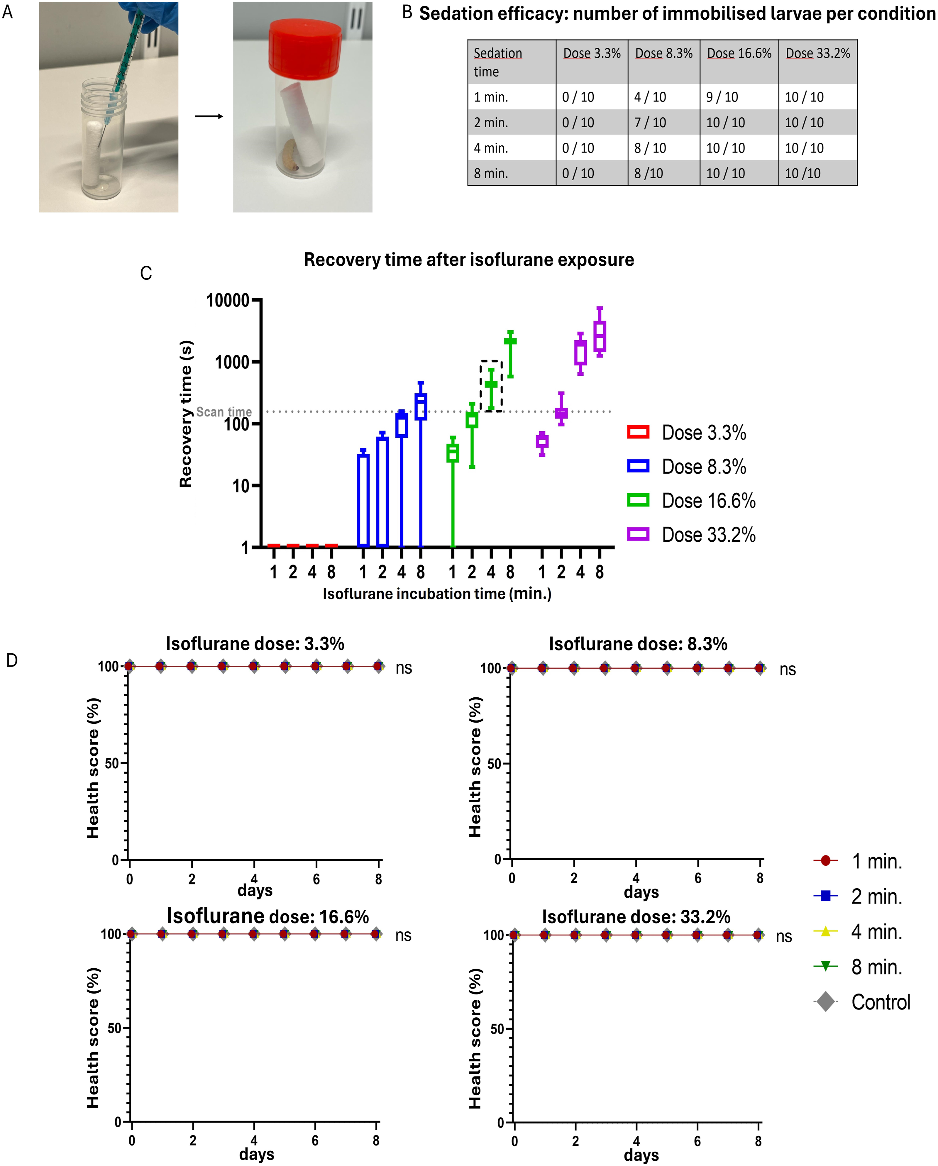
Micro-computed tomography (micro-CT)
Isolated samples of tissue or sedated larvae were positioned in a supine orientation on the micro-CT bed, centred within the field of view by using a polystyrene foam block and secured in place with paper tape. Micro-CT scans were performed with an in vivo Skyscan 1278 (Bruker μCT, Kontich, Belgium), employing the following scanning parameters: source voltage 65 kV combined with a 1 mm Al-filter; source current 770 μA; pixel size 50 μm; frame averaging 3; and exposure time 40 ms. For imaging, three contrast agents were used: Visipaque™ (320 mg iodine/ml; GE Healthcare, Machelen, Belgium), Fenestra™ HDVC (Medilumine Inc., Québec, Canada) and ExiTron™ nano 12000™ (Viscover, Berlin, Germany). Contrast agents were administered immediately before micro-CT imaging. Sedated larvae were injected in the right-hind proleg using a Hamilton syringe, before being positioned in a supine orientation on the micro-CT bed. Micro-CT data were reconstructed and subsequently visualised with software provided by the manufacturer (NRecon, DataViewer, CTVox, and CTan).
Tolerability testing
The health index scoring system used to assess tolerance of the G. mellonella larvae.
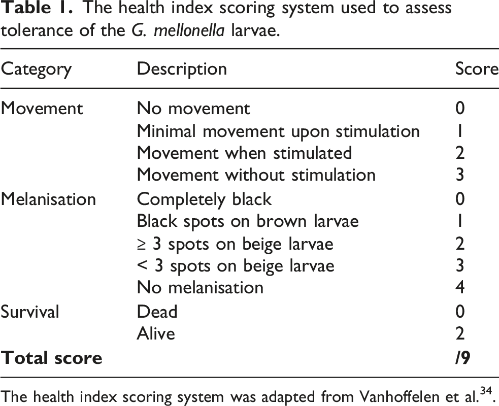
The health index scoring system was adapted from Vanhoffelen et al. 34 .
Statistical analysis
Statistical analyses were performed by using GraphPad Prism (Version 8.0.2; GraphPad Software, USA). Longitudinal health index scores were analysed by mixed-effects two-way ANOVA, to detect significant differences within an experiment and between groups at defined time points (post-hoc Sidak’s multiple comparisons test). Kaplan–Meier survival curves were analysed using the log-rank (Mantel–Cox) test. Statistical differences were considered significant at p < 0.05; n values represent the number of animals in each group.
Results
Establishing an isoflurane gas anaesthesia protocol that effectively immobilises the G. mellonella larvae and is well-tolerated
To be able to evaluate the in vivo functionality and tolerability of contrast agents in G. mellonella, we first required a reliable imaging protocol that would allow for repeated high-resolution micro-CT scanning and sedation without affecting larval health. To this end, we initially compared several sedation strategies and established a robust protocol that ensured complete immobilisation during the entire scan duration (158 seconds), effectively preventing motion artefacts and preserving high-resolution image quality. First, we sedated the larvae by placing them on ice, as commonly practised in existing protocols.27,45 While this method immobilised the larvae, it did not fully suppress subtle movements, such as dorsal body wall contractions and head twitches. As a result, reconstructed micro-CT images showed noticeable blurring, making this approach unsuitable for high-resolution micro-CT scanning. Subsequently, we tested gas anaesthesia with isoflurane vapour, delivered via an isoflurane-impregnated cotton swab placed in sealed air-filled containers (Figure 1(a)). To identify optimal and well-tolerated sedation conditions, the larvae were exposed to varying isoflurane concentrations (3.3%, 8.3%, 16.6% and 33.2%) and durations (1, 2, 4 and 8 minutes).
The results showed that the two lowest doses did not reliably sedate all larvae at any of the tested incubation times for anaesthesia induction (Figure 1(b)). At the lowest concentration (3.3%), no larvae were sedated after any of the incubation times. In contrast, for the two highest concentrations (i.e. 16.6% and 33.2%), an incubation time of at least 2 minutes consistently achieved full immobilisation for all larvae (10/10). Higher doses, or extended anaesthesia induction times, also resulted in the sedation of all larvae (10/10), with no immediate observable short-term impact on larval health across all tested conditions.
Next, we observed the recovery time (time until first movement) following incubation with the isoflurane-impregnated cotton swab. For both 16.6% and 33.2% isoflurane concentrations and incubation times over 4 minutes, all larvae were fully immobilised for longer (180–7380 seconds) than the average micro-CT scanning time of the larvae (158 seconds) (Figure 1(c)). Moreover, increasing the incubation time resulted in longer immobilisation. At lower doses and shorter incubation times, the larvae recovered faster than the scanning duration, resulting in unwanted movement of the larvae during scanning.
To assess long-term tolerance of the larvae to isoflurane gas anaesthesia, we evaluated the survival and health of the larvae over a further 7-day period, during which the larvae were sedated daily (Figure 1(d) and Figure 2). This was performed to simulate the conditions of longitudinal micro-CT studies, where daily repeated imaging sessions are needed to evaluate the functionality of contrast agents over time without affecting larval viability. Over the further seven days, none of the tested isoflurane doses, or anaesthesia induction durations, led to death or to significant changes in health index scores compared to the positive PBS control (Figure 1(d) and Figure 2(a)). These findings indicate that daily isoflurane-based gas anaesthesia was well-tolerated and suitable for daily repeated use in longitudinal imaging experiments of up to eight days. Survival of G. mellonella larvae after isoflurane sedation, repeated anaesthesia, repeated micro-CT exposure and micro-CT contrast agent administration. (a) Kaplan–Meier survival curves of larvae (n = 10 per group) following a single isoflurane sedation at different doses (3.3%, 8.3%, 16.6% and 33.2%) and incubation times (1, 2, 4 or 8 minutes). No significant differences were observed compared to the unhandled controls. (b) Survival following prolonged and repeated exposure to isoflurane (sedation once or twice daily; 16.6% isoflurane in air for 4 minutes) or repeated micro-CT scanning (once or twice daily). Repeated isoflurane exposure twice daily significantly reduced survival compared to the unhandled controls, whereas repeated micro-CT exposure alone did not affect survival. (c) Survival of larvae injected with micro-CT contrast agents compared to the PBS and 35% DMSO controls. All contrast agents resulted in post-exposure survival comparable to that of the PBS control, while DMSO significantly reduced survival. Data are presented as the mean ± SD, and analysed by using the log-rank (Mantel–Cox) test; ns = non-significant, *p < 0.05, **p < 0.01, ***p < 0.001.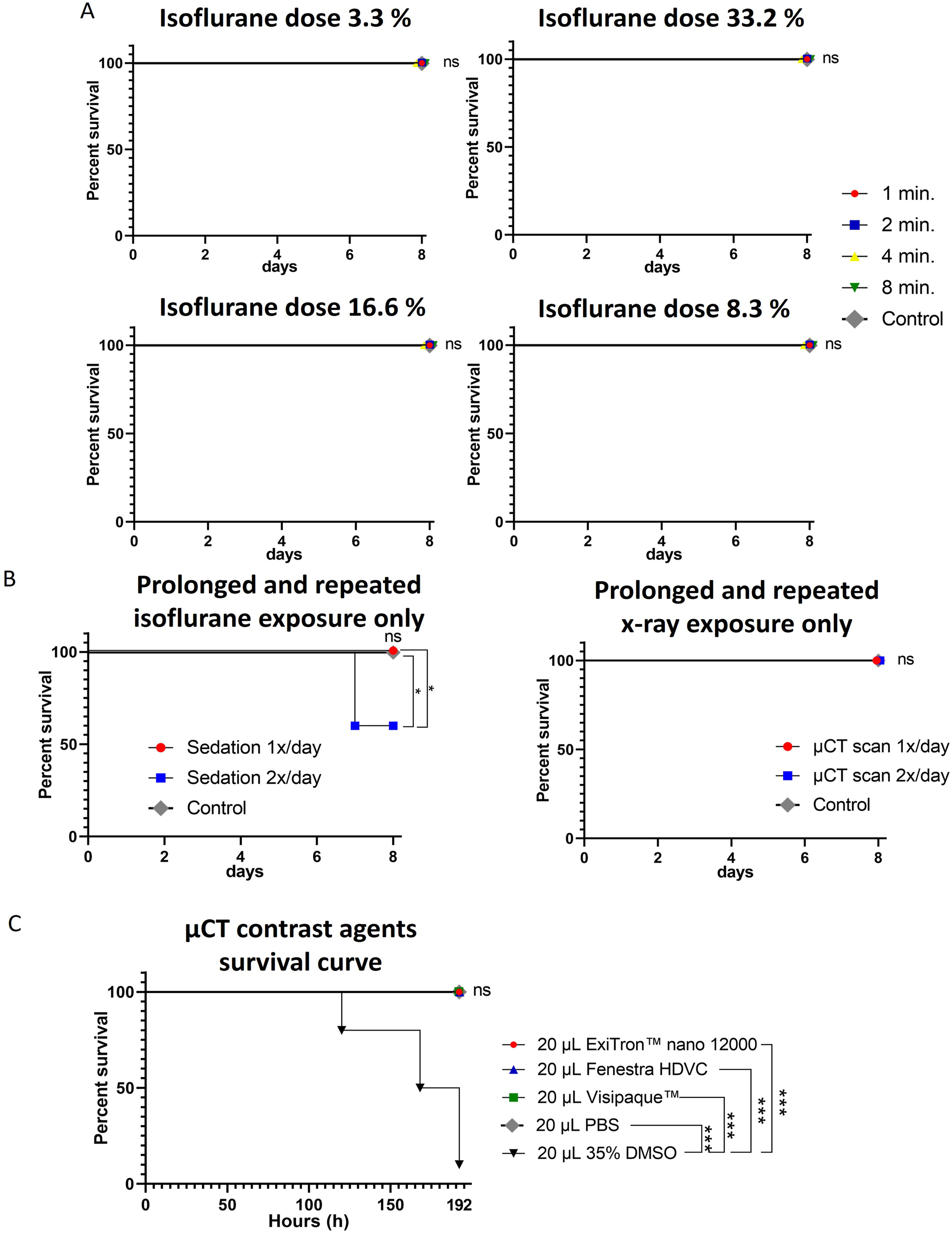
In summary, we established an isoflurane-based sedation method that effectively and reproducibly immobilises G. mellonella larvae for durations ranging from 180 seconds to 7380 seconds, fully covering the time requirements for positioning the animal and performing the high-resolution micro-CT scanning. For subsequent experiments, the 16.6% isoflurane dose with a 4-minute incubation time was selected, as this condition consistently provided a recovery duration exceeding the scanning time (i.e. 158 seconds), while representing the lowest effective concentration, thereby minimising any potential impact on the larvae. Importantly, no adverse health effects were observed following this sedation protocol, either immediately or over the further 7-day period involving daily sedation. This observed absence of impact on larval health confirms that our sedation method does not interfere with health-based readouts, making it a reliable approach for in vivo tolerability and functionality testing of contrast agents in G. mellonella.
The effects of repeated isoflurane administration and cumulative X-ray exposure on larval health
To assess whether G. mellonella larvae can serve as a reliable in vivo model for longitudinal micro-CT studies evaluating long-term contrast agent functionality and tolerability, we next examined the impact of cumulative daily exposure to sedation and ionising radiation on larval health over time. Given that time resolution, i.e. the number of imaging time points, is a key parameter in longitudinal studies, we tested both once-daily and twice-daily exposure to isoflurane and ionising radiation to determine the maximum feasible scanning frequency that does not affect larval health. To isolate the effects of isoflurane gas anaesthesia, we performed a 7-day follow-up experiment under optimised conditions (16.6% isoflurane in air for 4 minutes of anaesthesia induction), where larvae (n = 10) were sedated either once daily, or twice daily with an 8-hour interval between sedations (Figure 3(b)). Health index scores were recorded daily, and unhandled larvae served as healthy controls to assess tolerability. Sedation once per day had no significant effect on health compared to controls, supporting its use in longitudinal protocols. In contrast, twice-daily sedation led to a significant reduction in health index scores by Day 8 (Figure 3(b)), indicating that this frequency may compromise larval health and should therefore be avoided in tolerability studies. However, it may still be acceptable for short-term imaging protocols where increased temporal resolution is critical for assessing contrast agent functionality. Effects of prolonged isoflurane and X-ray exposure on the health of last-instar larvae of G. mellonella. (a) Illustrative image showing the placement of a G. mellonella larva in the micro-CT scanning bed. (b) Tolerance of the larvae (n = 10) to once-daily and twice-daily isoflurane sedation (16.6% isoflurane in air for 4 minutes), assessed via the health index scoring system; unhandled larvae served as the control. (c) Tolerance of the larvae (n = 10) to once-daily and twice-daily X-ray exposure from the micro-CT scan, assessed via the health index scoring system; unhandled larvae served as the control. Data are presented as the mean, with only the negative SD shown. ****p < 0.0001; ns = non-significant.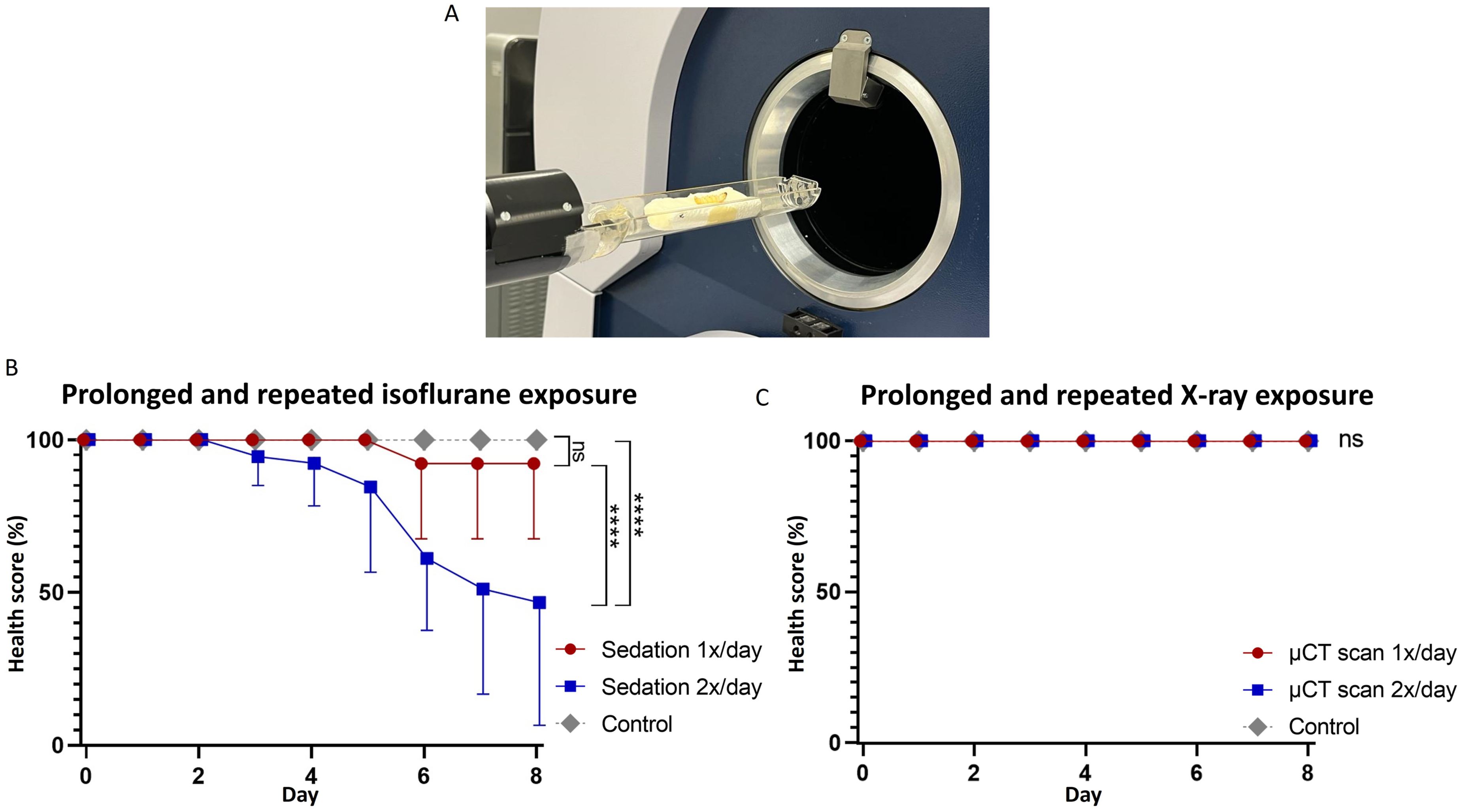
In parallel, we assessed whether repeated exposure to ionising radiation alone affected larval health. To this end, to isolate the effect of X-ray exposure, non-sedated larvae were scanned once or twice daily, using the same time intervals, and compared to unhandled controls. Health scoring revealed no measurable impact across groups, indicating that ionising radiation, even when applied multiple times daily, had no effect on larval health (Figure 3(c) and Figure 2(b)).
Investigating the biodistribution of an iodine-based molecular micro-CT contrast agent
Systemic distribution is a key requirement for establishing G. mellonella as a suitable in vivo functionality and tolerability screening model for blood pool contrast agents. Thus, we first investigated whether contrast agents injected into the right-hind proleg effectively enter the haemocoel network and circulate throughout the larval body. For this, we slowly injected a commonly used iodine-based molecular contrast agent (Visipaque 320) into the right-hind proleg of the larvae, while simultaneously acquiring a time-lapse series of 2D X-ray projection images. The resulting images confirmed that the contrast agent entered the haemocoel network upon injection and subsequently dispersed entirely throughout the body within a timeframe of 30 seconds (Figure 4(b)), thereby providing sufficient contrast enhancement to distinguish various internal anatomical structures, including the gastrointestinal tract, silk glands and tracheal airways (Figure 4(c)). Evaluating G. mellonella as an in vivo model, for use in assessing the functionality, biodistribution and tolerability of CT contrast agents. (a) Representative image of intrahaemocoelic injection of G. mellonella larvae via the right-hind proleg. (b) Time-lapse X-ray projection images of time-resolved micro-CT scans after injection of CT contrast agent (Visipaque) in the right-hind proleg showing hyperintense contrast enhancement from the needle into the haemocoel network of the larvae. (c) Reconstructed micro-CT image of the top view of a G. mellonella larva after intrahaemocoelic injection of contrast agent (Visipaque), revealing several anatomical structures, namely: the gastrointestinal system (GI), air trachea, silk glands and rectum. (d) Micro-CT reconstructed images of the larvae (n = 3) on seven consecutive days, demonstrating the biodistribution of the three different contrast agents (i.e. Visipaque, ExiTron nano 12000 and Fenestra HDVC) after intrahaemocoelic injection. (e) Tolerance of the larvae (n = 10) to the three commercially available contrast agents over a 192-hour follow-up, assessed via the health index scoring system. (f) Micro-CT reconstructed images of the larval faeces collected eight days after injection of the radiopaque contrast agents. Data are presented as the mean ± standard deviation (SD); ****p < 0.0001.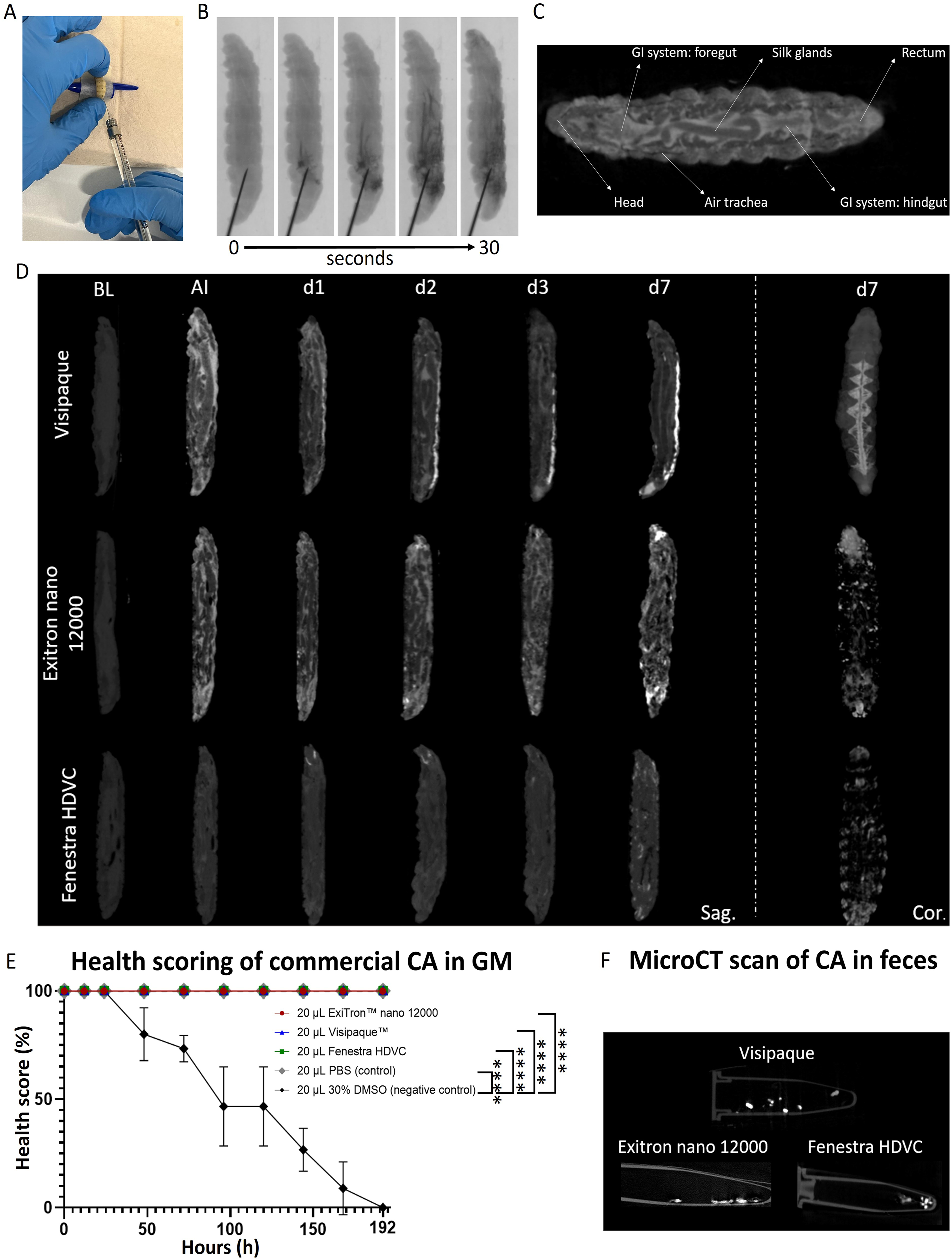
Evaluating three commercially available micro-CT contrast agents in the G. mellonella model
Once the systemic distribution was confirmed, we then investigated the potential of G. mellonella for use as a functional in vivo model to evaluate the performance and clearance of micro-CT contrast agents. To this end, we compared the biodistribution, in vivo functionality and excretion of three commercially available contrast agents with well-established in vivo safety and biocompatibility profiles, namely: — Visipaque 320 (320 mg iodine/ml), a clinically approved, iodine-based molecular contrast agent developed for rapid renal clearance;
46
— ExiTron nano 12000, a 110 nm metal-based iodinated nanoparticulate contrast agent, specifically designed for prolonged vascular contrast enhancement in small animal imaging; and — Fenestra HDVC, an iodine-based nanoemulsion (100 mg iodine/ml), developed for liver and pancreatic cancer research, where prolonged retention and organ-specific accumulation are critical.47,48
These three contrast agents were selected as reference compounds, based on their characterised pharmacokinetics and performance in validated in vivo models. By injecting these contrast agents into the larvae, we aimed to confirm whether their expected contrast enhancement and biodistribution profiles were observable in this model. Specifically, we sought to determine whether known differences in clearance kinetics, such as the rapid excretion of molecular contrast agents versus the extended retention of nanoparticulate formulations, could be distinguished. As such, we injected larvae with either one of the three contrast agents on Day 0, scanned them daily using micro-CT, and evaluated their health index scores for a total of eight days.
Biodistribution and in vivo functionality of the different agents in the model
For Visipaque, immediate contrast enhancement was observed throughout the entire larval body upon injection, confirming effective staining of the larval circulatory system (Figure 4(d)). By Day 1, contrast agent began localising within the dorsal vessel and rectum, with signal intensity increasing progressively over time in this area. On Day 7, the dorsal coronal section still revealed contrast enhancement in the haemocoel network of the larvae.
Both nanoparticulate contrast agents (ExiTron nano and Fenestra HDVC), showed similar systemic distribution throughout the larval body immediately after injection. However, unlike the molecular contrast agent (i.e. Visipaque), from Day 2 onward, the signal gradually became more localised in peripheral regions instead of the dorsal vessel, with the most prominent enhancement seen in the head and rectum by Day 7 (Figure 4(d) and Figure 5(b)). To further investigate this tissue-specific accumulation, larvae were frozen on Day 7 post-injection, after which the fat body (a metabolically active organ functionally analogous to the mammalian liver) was dissected and analysed by ex vivo micro-CT. For both nanoparticulate contrast agents, a clear signal was detected in the fat tissue, indicating accumulation, whereas no detectable signal was observed in the fat body of larvae treated with Visipaque or the PBS control (Figure 5(a)). Pharmacokinetic profiling and tissue-specific accumulation of micro-CT contrast agents in G. mellonella. (a) Tissue-specific accumulation of contrast agents on Day 7 post-injection. The image shows representative photographs (top panel) and corresponding ex vivo micro-CT images (bottom panel) of dissected head and fat body tissue from larvae injected with PBS (control), Visipaque, Fenestra HDVC or ExiTron nano 12000. The nanoparticulate contrast agents (Fenestra HDVC and ExiTron nano 12000) show clear accumulation (elevated contrast in white) in the head and fat tissue, whereas the molecular contrast agent (Visipaque) and PBS control did not lead to detectable signal in these tissues. (b) Pharmacokinetic analysis of ExiTron nano 12000, Visipaque and Fenestra HDVC following intrahaemocoelic injection. At predefined time points after the injection of 20 μl of contrast agents, the larvae were bled to collect haemolymph, which was subsequently analysed ex vivo by micro-CT to quantify attenuation values (Hounsfield units). All contrast agents remained detectable for over eight days, with gradually decreasing attenuation values over time. PBS-injected larvae served as the negative control. Data are presented as the mean ± SD; ****p < 0.0001.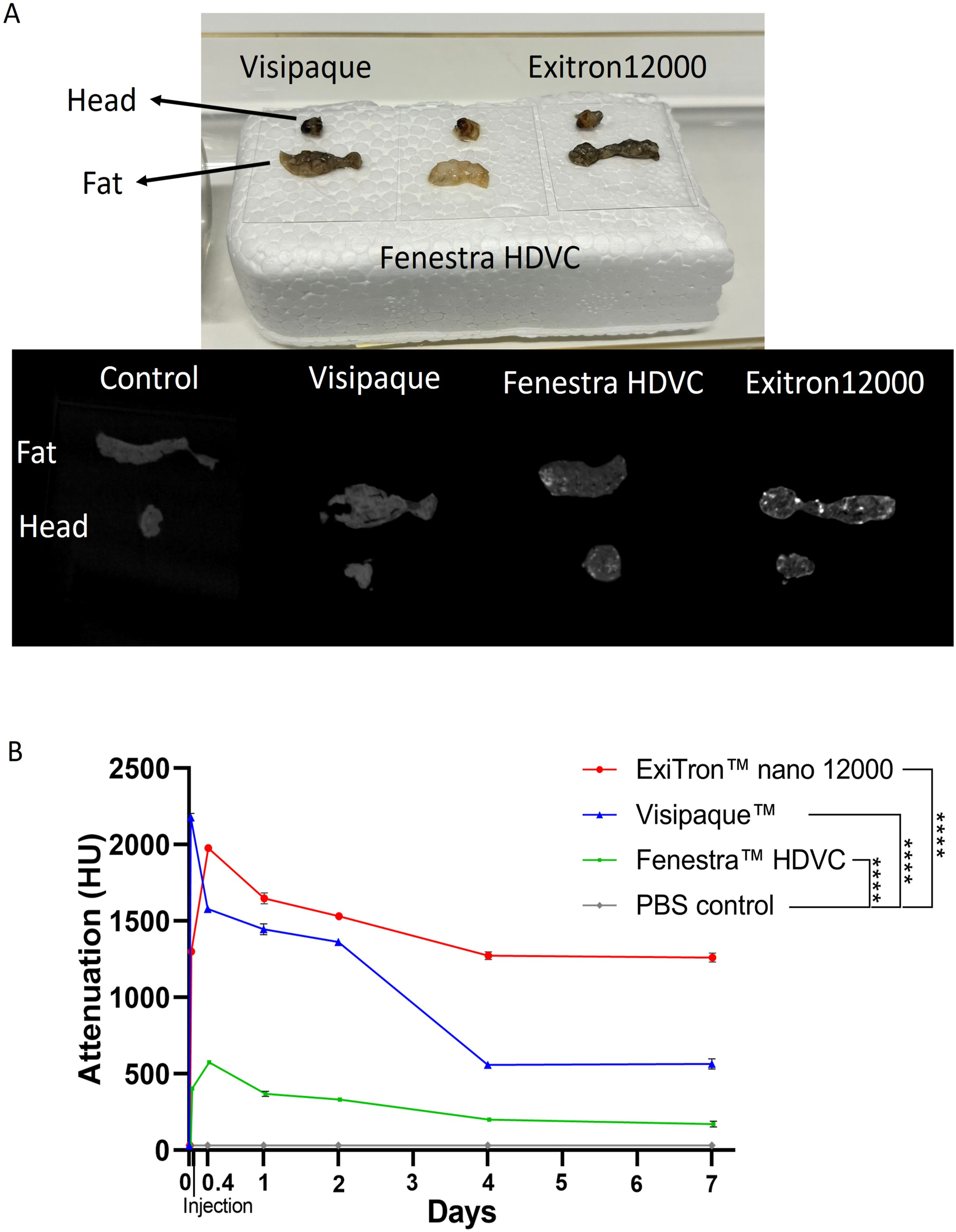
To specifically evaluate this systemic persistence in vivo of all three contrast agents, full haemolymph samples were collected from the larvae at predetermined time points post-injection and analysed ex vivo by using micro-CT, to determine attenuation values (Figure 5(b)). The resulting pharmacokinetic curves showed that all three contrast agents remained detectable in the haemolymph throughout the 8-day observation period. Although haemolymph attenuation gradually decreased for all agents, in parallel with increased attenuation in the rectum consistent with progressive contrast agent accumulation, the haemolymph attenuation on Day 7 remained significantly higher than that of the PBS control group, thus confirming sustained systemic presence of all contrast agents (Figure 5(b)).
Among the three agents, we observed that Fenestra HDVC yielded the lowest overall contrast enhancement, likely due to its lower iodine concentration relative to the other agents. In addition, all three contrast agents were well-tolerated, with health index scores equal to the non-toxic PBS control in each case, thus indicating that there were no significant effects on larval health; only the DMSO-treated control group (for negative tolerance/toxicity) gave significantly lower scores compared to the other groups (Figure 4(e) and Figure 2(c)). This confirms that the G. mellonella model can reliably be used to assess the biocompatibility and tolerability of micro-CT contrast agents in vivo.
Analysis of the contrast agent excretion pathways
The two different types of contrast agent used in this study typically follow different excretion pathways in rodent models, i.e. rapid renal clearance for the molecular contrast agent (Visipaque) and prolonged circulation with hepatic clearance for the nanoparticulate formulations (ExiTron nano and Fenestra HDVC).49–51 However, in the current study, we observed that all three agents consistently accumulated in the rectum of G. mellonella from Day 2, suggesting a shared gastrointestinal elimination pathway in this organism, regardless of formulation type.
To gain further insight into their elimination pathways, we investigated whether the contrast agents were excreted in the faeces of the larvae. We tracked the contrast agents, from injection through systemic distribution to excretion, by systematically collecting all faeces accumulated in the recovery jars over the 7-day period. Subsequent micro-CT scans of the faecal material revealed clear contrast enhancement for all three agents, confirming their passage through the larval gastrointestinal system and final elimination via the faeces (Figure 4(f)). The gradual decrease in haemolymph attenuation values over time corresponds to the faecal excretion observed, indicating continuous clearance, while showing that complete elimination is not achieved within the 7-day period (Figure 5(b)).
Overall results summary
The results show that the spatial biodistribution profiles of the iodine-based molecular contrast agent and the two preclinical nanoparticulate formulations were distinguishable in the G. mellonella model. While all three agents showed rapid systemic distribution following injection, no marked differences in initial extravasation or circulation time were observed between the molecular and nanoparticulate contrast agents during the 7-day follow-up period.50,52 Although nanoparticulate micro-CT contrast agents are often formulated to extend the blood-pool imaging window relative to small molecular iodinated agents, the reported intravascular persistence in mammalian models is formulation-dependent and generally occurs over a time-frame of a few hours to approximately one day. In the present 7-day follow-up, prolonged initial circulation could not be clearly visualised in this invertebrate system (Figure 4).50,52
We confirmed the distribution of the three agents via the haemocoel, the subsequent dispersion of the agents throughout the larval body, and their accumulation in the rectum by Day 7. Quantitative pharmacokinetic analysis of haemolymph samples showed that all agents remained systemically detectable over the total eight days. Attenuation values gradually declined over time, consistent with the continuous rectal accumulation via the gastrointestinal pathway. However, levels remained significantly elevated compared to PBS controls at the final time point (Day 7). All three contrast agents were well-tolerated by the larvae, with health index scores remaining comparable to non-toxic controls throughout the 7-day follow-up period.
We showed that differences in biodistribution of the clinically approved molecular contrast agent and the two nanoparticulate formulations could be effectively observed in the model (e.g. dorsal vessel versus peripheral accumulation in fat tissue, respectively), even though all three agents ultimately shared the same elimination pathway via accumulation in the rectum and faecal excretion. Subsequent micro-CT scans of faecal samples confirmed the presence of contrast agents, indicating their passage through, and elimination from, the organism.
Discussion
In this study, we evaluated the use of G. mellonella as a robust and scalable in vivo model that serves as an alternative to traditional mammalian species. This non-vertebrate model represents an intermediate platform, bridging in vitro assays and in vivo rodent studies for screening the safety, functionality and biodistribution of novel preclinical and clinical micro-CT contrast agents in a cost-effective and ethically favourable manner. To this end, we present a novel standardised immobilisation and micro-CT imaging protocol for G. mellonella larvae. We validated the feasibility and reliability of this approach by focusing on several critical micro-CT scanning aspects, including effective larval sedation parameters and larval X-ray exposure safety. Once established, we used the novel protocol to assess the safety, biocompatibility and in vivo functionality of three commercially available contrast agents in the G. mellonella model, to evaluate its potential use in early-stage functionality and biocompatibility screening during CT contrast agent development.
The optimised isoflurane gas anaesthesia protocol effectively immobilises Galleria mellonella larvae and is well-tolerated
We achieved a reliable and reproducible method for the immobilisation of G. mellonella larvae, which is essential for high-resolution micro-CT, as even minimal motion can introduce artefacts and thus compromise the quality of 3D image reconstructions. We established that current immobilisation approaches, which typically rely on cold-induced sedation,53,54 did not suffice in terms of ensuring complete stillness throughout the scanning protocol. Despite reducing larval activity, subtle movements such as dorsal contractions remained observable, thus limiting the suitability of this method for high-precision micro-CT imaging. We overcame this limitation by introducing an isoflurane-based gas anaesthesia protocol for G. mellonella that provided a consistent and prolonged immobilisation across all larvae. While this approach has been used in other larval insects, such as Manduca sexta, 55 to our best knowledge, its application to G. mellonella has not been previously explored. This optimised sedation strategy not only enabled artefact-free image acquisition for micro-CT, but also broadens the potential of G. mellonella for use as a versatile and accessible model in a range of imaging modalities in preclinical and biomedical imaging research that require complete immobilisation — for example, SPECT, micro-PET, MRI, ultrasound and high-resolution optical imaging.56–59
Repeated isoflurane administration and cumulative X-ray exposure do not affect larval health
We demonstrated that G. mellonella larvae can undergo repeated micro-CT imaging over an 8-day period with no observable impact on health, if sedation is limited to once daily. This establishes the model’s suitability for longitudinal contrast-enhanced studies. By evaluating the impact of repeated exposure to both ionising radiation and isoflurane anaesthesia, we confirmed that both were well-tolerated when administered separately, once daily. Because radiation alone had no observable effect on larval health, we can reasonably expect full tolerability when both procedures are applied once daily. With larval health remaining stable throughout the follow-up period, this validated the safety of our imaging protocol. Importantly, this resilience is crucial, as more and more contrast agents with prolonged serum residence times are being developed, requiring longer observational windows to evaluate their tolerability, biodistribution and functionality.60,61 Hence, the ability of G. mellonella larvae to withstand repeated imaging procedures without adverse effects strengthens their applicability as a robust and reliable non-vertebrate in vivo model for the assessment of (micro-)CT contrast agents (i.e. both preclinical micro-CT and clinical CT contrast agents). The 8-day imaging window we established offers a practical and physiologically relevant timeframe that broadly accommodates the needs of most early-phase tolerability, biodistribution and functionality studies of contrast agents.
The biodistribution and in vivo functionality of three commercially available micro-CT contrast agents in the Galleria mellonella model
The goal of the current study was to establish the suitability of G. mellonella for use as an in vivo model to assess the functionality, tolerability and safety of novel contrast agents. We achieved this goal by using the model to evaluate three commercially available contrast agents that are either clinically approved or preclinically validated, and have been proven safe for use in humans and/or mice. All three agents were well-tolerated, with no observed adverse effects on larval health over the 7-day follow-up period (in contrast to the toxic DMSO control), thereby confirming the model’s suitability for use in early-stage biocompatibility and safety screening. Moreover, through intrahaemocoelic injection and longitudinal micro-CT imaging, we achieved consistent systemic contrast enhancement for all agents, confirming effective distribution within the larval body.
Specifically, the molecular contrast agent (Visipaque 320) dispersed rapidly and accumulated in the dorsal vessel from Day 2, while the nanoparticulate contrast agents (ExiTron nano 12000 and Fenestra HDVC) also initially spread systemically but later localised in peripheral regions, including the head and the tail. These peripheral areas contain the fat body, a metabolically active tissue analogous to the mammalian liver, suggesting that this might be a potential site for nanoparticle accumulation.62–64 This was also observed by ex vivo micro-CT analysis of the isolated fat body, which showed a contrast signal only in the fat tissue of larvae treated with the nanoparticulate agents. The observed differences in distribution between molecular and nanoparticulate formulations suggest that the larval system can distinguish between agents with distinct physicochemical properties. This capability further supports the suitability of G. mellonella for use in investigating biodistribution patterns and early toxic responses to novel contrast agents. Furthermore, pharmacokinetic data demonstrated the sustained presence of all three contrast agents over eight days in the haemolymph. Although attenuation gradually decreased in line with progressive rectal accumulation, levels remained significantly elevated compared to controls at the final time point, indicating incomplete clearance. Notably, although the molecular contrast agent is designed for rapid renal clearance in mammalian systems, such accelerated excretion was not observed in the larval model.
Excretion pathways of the three commercially available micro-CT contrast agents
While differences in biodistribution profiles could be distinguished in the larval model, specifically the accumulation of nanoparticulate contrast agents in the fat tissue, all three agents ultimately followed the same excretion route, with evident accumulation in the rectum and elimination through the faeces. In insects, excretion occurs via the Malpighian tubules, which actively secrete water, ions and waste products from the haemolymph into the gut lumen. This primary urine is subsequently processed in the hindgut before final elimination through the faeces. Although this pathway shares certain functional similarities with mammalian renal tubular secretion, it does not reflect the compartmentalised glomerular filtration and hepatobiliary processing characteristic of vertebrate physiology. Importantly, for nanoparticulate contrast agents larger than 8 nm, this difference becomes even more relevant, as, in mammals, such particles are predominantly taken up and cleared by the liver and spleen, rather than through direct renal elimination. 65
The fact that all formulations, independent of formulation type, ultimately shared the same elimination pathway in G. mellonella highlights the need for follow-up studies to evaluate mammalian-specific excretion pathways, such as renal or hepatic clearance. These studies could include physiologically relevant human-based models, such as human tissue or organ-on-chip systems, where appropriate. Therefore, while G. mellonella offers an efficient and ethically favourable model for evaluating the safety, tolerability and early in vivo functionality of contrast agents, detailed pharmacokinetic and pharmacodynamic assessments will still require vertebrate models. Ultimately, this positions G. mellonella as an ideal intermediate in vivo model to bridge the gap between in vitro screening and mammalian validation in the development pipeline of novel (micro-)CT contrast agents. Hence, by enabling initial in vivo screening without the need for vertebrate animal use, this model contributes to reducing animal use, in line with the principles of the Three Rs.
Advantages of the model
This model offers a physiologically active system with an open circulatory network — as such, it enables significant advantages compared to conventional in vitro assays for testing the functionality and distribution of novel contrast agents. Traditional in vitro models for evaluating contrast agents include phantoms that mimic tissue contrast densities, as well as spheroids and organoids.66–69 However, these models lack dynamic circulation and therefore cannot replicate the systemic biodistribution or time-resolved behaviour of contrast agents in vivo.
In contrast, due to their open circulatory network, G. mellonella larvae can be used for the longitudinal tracking of contrast agents across tissues over time, thus permitting assessment under whole-organism conditions. This makes the model particularly well-suited for early-stage evaluation of both the biological behaviour and imaging performance of novel contrast agents. Moreover, owing to the ease of maintenance and minimal ethical restrictions, this model can be implemented directly in research environments that lack access to dedicated animal facilities, where testing would otherwise be limited to non-physiological in vitro systems that lack a host response. Accordingly, our model offers a valuable platform for screening contrast agents being developed for emerging contrast-enhanced CT technologies, including spectral and photon-counting CT, where biodistribution and imaging characteristics are critical to agent design.
Beyond contrast agent development, CT in G. mellonella offers advanced applications such as the investigation of implant-associated infections and the visualisation of bacterial biofilms on biomaterial surfaces.35,36 Micro-CT enables non-destructive and longitudinal assessment of infection progression and implant–host interactions, applications in which contrast performance, biodistribution and tolerability are critical.35,36,70,71 Additionally, the model may support the evaluation of radiopaque or contrast-doped biomaterials and implant coating, allowing the simultaneous assessment of material visibility, host response and infection progression in vivo.72,73 In this context, an ethically favourable, imaging-compatible in vivo screening platform such as the one we present here, could facilitate the early-stage evaluation of novel contrast agents intended for a plethora of biomedically relevant CT applications, before their further testing and validation in vertebrate models.
Conclusions
Our findings establish G. mellonella as a reliable and ethically favourable intermediate in vivo model for use in the early-stage screening of (micro-)CT contrast agents. This opens the door toward the model’s use in a broad range of advanced CT-supported biomedical research applications. By offering both physiological relevance and technical feasibility, the model enables the in vivo assessment of safety, tolerability and functionality under whole-organism conditions, without the ethical and logistical constraints that are commonly associated with the use of vertebrate models. Its unique advantages, including a systemic circulation, compatibility with longitudinal imaging and ease of implementation, make it ideal to support the development of novel contrast agents for next-generation CT modalities. As such, this approach not only complements existing in vitro and rodent-based studies, but also directly contributes to advancing the principles of the Three Rs through the reduction and potential replacement of vertebrate model use in preclinical imaging experiments.
Footnotes
Acknowledgements
We gratefully acknowledge the contributions of Eliane Vanhoffelen, Tine Van Win, Lauren Michiels, and Kasia Błażejczyk for their assistance in maintaining the Galleria mellonella colony throughout this study. We also thank Medilumine Inc. for kindly providing a sample of Fenestra™ HDVC contrast agent used in this research.
Ethical considerations
This article does not contain any studies with human or animal participants. Experiments were performed in compliance with European and local ethical guidelines, under which research on invertebrates like insects does not require prior ethical approval.
Author contributions
Conceptualisation: GVV; Methodology: TM, BT, ARS; Validation: TM; Formal analysis: TM, BT; Investigation: TM; Resources: BT, ARS, GVV; Writing (original draft): TM; Writing (review & editing): BT, ARS, GVV; Visualisation: TM; Supervision: GVV; Project administration: ARS, GVV; Funding acquisition: BT, GVV.
Funding
The authors acknowledge funding from the European Union’s Horizon Europe research and innovation programme EIC grant agreement No. 101046894 (SynEry), the Flemish Research Foundation (FWO, infrastructure grant I006524N), the Department of Imaging and Pathology and KU Leuven Internal Funds (C3/23/005).
Declaration of conflicting interests
The authors have no conflicts of interest to declare.
Data Availability Statement
The data supporting the findings of this study are available from the corresponding authors upon reasonable request.