
Abstract
Orthopaedic implants commonly use titanium and its alloys because of their superior mechanical properties, corrosion resistance and biocompatibility. However, these metal implants have limited application, hindered by poor osseointegration and the potential risk of implant-associated infections. To overcome these obstacles, this study intends to address these issues by integrating haemocompatible and antibacterial coatings made from green synthesised zinc oxide nanoparticles, using Saraca asoca leaf extract (SA-ZnO). These nanoparticles may serve to improve the osteoconductivity and limit bacterial growth at the site of implant. The prepared nanoparticles (SA-ZnO) were characterized using various techniques and then integrated with polyvinyl alcohol (PVA), resulting in (SA-ZnO/PVA) nanocomposite. These prepared nanocomposites were further coated onto the Titanium alloy substrate (Ti-6Al-4V) using doctor's blade technique and morphologically characterized. Atomic force microscopy analysis demonstrated that SA-ZnO/PVA nanocomposite coated substrate exhibited favourable micro-roughness, enhancing cell attachment and promoting protein adsorption, which is crucial for effective osseointegration. Additionally, haemocompatibility tests demonstrated no adverse effects on blood cells, indicating the coating's compatibility with biological systems. Antimicrobial studies showed a significant zone of inhibition against both gram-negative and gram-positive bacteria, highlighting the antibacterial potential of SA-ZnO/PVA nanocomposite coatings. Thus, these findings imply that the green synthesised ZnO nanoparticles amalgamated with (PVA) serve as promising surface coating materials, that can be used effectively in orthopaedic implants to reduce bacterial adherence and growth, thus lowering the incidence of postoperative infections and enhancing overall implant integration success.
Introduction
Titanium (Ti) and its alloys are frequently used in orthopaedics due to its superior mechanical qualities, biocompatibility, and potential for osseointegration.1,2 Despite nearly a thousand tons of titanium being implanted annually, bonding with host bone remains challenging due to its bioinert nature. Implant-associated infections in orthopaedic surgeries pose serious risks, such as revision procedures, implant failures, amputations, or death.3–5 Furthermore, implants prolonged hospitalization and increased costs, leading to patient suffering and anxiety. Their stiff surfaces promoted bacterial adherence and biofilm formation, complicating infection treatment. Although antibiotics were typically administered post-surgery, the development of antibiotic resistance required alternative treatments. 6 Developing antimicrobial coatings for implants helps prevent bacterial adhesion and reduce infection risk. Surface modifications, including biomimetic coatings and topographical changes, enhance cytocompatibility and osseointegration. Antibacterial-loaded nanoparticles on implant surfaces promote bone formation, minimize resorption, and support cell adhesion and proliferation.7–9
Regarding the fabrication of nanoparticles, Green synthesis techniques provide a simple, rapid, efficient, and eco-friendly method, making nanoparticles valuable for both therapeutic and industrial research. 10 Plant-derived agents provide effective antimicrobial properties with fewer side effects compared to conventional antibiotics. 11 The phytochemical components of the plant extract such as flavonoids and phenols play a crucial role as reducing agents, enabling the transformation of metal precursors into metal nanoparticles. These components, free from toxic substances, also serve as capping agents to stabilise the nanoparticles by preventing aggregation.12,13 A schematic representation of the role of phytoconstituents in the green synthesis of metal nanoparticles has been shown in (Figure 1). Saraca asoca, traditionally used in Ayurvedic medicine, is rich in various phytocompounds like flavonoids, phenols, etc. which may play a crucial role in the eco-friendly green synthesis of nanoparticles, enhancing antibacterial activity. 14
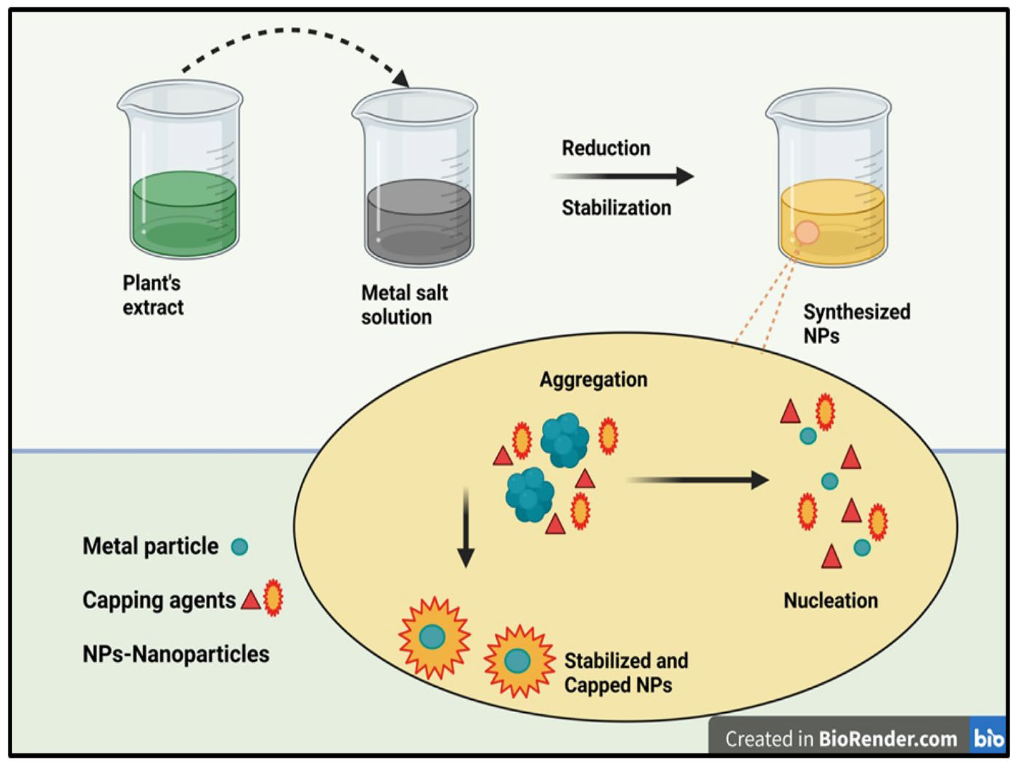
Schematic representation of the green synthesis mechanism for metal oxide nanoparticles.
Metal oxide nanoparticles, particularly Zinc oxide, are renowned for their stability and strong antimicrobial properties. ZnO nanoparticles show great promise in drug delivery, biological sensing, gene transfer, and nanomedicine. 15 ZnO nanoparticles have been used in medical implant coatings due to their antibacterial and osteoconductive properties, promoting bone regeneration and implant integration. They have also been used to support muscle repair and growth by enhancing cell proliferation and differentiation, while being safe and dissolving slowly in physiological conditions as they generate reactive oxygen species (ROS) upon interaction with bacterial cells.16,17 Cuypers et al., developed zinc-doped hydroxyapatite nanoparticles and concluded that zinc ions exhibit antibacterial activity at non-toxic levels without harming pre-osteoblast cells. 18 Similarly, other study using zinc oxide and manganese-doped ZnO nanoparticles demonstrated their antimicrobial potential due to sustained ion release. 19 Thus, ZnO nanoparticles, due to their excellent antibacterial effects, may prove to be more effective for implant-associated infections.
The current research trend has shifted towards coating techniques where green synthesised nanoparticles have found their effectivity with minimal side effects. Green synthesis of ZnO nanoparticles using Saraca asoca extract for titanium implant coatings has not been explored. Therefore, the potential of ZnO-incorporated coatings to enhance antibacterial activity, durability, and osseointegration may present a promising avenue for improving the longevity and performance of orthopaedic implants. Furthermore, PVA is a biodegradable, synthetic polymer that dissolves easily in water, making it ideal for applications in orthopaedic and cartilage repair. It is used to create artificial cartilage for implants, with hydrogels that mimic articular cartilage's porous structure, being biocompatible and non-toxic. When combined with ZnO nanoparticles, the resulting nanocomposites can enhance various properties, offering a more effective and environmentally friendly material for coating, with potential uses across diverse fields. 20
Thus, the purpose of this study is to develop and characterize Saraca asoca extract-mediated ZnO/PVA nanocomposites, exploring their potential as effective antibacterial coatings for orthopaedic implants which would resolve the issue of antimicrobial resistance, a major concern in the current scenario. This work aims to combine the reducing potential of Saraca asoca extract in the green synthesis of biocompatible ZnO nanoparticles with the processability of PVA to create a multifunctional coating that can reduce the risk of infection and promote faster recovery in patients with orthopaedic implants.
Materials and methods
Materials
PVA (purity 98%) was obtained from Sigma-Aldrich, Zinc acetate dihydrate (purity 99%), Sodium hydroxide, Nutrient agar, Triton-X 100 and other chemicals were obtained from Merck, Mumbai, India etc.
Preparation and preliminary phytochemical screening of Saraca asoca (SA) leaf extract
Fresh Saraca asoca leaves (25 g) were washed, chopped, and boiled with 100 ml water for 30 min. The extract was filtered (Whatman No. 1) and stored at 4°C for further studies. The prepared Saraca asoca leaf extract was subjected to various qualitative tests to identify the presence of different phytochemicals. Additionally, the total phenolic content and total flavonoid content were determined using standard protocols to evaluate the plant's bioactive potential. 21
Green synthesis of ZnO nanoparticles using Saraca asoca leaf extract (SA-ZnO)
Twenty millilitres of plant extract was heated at 50°C for 10 min, then 50 ml of 91 mM zinc acetate solution was added dropwise with stirring, forming yellowish cream-colored zinc hydroxide precipitate, which was enabled to react for 30 min. The precipitate was then collected by centrifugation at 10,000 rpm for 10 min at 4°C, followed by vacuum drying at 30°C for 24 h to carefully remove moisture without altering the structural or functional properties of the SA-ZnO nanoparticles, ensuring their stability. The resulting nanoparticles were stored for further studies. 22
Characterization SA-ZnO nanoparticles
The synthesized SA-ZnO nanoparticles were characterized to evaluate properties such as particle size, crystalline structure, and morphology. Dynamic light scattering (DLS) was used to determine the particle size distribution
23
with a Malvern Zetasizer HPPS-5001. TGA analysis of thermal transformation was performed using the Netzsch TG209 F1 Iris instrument. The crystalline structure was examined via X-ray diffraction, and surface structure was studied using Field Emission Scanning Electron Microscopy (FESEM). Fourier-transform infrared (FT-IR) spectroscopy identified the functional groups involved in the reduction and capping processes (FTIR-8400 S, Shimadzu, Japan)
Preparation of SA-ZnO/PVA nanocomposite coated titanium alloy
SA-ZnO/PVA nanocomposites for coating were prepared by dissolving 2 wt.% polyvinyl alcohol in 50 mL distilled water at 70–80°C, dispersing SA-ZnO nanoparticles (3:1), and stirring vigorously for 1 h to prevent agglomeration. The prepared SA-ZnO/PVA nanocomposites were characterized and coated on Ti-6AL-4V surfaces using the doctor blade technique, allowing precise control over layer thickness and ensuring uniform application.27,28 The adhesion quality of the coating was assessed using the cross-cut tape test following ASTM D3359 standard. 29 Further, the coated samples were analysed in terms of morphology, haemocompatibility and antibacterial potential.
Morphological characterization of SA-ZnO/PVA coated titanium alloy
Atomic Force Microscopy (AFM) analysis was conducted to investigate the surface topography and crystalline structure of pure and SA-ZnO/PVA coated titanium alloy substrates. The roughness and morphology of the samples were assessed for highlighting the granularity distribution of the coating. Roughness, a topographical property of implant surfaces, may hinder cellular interactions at macrolevels or offer significant advantages for bone implants by enhancing cell attachment at micro-topographic levels. 30
Haemocompatibility evaluation of SA-ZnO/PVA coated titanium alloy
The standard method for assessing haemolytic activity was utilised to evaluate the haemocompatibility of uncoated and SA-ZnO/PVA coated titanium alloy substrates. A red blood cell (RBC) suspension (2%) was prepared in phosphate buffered saline. The suspension was incubated with the coated and uncoated samples at 37 °C with moderate shaking for 2 h. A physiological saline solution served as a negative control, while a 10% Triton X-100 solution was employed as a positive control to accurately assess haemolytic activity. Saline maintains physiological conditions, while Triton X-100 facilitates RBC lysis, to evaluate cytotoxicity and ensuring reliable hemocompatibility results for the coated titanium alloy. Following the incubation period, the suspensions were centrifuged at 1000 rpm for 10 min. The supernatant was analysed for absorbance at 540 nm to measure the amount of released haemoglobin and the percentage haemolysis was calculated. 31
Evaluation of antibacterial potential of coated samples
To investigate the antimicrobial activity of the coatings, the zone of inhibition method was used against two aerobic bacteria: Gram-positive Staphylococcus aureus and Gram-negative Escherichia coli. Nutrient agar plates were prepared, and a bacterial suspension with turbidness equivalent to the 0.5 McFarland standard was spread across the agar surface. Sterilized uncoated and coated titanium alloy substrates were placed on the plates and incubated at 37°C for 24 h, after which inhibition zones were measured. 32
Results and discussion
Preliminary phytochemical screening
Phytochemical screening of the aqueous extract of Saraca asoca leaves showed the presence of various phytoconstituents including flavonoids, phenols, alkaloids, saponins, and glycosides etc. The presence of these phytoconstituents has been categorized as ‘+++’ (strong), ‘++’ (moderate), ‘+’ (weak), and ‘−’ (absent), as shown in Table 1. Further, the quantitative estimation of phenols and flavonoids in the extract demonstrated the total phenolic content of 7.04 mg GAE/g and total flavonoid content of 10.132 mg QE/g.
Preliminary phytochemical screening of Saraca asoca leaf extract.
Characterization of SA-Zno and SA-ZnO/PVA
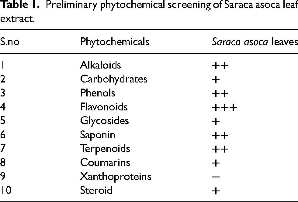
The average particle size of the biosynthesized ZnO nanoparticles was determined to be 131.5 nm, with a polydispersity index (PDI) of 0.167, indicating a narrow size distribution and relatively uniform particle size where as SA-ZnO/PVA nanocomposites, with an average size of 308.5 and PDI of 0.244 are still suitable for nanocomposites (Figure 2). The increase in particle size for SA-ZnO/PVA nanocomposites compared to SA-ZnO nanoparticles is attributed to the addition of PVA, which forms a coating or matrix around the nanoparticles. This suggests good stability, consistent performance, and predictable behaviour in nano-formulation applications, such as bone implant materials for cell attachment. These size range offers a balance between stability and biological activity, enhancing their potential for use in various applications, including biomedical and catalytic processes. 33
Particle size of SA-ZnO nanoparticles and SA-ZnO/PVA nanocomposites.
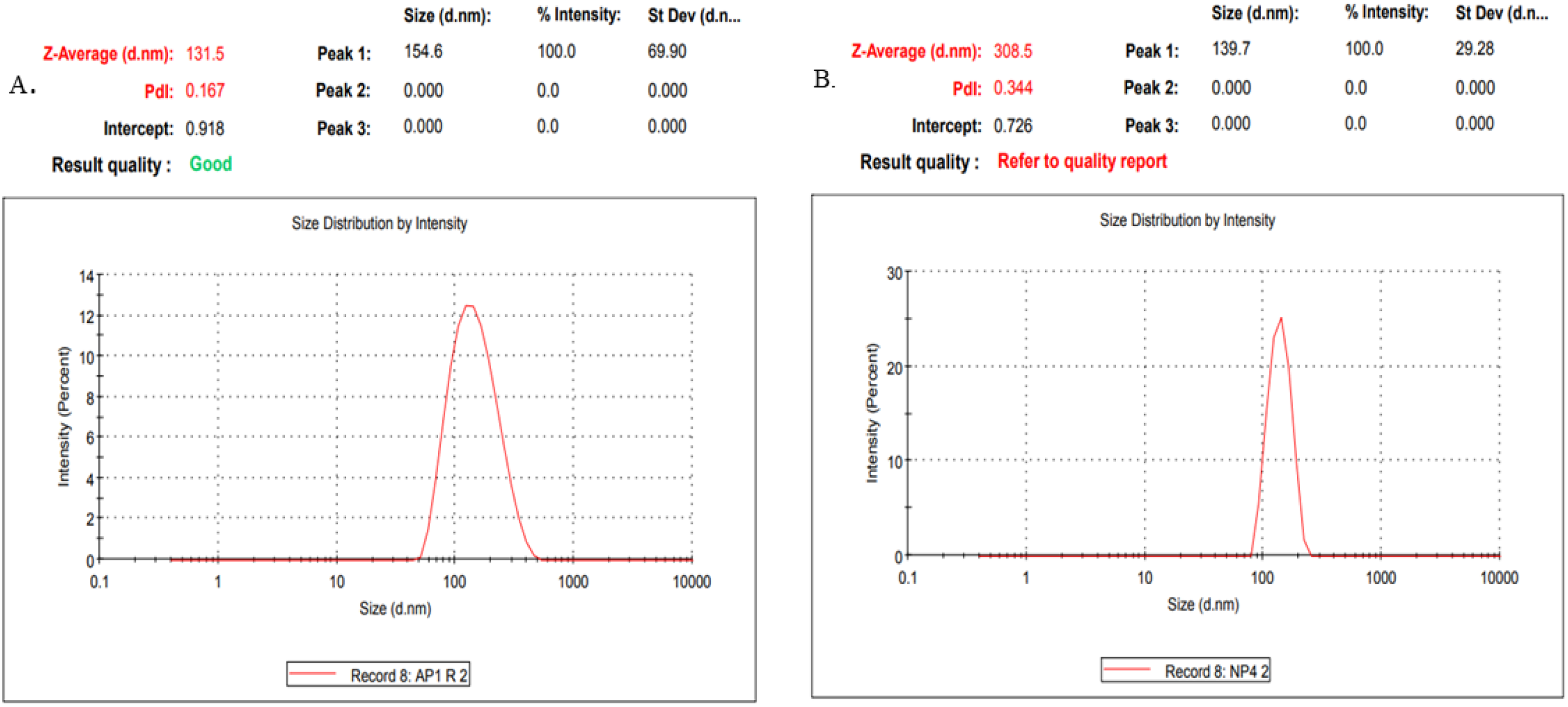
The thermal decomposition profile illustrates the effect of temperature on the stability of nanoparticles and the volatility of components present in the sample. The TGA (Thermogravimetric Analysis) of SA-ZnO demonstrates significant decomposition of the sample as the temperature increases, primarily due to various volatile components from the plant material. 30 The analysis shows notable weight loss at higher temperatures: at 201.57 °C, the weight loss was 0.810 mg, which is 19.764%, after 17.91 min; and at 495.00 °C, the total weight loss reached 3.063 mg, representing a substantial 74.778% after 47.24 min. For SA-ZnO/PVA nanocomposites, at 200.56 °C, the weight loss was 0.892 mg, corresponding to a 16.823% weight percent loss after 17.68 min; at 400.84 °C, the weight loss reached 3.817 mg, which is 71.966%, after 37.66 min; and at 793.62 °C, the total weight loss was 4.111 mg, representing 77.510% after 76.91 min. The DSC curve of SA-ZnO nanoparticles shows a peak temperature of 83.57°C with a thermal enthalpy of 745.90 J/g and an onset of 57.91 °C indicates the loss of volatile surfactant molecules adsorbed on the surface during synthesis. A second peak occurs at an onset temperature of 254.49 °C and peaks at 313.60 °C, corresponding to the conversion of zinc hydroxide to zinc oxide nanoparticles, with a thermal enthalpy of 75.300 J/g. For SA-ZnO/PVA nanocomposites, the DSC curve shows a peak temperature of 79.09 °C with an enthalpy of 677.58 J/g and an onset at 55.98 °C. A second peak occurs at 449.35 °C with an enthalpy of 794.14 J/g and an onset at 396.79 °C. Overall, the results showed that both the nanoparticles and nanocomposites exhibited clear changes in thermal behaviour, which concluded their stability at higher temperatures and provided valuable insights into their decomposition patterns (Figure 3).
TGA analysis and DSC analysis of SA-ZnO nanoparticles (A and B, respectively) and SA-ZnO/PVA nanocomposites (C and D, respectively).
XRD analysis
The X-ray diffraction technique was employed to evaluate the phase purity and crystalline properties of SA-ZnO and SA-ZnO/PVA. The resulting XRD pattern confirmed the high crystalline quality of the nanoparticles, as indicated by distinct and intense peaks with narrow widths. This analysis also affirmed the purity of the zinc oxide phase, demonstrating the effectiveness of Saraca asoca leaves in producing highly crystalline nanoparticles without any amorphous characteristics. The XRD pattern exhibited peak broadening, indicating that the particles are in the nanoscale range. This phenomenon is commonly observed for nanoparticles, as smaller particles result in increased crystal defects and a reduced size, leading to broader diffraction peaks. 34 The diffraction peaks at 31.64, 34.13, 36.16, 47.5, 56.46, 62.74, and 67.64 correspond to the hexagonal wurtzite structure of ZnO, confirming the crystalline phase and high structural order of the nanoparticles. Similarly, the peaks for SA-ZnO/PVA nanocomposites appear at 31.88, 34.52, 36.36, 47.64, 56.68, 62.92, and 68.02 (Figure 4). The close alignment of these peak positions suggests that PVA interacts effectively with the zinc oxide nanoparticles.

XRD spectra of SA-ZnO nanoparticles and SA-ZnO/PVA nanocomposites.
FTIR analysis
The Fourier Transform Infrared (FTIR) spectra of SA-ZnO and SA-ZnO/PVA nanocomposites confirm the presence of functional groups that play a critical role in the green synthesis process. The spectra illustrate the interaction of SA-ZnO with the PVA polymer through the presence of overlapping functional groups. Peaks observed between 625–970 cm−1 indicate C-H bending out of the plane, characteristic of aromatic structures. The range of 1015–1300 cm−1 corresponds to C-O stretching vibrations, associated with hydroxyl groups involved in reducing metal ions. Peaks between 1300–1380 cm−1 represent CH₂ bending vibrations, indicating the presence of organic molecules. Carbonyl stretching vibrations (C=O) observed between 1550–1780cm−1 suggest the involvement of carbonyl groups in the reduction and stabilisation of nanoparticles. C-H stretching vibrations at 2912 and 2944 cm−1 further support the presence of aliphatic components while the broad band at 3282 cm−1 is associated with O-H hydrogen-bonded alcohols (Figure 5). These functional groups collectively indicate the active participation of biomolecules in reducing and stabilising nanoparticles. Pure PVA shows peaks at 2941, 2900, 1775, 1096, and 880 cm−1 in FTIR spectrum. 35 The shifting of peak to 837 cm−1 in the prepared nanocomposite may be due to interaction with SA-ZnO nanoparticles. The spectral shifts and the appearance of new peaks in the nanocomposites confirm the effective integration of the PVA polymer with the SA-ZnO nanoparticles.
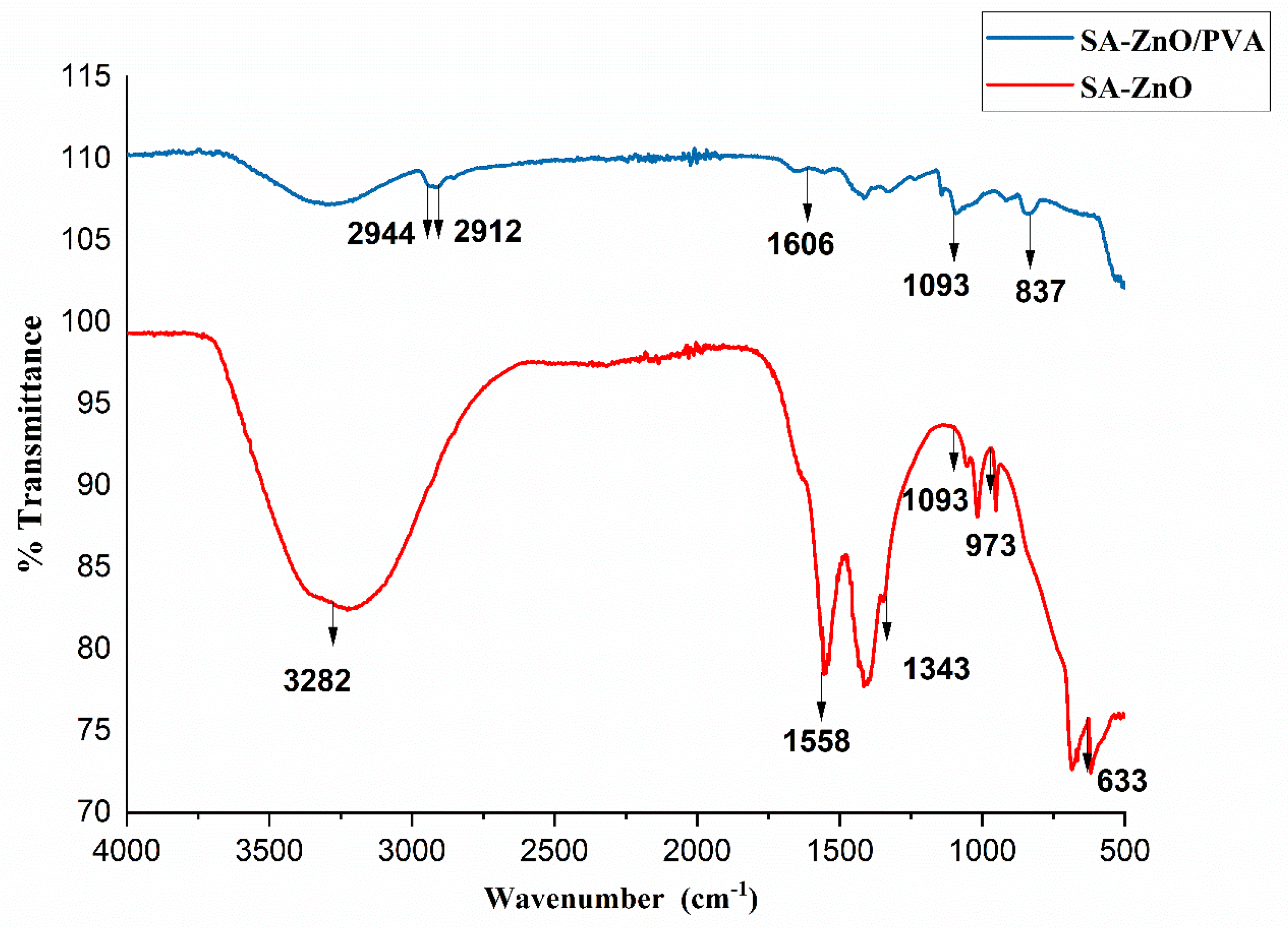
FTIR spectra of SA-ZnO nanoparticles and SA-ZnO/PVA nanocomposites.
FESEM analysis
FESEM was employed to analyse the surface morphology of the synthesised nanoparticles, confirming their agglomerated state. This analysis highlights the influence of surface morphology on the synergistic properties of zinc oxide nanoparticles. The compatibility between components of the polymer composites was also studied, demonstrating phase separations and interfaces that significantly affect the physical properties of the composites. The FESEM images of SA-ZnO surface morphology analysis confirms the presence of nanoparticles in an agglomerated form. This result is steady with many studies in the works that discuss the relationship between surface morphology and the synergistic properties of Zinc oxide. These properties, which are crucial for various applications, are often influenced by the arrangement and size of the nanoparticles, as these factors play a key role in enhancing the material's performance where as SA-ZnO/PVA demonstrated a uniform density of grain distribution on the surface, with slight aggregation observed, highlighting the effective dispersion of nanoparticles in the composite as shown in (Figure 6). The morphology of the ZnO nanoparticles remains consistent in the membrane. The FESEM images of SA-ZnO/PVA nanocomposite displayed a uniform distribution of ZnO nanoparticles, which formed a structure resembling barbed wire.
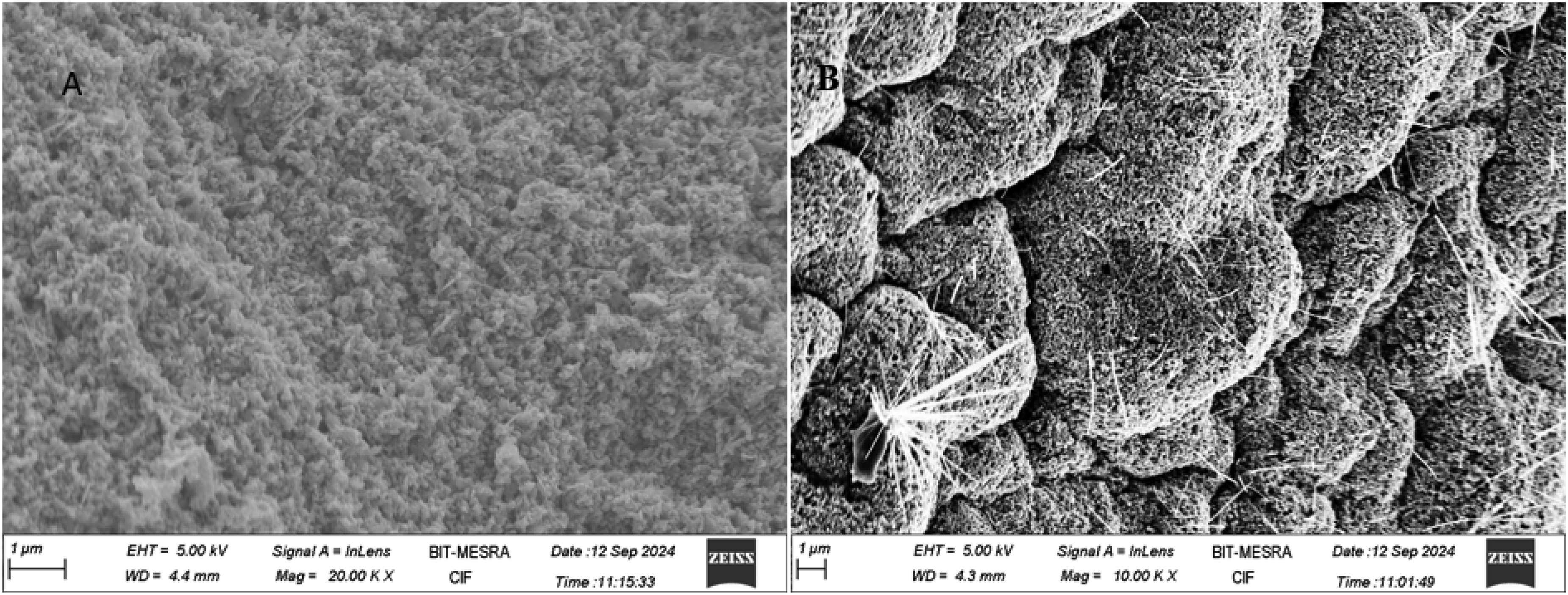
FESEM images of SA-ZnO nanoparticles and SA-ZnO/PVA nanocomposites.
Adhesion quality of SA-ZnO/PVA coated titanium alloy
To determine the adhesive strength of the SA-ZnO/PVA coating on Ti-6Al-4V, a cross-cut tape test was performed. A grid pattern with small squares was cut through the coating to the substrate, adhesive tape was pressed over the cut area, and then peeled off and adhesion was classified on the ASTM scale from 0B (poor) to 5B (excellent) based on the coating removed.
29
The adhesion strength of the SA-ZnO/PVA coating on Ti-6Al-4V was rated 3B in the cross-cut tape test, indicating adhesion with minimal coating removal (Figure 7
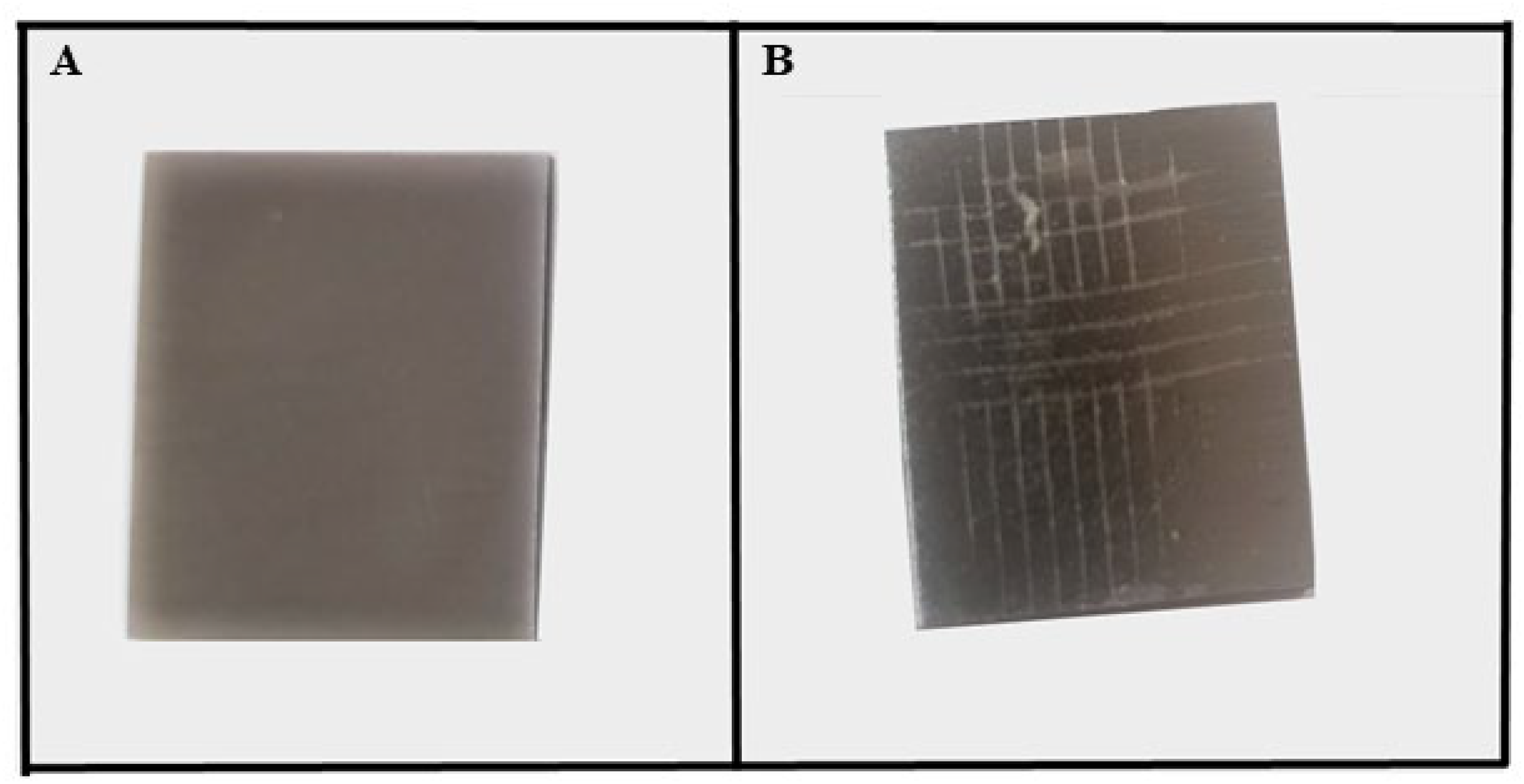
SA-ZnO/PVA coated titanium alloy substrate (A), and Adhesion test result depicting the cross-cut pattern and tape removal area (B).
Haemocompatibility evaluation of SA-ZnO/PVA coated titanium alloy
The results of haemolysis assay indicate that coated titanium alloy exhibited negligible haemolysis, with a haemolytic index of 0.581%, compared to bare titanium alloy, which showed a higher index of 4.069% (Figure 8). These values are below the standard limit of 5%, which classifies them as haemocompatible. However, the SA-ZnO/PVA coated titanium alloy demonstrated significantly better results, highlighting its superior haemocompatibility. These findings suggest that SA-ZnO/PVA coating enhances the biocompatibility of titanium for potential implant applications.
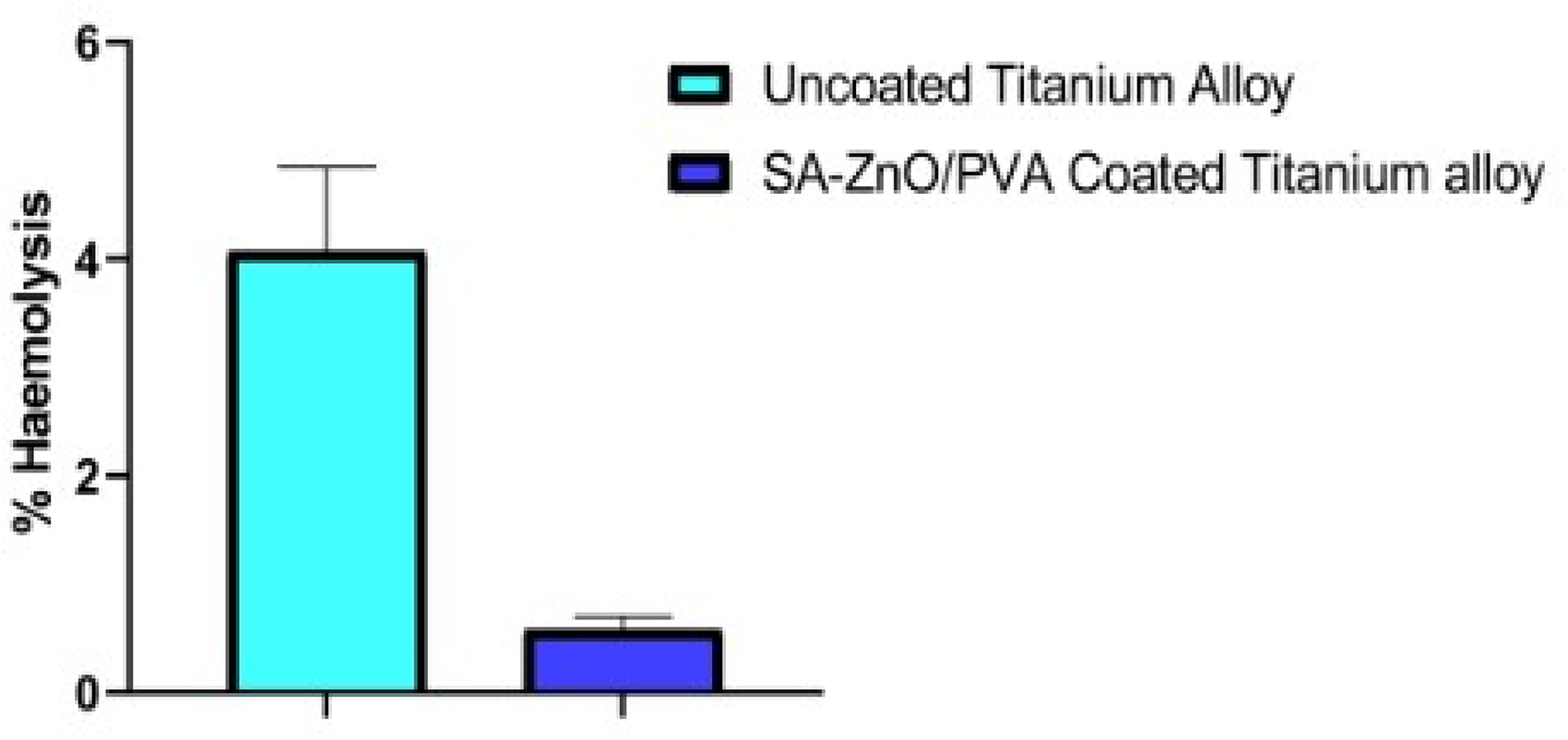
Haemocompatibility analysis of uncoated and SA-ZnO/PVA coated titanium alloy substrates.
Atomic force microscopy analysis of SA-ZnO/PVA coated titanium alloy
The AFM analysis of uncoated and SA-ZnO/PVA coated titanium alloy demonstrated important insights into their surface topography and crystalline structure. The 3D AFM images illustrate the roughness and morphology of the surfaces, highlighting the granularity distribution of the SA-ZnO/PVA coating (Figure 9). The average roughness of the coated titanium alloy is observed to be 10.6114 nm, with a root mean square (RMS) value of 13.6947 nm, while uncoated titanium alloy exhibits a higher average roughness of 22.8995 nm and RMS of 27.7895 nm. Roughness, a topographical property of implant surfaces, is classified into three levels: macro-topographic (Sa > 10 μm), micro-topographic (1 μm ≤ Sa < 10 μm), and nano-level (<1 μm). The reduced roughness of the SA-ZnO/PVA coated alloy may lead to improved osteoblast adhesion, protein adsorption, and osseointegration and effective in optimising osteoclast-material interactions. Thus, a balance between osteoclastogenesis and osteogenesis is crucial for maintaining bone quality, as disruption of this balance can lead to bone fragility.30,36,37
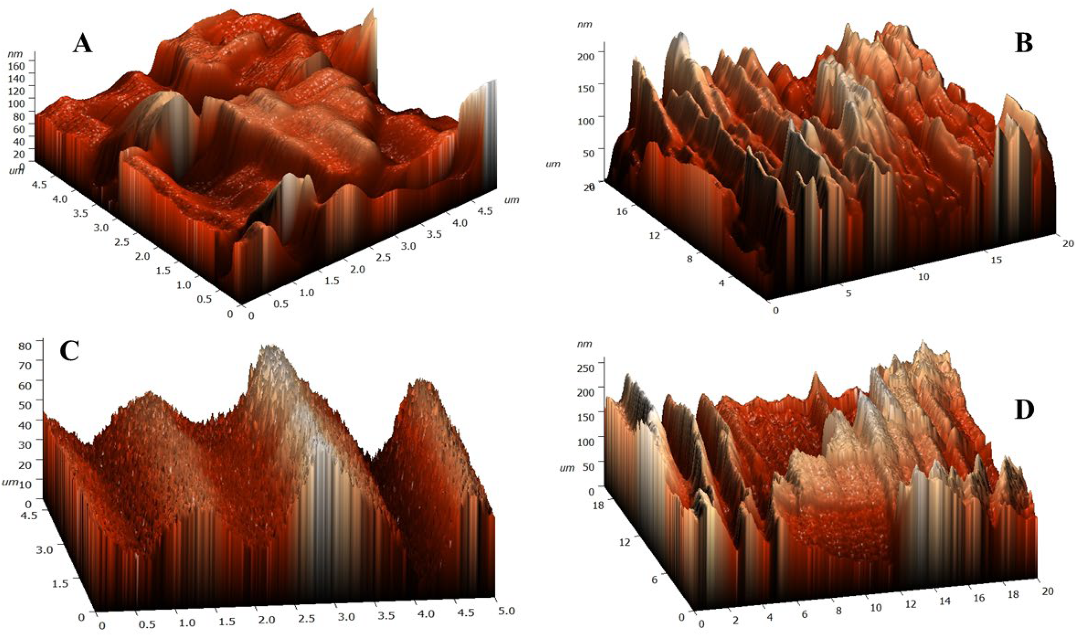
AFM images of uncoated titanium alloy at 5 µm (A), at 20 µm scale (B) and SA-ZnO/PVA coated titanium alloy at 5 µm (C), at 20 µm (D).
Antibacterial potential of SA-ZnO/PVA coated titanium alloy
The results of antibacterial study revealed a greater zone of inhibition against S. aureus and E. coli with SA-ZnO/PVA coated samples. Specifically, the coated samples exhibited a zone of inhibition of 1.5 cm against the gram-positive bacteria, compared to the uncoated samples. Moreover, for the gram-negative strain, the coated samples demonstrated a zone of inhibition of 1.2 cm, while the uncoated samples showed negligible activity (Figure 10). This significant increase in antibacterial activity illustrates the effectiveness of the SA-ZnO/PVA coating in preventing infections, which is a common concern following bone implant surgeries, often leading to complications such as osteomyelitis or implant failure. These findings suggest that SA-ZnO/PVA coatings may not only enhance antimicrobial action but may also provide a promising solution to mitigate the risk of post-surgical infections, which can complicate recovery and lead to the need for systemic antibiotics potentially causing further internal damage. Overall, this study highlights the significant potential of SA-ZnO/PVA coated implants to overcome infection-related challenges in orthopaedic applications, offering a pathway toward safer and more effective medical implants.
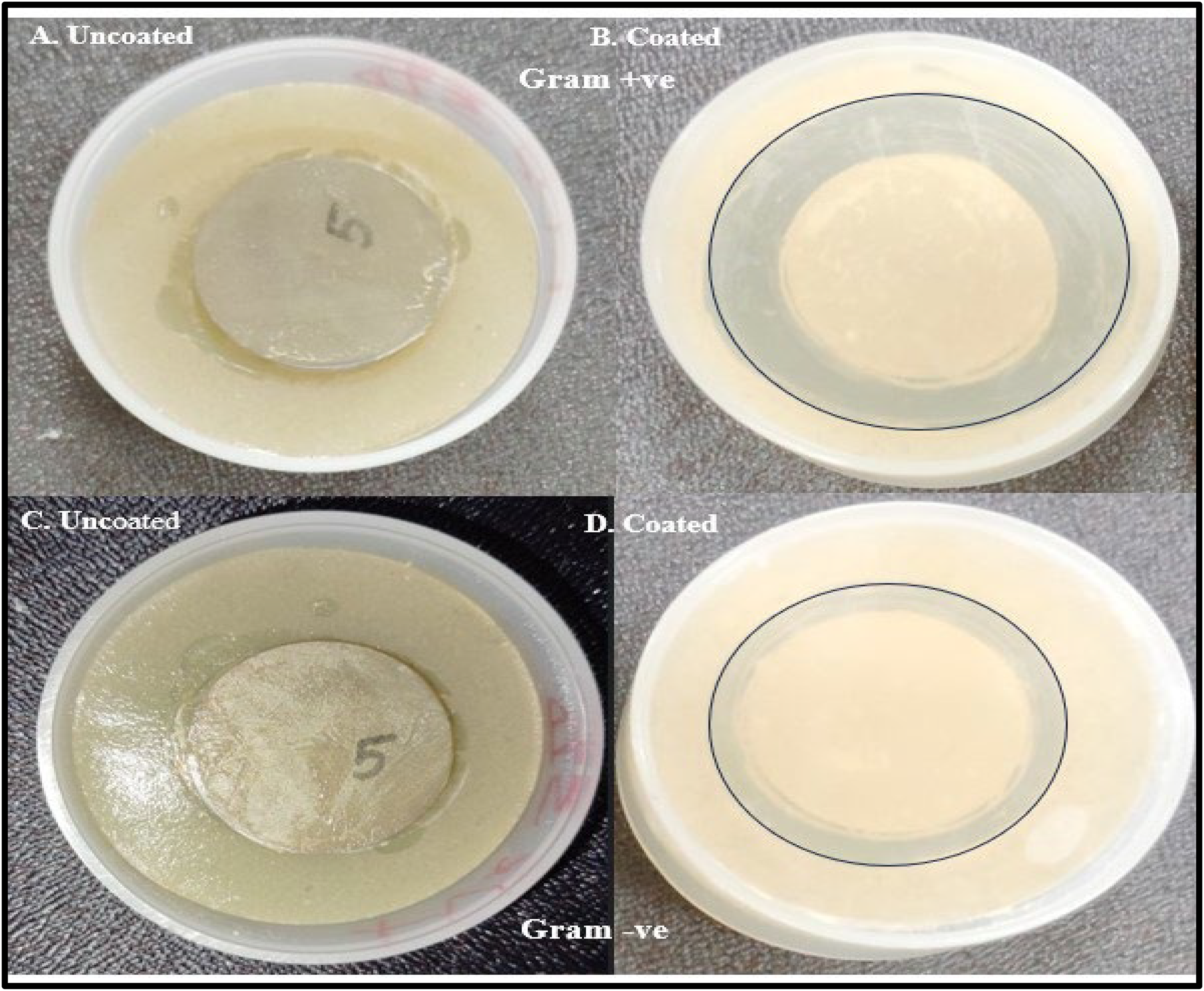
Antibacterial activity of uncoated Titanium alloy substrate (A) and SA-ZnO/PVA coated titanium alloy substrate (B) against S. aureus (Gram + ve); uncoated Titanium alloy substrate (C) and SA-ZnO/PVA coated titanium alloy substrate (D) against E. coli (Gram -ve) bacteria.
Conclusion
The present investigation demonstrates the promising potential of Saraca asoca extract-mediated ZnO/PVA nanocomposites as antibacterial coatings for orthopaedic applications. The aqueous extract of Saraca asoca, rich in flavonoids (10.132 mg QE/g) and phenolics (7.04 mg GAE/g), acted as a reducing and capping agent for green synthesis of ZnO nanoparticles. These SA-ZnO nanoparticles (131.5 nm, PDI 0.167) and SA-ZnO/PVA nanocomposites (308.5 nm, PDI 0.244) exhibited uniform size, stability, and dispersion that is suitable for biomedical applications. Thermal analysis via TGA and DSC confirmed high thermal stability, with TGA showing 77.510% weight loss at 793.62°C for nanocomposites. DSC indicated the conversion of Zn (OH)₂ to ZnO between 83.57°C and 313.60°C. These results ensure durability under physiological conditions, making the nanocomposites suitable for long-term use in orthopaedic applications. The diffraction peaks of SA-ZnO/PVA nanocomposites in XRD exhibited at 31.88°, 34.52°, 36.36°, 47.64°, 56.68°, 62.92°, and 68.02°, closely aligning with SA-ZnO peaks, indicating effective interaction between PVA and ZnO nanoparticles and their high crystallinity and phase purity. FTIR analysis showed strong ZnO-PVA interactions, enhancing the structural integrity of the composite material. FESEM analysis revealed uniform dispersion of SA-ZnO nanoparticles in PVA, which is critical for osteoblast attachment. Haemolysis testing showed improved biocompatibility (0.581% haemolytic index) and antibacterial study confirmed the effective inhibition of S. aureus and E. coli.
Thus, the results support the potential use of SA-ZnO/PVA nanocomposites in both biomedical and environmental applications due to their key properties. The composite shows strong antibacterial activity, making it effective for preventing infections in medical devices, while its ability to reduce implant-related infections and failure ensures safety in implants. The eco-friendly synthesis of SA-ZnO/PVA nanocomposite using Saraca asoca plant extract supports their antibacterial properties that may control microbes in water treatment or may degrade organic pollutants. Further preclinical studies may be carried out to evaluate the effectiveness and safety of SA-ZnO/PVA nanocomposites coated implants. Future research may also focus on scaling production, studying long-term corrosion resistance, and exploring interactions with different cell lines, and developing multi-functional coatings for combined antibacterial, anti-inflammatory, and osteogenic properties.
Footnotes
Acknowledgments
I would like to express my gratitude to BIT Mesra, Ranchi, for the continuous support for providing the research facilities.
Authorship contribution
Experimentation and writing original draft, Apurva Singh; designing, editing and supervision, Neelima Sharma. Both have read and approved the final version of the manuscript.
Declaration of conflicting interests
The authors declared no potential conflicts of interest with respect to the research, authorship, and/or publication of this article.
Funding
The author(s) disclosed receipt of the following financial support for the research, authorship, and/or publication of this article: This research work is funded by DST Inspire Fellowship, Government of India, (grant number IF230156).