
Abstract
In this study, a TiO2, Ag−doped or Cu−doped TiO2 film was deposited on a TC4 alloy, and the effects of the dopants (i.e., Ag and Cu) on the photoelectrochemical response, biofilm suppression as well as corrosion resistance of the TiO2−coated alloy were systematically investigated. It can be found that the Ag doping in TiO2 can bring about the increased concentration of oxygen defects, thereby elevating the charge carrier density and enhancing the photoelectric and corrosion current responses, which can promote the photoelectrochemical response but concurrently
Introduction
TC4 titanium alloys exhibit significant application potential as base materials of solar interfacial evaporators owing to their high specific strength and outstanding corrosion resistance.1–3 However, because the base materials of solar interfacial evaporators are generally prone to water−induced corrosion and contamination by organic matter and microorganisms, it is essential to enhance their resistance to contamination. Among these issues, biofilm accumulation on evaporator surfaces can reduce light absorption efficiency, obstruct pore channels, and ultimately decrease evaporation performance. In addition, biofilms can promote localized corrosion and structural degradation of the substrate. Therefore, resistance to surface biofilm formation is critical for maintaining long-term operational efficiency and durability.
Due to the merits of superior photoelectrochemical response, biofilm suppression performance as well as corrosion resistance, TiO2 films have been extensively adopted in multiple applications such as development of self−cleaning glasses,4,5 biofilm suppression performance,6,7 and photovoltaics.8,9 Their photocatalytic and biofilm suppression properties stem from the semiconducting property of TiO2. TiO2 generates superoxide radicals upon photoirradiation, which degrade and decompose organic substances into small inorganic molecules such as carbon dioxide and water.
10
The biofilm suppression performance of TiO2 is ascribed to the
The biofilm suppression activity of TiO2 films can be enhanced by doping with metals, such as Ag or Cu, and the primary mechanism involves the release of metal ions that disrupt microbial cellular structures and metabolic functions.12,13 Ag−, Cu−doped TiO2 films are generally characterized by distinct optical transmittances and bandgap energies, which can directly affect the generation of photoelectrons. Moreover, the dopants within the film cause the occurrence of lattice distortions and thus the crystallinity of TiO2 is reduced, leading to the diminished charge−carrier mobility, which may promote the increase in carrier concentration and decrease in mobility. 14 The corrosion resistance of a semiconductive film is directly affected by its conductivity, i.e., its charge−carrier concentration and mobility.
In this study, pristine, Ag−, Cu−doped TiO2 films were prepared through magnetron sputtering to investigate the effects of the Ag and Cu dopants on their multiple functions, including photoelectrochemical behavior related to photoelectrochemical response and biofilm suppression activities and corrosion resistance.
Materials and methods
TC4 plates were used as the substrate, and 99.95% pure titanium and 1 at.% Ag−doped or Cu−doped titanium plates were used as the targets for reactive magnetron sputtering. The TiO2 films were prepared with background vacuum and deposition pressure of 3 × 10⁻³ and 0.14 Pa, respectively. A pulsed−target power supply and sputtering current of 1 A, along with a substrate bias voltage and oxygen flow rate of −60 V and 3 sccm, respectively, were employed. A deposition time of 3 h resulted in a film thickness of 250 nm.
The film thickness was measured using a profilometer (Alpha−Step D−100, KLA, USA), with a stylus force of 0.03 mg and scanning step of 2 mm. The surface morphologies of the TiO2 films were characterised through atomic force microscopy (AFM, CSPM 5500, Guangzhou, China) in contact mode at a scanning frequency of 1.8 Hz; the needle insertion voltage was between −15 and −20 V. The roughness and three−dimensional morphology of the films were analysed with the Imager analysis software. The crystal structures of the TiO2 films were analysed through X−ray diffraction (XRD, D8 ADVANCE, Bruker, Germany); the scanning mode was continuous PHD fast, the scanning step size was 0.117 s, and the phases of the films were characterised using the High Score Plus software. Further, the valence states of the surface elements were determined through X−ray photoelectron spectroscopy (XPS, ESCALAB 250Xi, USA), conducted at a test passing energy of 20 eV and step size of 0.1 eV. The electrical parameters of the TiO2 films were evaluated using a Hall−effect tester (Swin Hall 8000, Taiwan, China), applying an input current of 10−6 A, a voltage of 10 V, and a magnetic field strength of 4000 Gs. The carrier concentration and electron mobility of the film were output based on the ‘four−point probe’ method. Photocurrent studies were conducted in a 0.5 mol/L Na2SO4 solution with an electrochemical workstation (Vertex C) under simulated sunlight (100 mW/cm2) obtained using a CEL−HXUV xenon lamp (Zhongjiao Jinyuan Company). The test sample was subjected to light switching every 10 s to evaluate its photocurrent density. To avoid randomness, multiple samples of the same group were soaked in the test solution in biological contamination experiments. The samples were inoculated in the prepared sulfate−reducing bacteria (SRB) suspension for 7 days; thereafter, the biofilms were fixed with a 5% glutaraldehyde solution and finally subjected to gradient dehydration with ethanol solutions, followed by dry storage for SRB observation by scanning electron microscopy (SEM, Apreo 2, USA), which was performed at a working voltage of 1–3 kV to achieve good imaging contrast. For quantitative analysis, three randomly selected regions from each sample were observed by SEM to ensure the statistically representative data. The biofilm areas were calculated using ImageJ software, and the coverage percentage was defined as (biofilm area / field area) × 100%. The reported coverage percentage was the average data of three independent measurements with standard deviation less than 5% for each region. The doping contents of Ag and Cu were only 1 at.%, which
Results and discussion
Morphology and structure
Figures 1(a)−(c) and Table 1 show the AFM of the films. The TiO2 film exhibits a dense, fine morphology with a roughness of 2.93 nm. In contrast, the maximum roughness of 5.88 nm was observed in the TiO2(Ag) film, which is attributed to the formation of large clusters of grains induced by Ag doping. Moreover, the TiO2(Cu) film exhibits a larger roughness of 3.93 nm, because Cu doping deteriorates the uniformity of the grains.
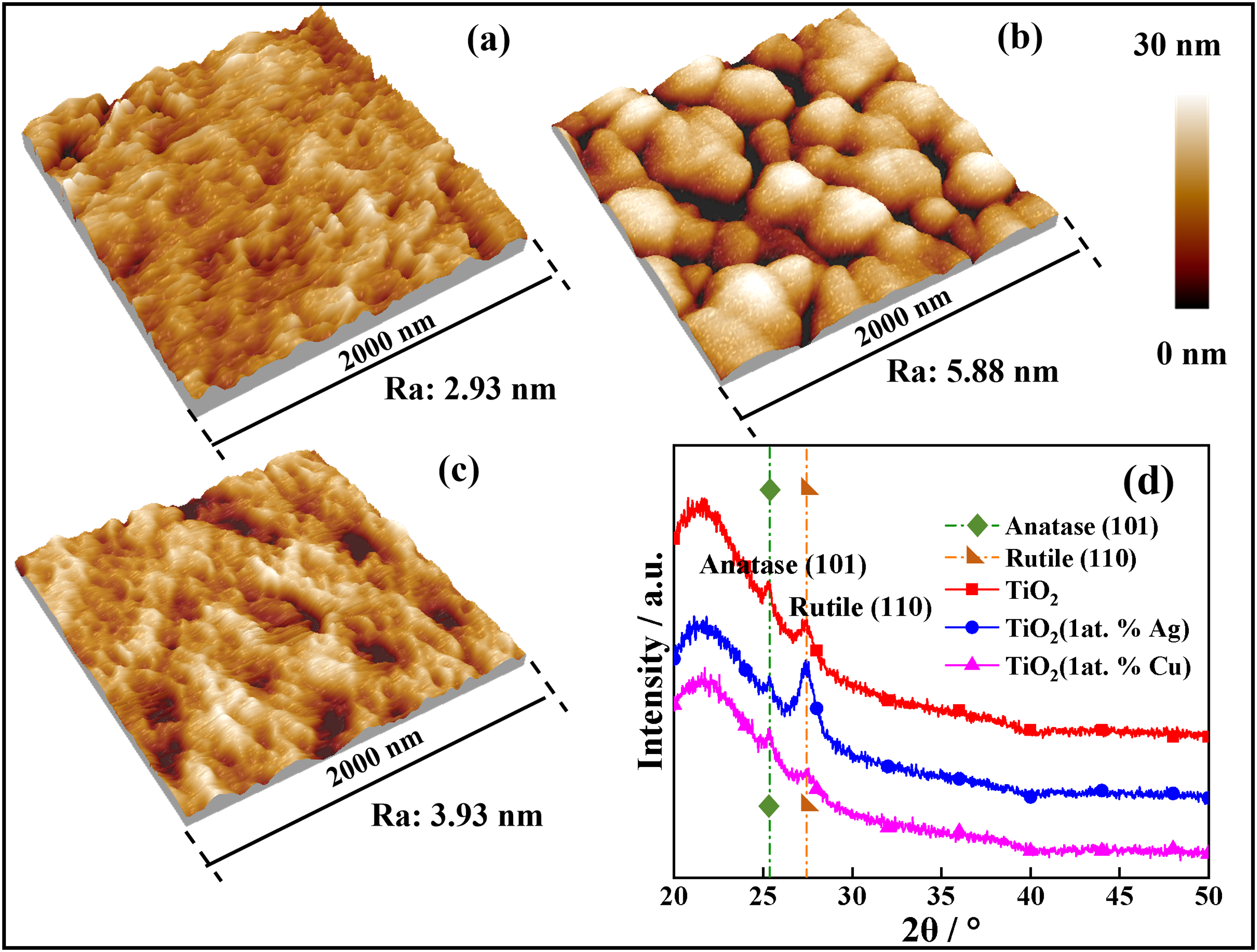
AFM morphologies of the (a) TiO2, (b) TiO2(Ag), and (c) TiO2(Cu) films, and (d) XRD patterns (PDF #: anatase, 01−073−1764; rutile, 00−034−0180).
Performance and parameters the pristine and Ag−/Cu−doped TiO2 films.
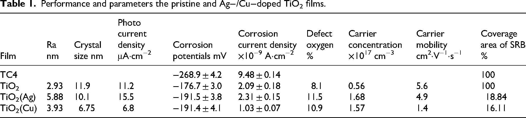
Figure 1(d) presents the XRD patterns of the films, which reveal that all samples are composed of mixed anatase and rutile phases. The crystallite size calculated from the rutile (110) peak is approximately 11.9 nm for the TiO2 film, according to the equation of D = kλ/βcosθ. 15 For the TiO2(Ag) film, the intensity of the rutile (110) peak is enhanced, this is because Ag may be incorporated into the TiO₂ film either through partial lattice incorporation or as segregated metallic/oxide species, resulting in the formation of unsaturated covalent bonds. Additionally, the crystallite size is slightly reduced to 10.1 nm after Ag doping, indicating the lattice distortion caused by the difference in the ionic radii between Ag+ (115 pm) and Ti4+ (74.5 pm), 16 consistent with the AFM in Figure 1(b). The diffraction peaks of the TiO2(Cu) film are weak, and the crystal size of TiO2 is reduced to 6.75 nm, indicating that the crystallisation is suppressed by the Cu dopant. Unlike in the case of Ag+ doping, the ionic radius of Cu2+ (73 pm) is comparable to that of Ti4+, leading to relatively minimal lattice distortion and a smaller change in the valence bonding. 17
Figures 2(a)−(d) show the XPS survey spectra and Ag3d, Cu2p, Ti2p, O1 s XPS spectra of the TiO2 films. The survey spectra in Figure 2(a) and the Ag3d and Cu2p spectra of the doped samples in Figure 2(b) confirm the presence of the Cu and Ag dopants in the corresponding doped TiO2 films. The Ag3d spectrum of the Ag−doped film is characterized by Ag3d3/2 and Ag3d5/2 peaks. 18 Similarly, the Cu2p spectrum of the Cu−doped film exhibits Cu2p1/2 and Cu2p3/2 peaks. 19 The intensity difference of the Ag and Cu peaks is mainly derived from the different ionisation cross−sections resulting from the different radii of the Ag3d and Cu2p orbitals. 20 The Ti2p spectra of the films show pronounced Ti2p3/2 and Ti2p1/2 peaks, accompanied by satellite peaks. 21 The shock−induced satellite peaks of Ti2p are a typical feature of the XPS spectra of TiO2 films. 22 The O1 s spectra of the films could be deconvoluted into peaks representing lattice (Oα), defect (Oβ), and adsorbed oxygen (Oγ) atoms. 23 Notably, Oβ is extensively recognized as a critical contributor to photocatalytic activity because of its role in promoting the charge−carrier separation or delay the electron−hole recombination. 24 The proportion of Oβ within the TiO2 film was 8.1%, which is increased to 11.5 and 10.9% in the TiO2(Ag) and TiO2(Cu) films, respectively. This increase is attributed to the substitution of lattice Ti4+ with the Ag+ and Cu2+ species, resulting in the generation of charge imbalance and oxygen−related defects.
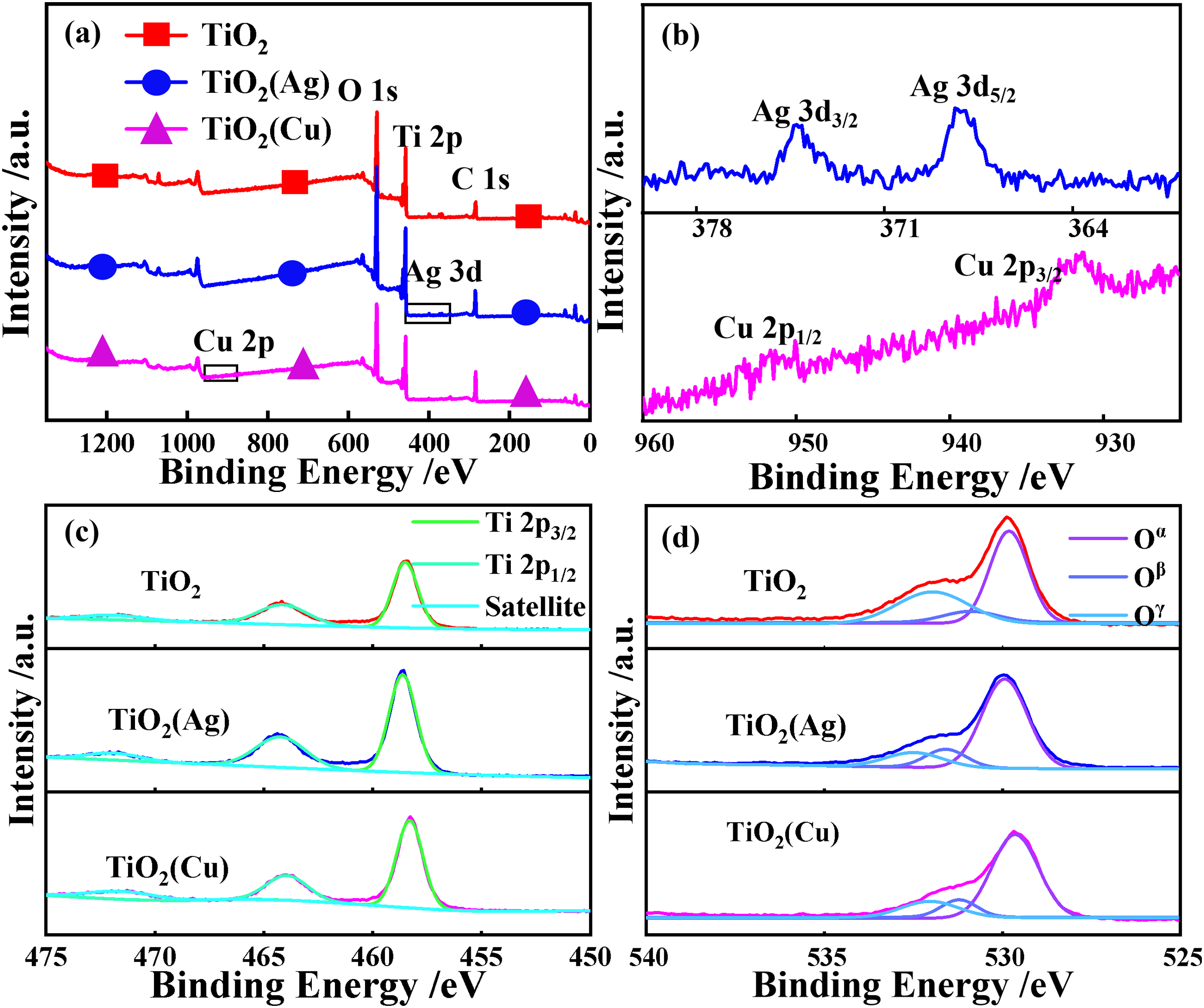
X−ray photoelectron spectroscopy of the TiO2 films. (a) survey spectra, high−resolution spectra of (b) Ag and Cu, (c) Ti, and (d) O.
Functionality
Due to the limited specific surface area of the films, a quantitative comparison of their degradation efficiencies could not be reliably conducted. Nevertheless, earlier studies have demonstrated photocurrent reflect photoinduced charge separation behavior and serve as an indirect indicator of potential photocatalytic response.25,26 Therefore, the photocurrent density of the films
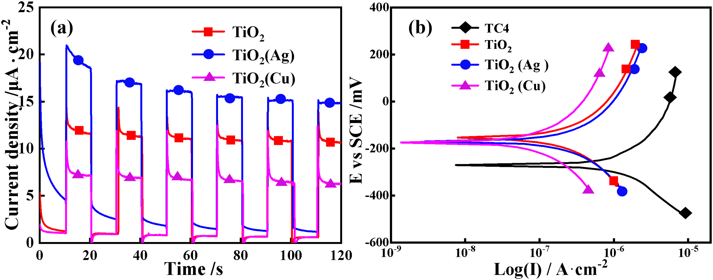
(a) Photocurrent densities, (b) polarisation curves of the TiO2 films.
As mentioned in the introduction, the application of this study is the solar interfacial evaporator. The critical biofilm suppression requirement is to suppression of biofilm formation on the sample surface, in order to prevent reducing the light absorption efficiency and the intensifying corrosion, rather than inhibiting bacteria in the surrounding environment. Hence, the biofilm suppression performance of the films 
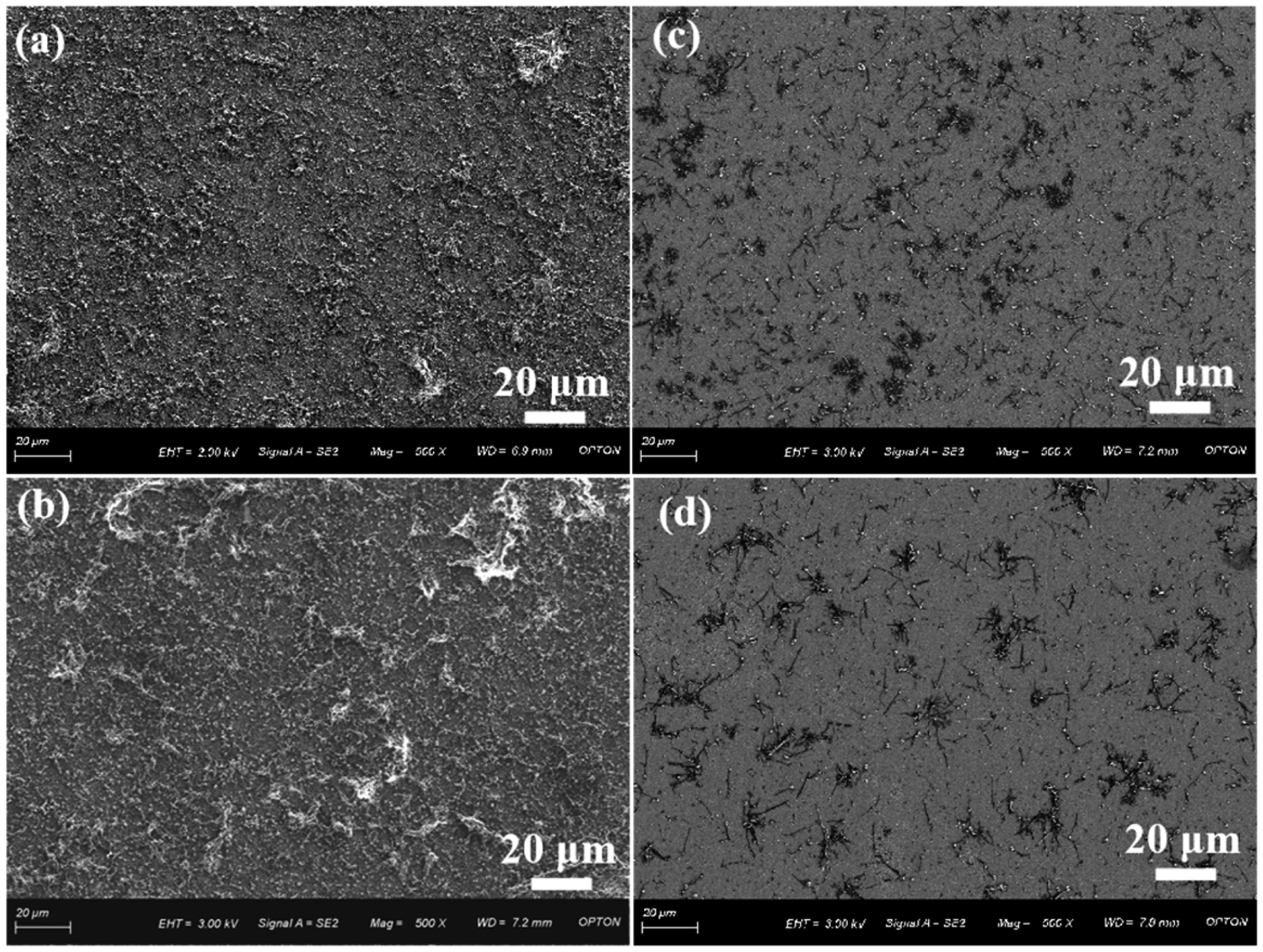
Biofilm−resistant morphologies of the (a) TC4 alloy, and (b) TiO2, (c) TiO2(Ag), and (d) TiO2(Cu) films.
The corrosion current and photocurrent densities of the films are related to their crystallinity and Oβ proportion, as well as the concentration and mobility of the free charge carriers. As shown in Figure 5, a significantly increased Oβ proportion can be detected in the TiO2(Ag) and TiO2(Cu) films (11.5 and 10.9%, respectively), which is attributed to the different bonding interactions of Ag+, Cu2+, and Ti4+ with oxygen, leading to an increase in the concentration of free charge carriers within the doped films. Specifically, the free charge−carrier concentration is enhanced from the order of 1016 in the TiO2 film to 1017 in the doped films, as shown in Figure 5(a) and Table 1. The Oβ proportion and free carrier concentration can be measured as 8.1% and 0.56 × 1017 cm−3 in the TiO2 film, 10.9% and 1.57 × 1017 cm−3 in the Cu−doped film, and 11.5% and 1.68 × 1017 cm−3 in the Ag−doped film, respectively. Cu2+ induces slightly greater changes than Ag+ in covalently bonding with O2–, and the Oβ proportion and free carrier concentration of the TiO2(Cu) film are slightly lower than those of the Ag−doped film. Further, the TiO2(Ag) and TiO2(Cu) films exhibit smaller crystallite sizes (6.75–10.1 nm) than the TiO2 film (11.9 nm). Hence, the mobility of the free charge carriers is decreased from 5.6 cm2·V−1·s−1 in the TiO2 film to 4.9 and 1.4 cm2·V−1·s−1 in the TiO2(Ag) and TiO2(Cu) films, respectively. In particular, the TiO2(Cu) film with an almost amorphous structure (Figure 1(d)) exhibits a low carrier mobility 29 (Figure 5(b) and Table 1). In contrast, the TiO2(Ag) film is characterized by a substantially higher carrier concentration and relatively high mobility, and the photocurrent density of 15.5 μA·cm−2. 30 The relatively large crystallite size and excellent semiconductive properties of TiO2(Ag) can provide interfacial boundaries that facilitate the formation of galvanic cells and the participation of charge carriers during the electrochemical corrosion process. Consequently, the TiO2(Ag) film exhibits a higher corrosion current density (2.31 × 10⁻⁷ A/cm2) than the TiO2 film (Figure 5(c) and Table 1), indicating that the enhanced electrochemical activity is closely associated with its higher carrier density and mobility. In contrast, the extremely low carrier mobility within the TiO2(Cu) film restricts the charge motion, and thereby the generation of photoelectrons is suppressed, resulting in a relatively low photocurrent density (6.8 μA·cm−2). Besides, the almost amorphous structure of the TiO2(Cu) film confines the corrosion process at the interface, leading to a lower corrosion current density (1.03 × 10⁻⁷ A/cm2) than the TiO2 film.
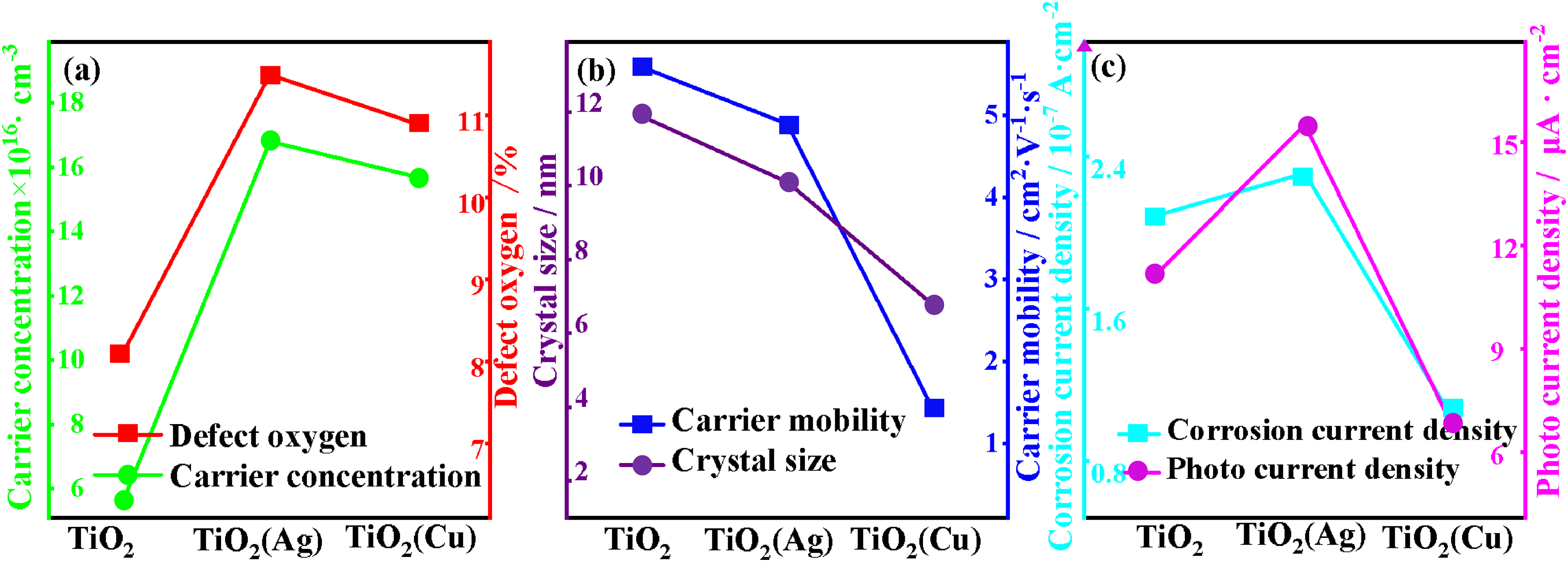
(a) Carrier concentration and proportion of defect oxygen, (b) carrier mobility and crystal size, (c) corrosion current and photocurrent densities.
The TiO2(Ag) film exhibits a better photoelectrochemical response than the TiO2 film. This behaviour is ascribed to the combined effect of high concentration of free charge carriers, a high mobility and relatively large crystallite sizes. In contrast, the TiO2(Cu) film displays lower photoelectrochemical response than the TiO2 film, which is derived from the low mobility of the free charge carriers and its almost amorphous structure. The deposition of TiO2 films on the TC4 alloy can enhance the corrosion resistance of the substrate. It should be noted that the improved biofilm suppression may be related to the presence and possible release of Ag- or Cu-containing species.
Conclusions
In this study, the TC4 alloy was imparted with photoelectrochemical response, corrosion resistance, and biofilm suppression functions through the deposition of TiO2, TiO2(Ag), and TiO2(Cu) films. It can be found that compared with the TiO2 and TiO2(Ag) films, the Ag−doped TiO2 film exhibits superior photoelectrochemical response. The intrinsic and doped TiO2 films on the TC4 alloy significantly enhanced the corrosion resistance of the substrate. Particularly, both the Ag and Cu dopants can remarkably improve the biofilm suppression activity of TiO2 films. This outstanding comprehensive performance can play a significant role in the application of the TiO2−coated TC4 alloy as a solar interfacial evaporator for water evaporation, protecting against corrosion, bacterial growth, and organic matter adsorption.
Footnotes
Acknowledgements
This work is supported by the National Natural Science Foundation of China in Nos. of 52271056, the education department of Liaoning in Nos. of LJ212410146060, LJ232510146002, the research fund of national key laboratory of marine corrosion and protection of luoyang ship material research institute under the contract No. KJS2403.
Author contribution(s)
Funding
The authors disclosed receipt of the following financial support for the research, authorship, and/or publication of this article: This work was supported by the National Natural Science Foundation of China, education department of Liaoning, (grant number 5227105, LJ212410146060, LJ232510146002).
Declaration of conflicting interests
The authors declared no potential conflicts of interest with respect to the research, authorship, and/or publication of this article.