
Abstract
Laser surface modification of Ti6Al4 V alloy plays an essential role in improving its bioactivity and fitness for bone implant applications. In the present work, different surface structures, including untextured, linear groove, crisscross, and wavy groove patterns, were created and assessed for surface roughness, wettability, protein adsorption, and calcium mineralisation. The textured samples exhibited higher roughness than the untextured specimens, with the wavy-textured surface showing the highest roughness. Contact angle measurements showed a change from hydrophilic to superhydrophobic behaviour, strongly influenced by surface topography. Protein adsorption studies indicated enhanced biomolecular interaction on textured surfaces, with the wavy pattern demonstrating the highest adsorption. Similarly, calcium mineralisation results showed a substantial increase in calcium deposition for textured samples, with the wavy surface exhibiting superior behaviour. The enhanced biological response is attributed to the combined effect of hierarchical roughness and modified contact angle.
Introduction
Total hip replacement is a procedure that replaces the hip joint with a biocompatible implant. The selection of the implant materials is based on their mechanical properties and biocompatibility. One of the most important mechanical phenomena to consider is stress shielding. Hence, titanium alloys such as Ti-6Al-4 V are the best materials for the femoral head, femoral stem and acetabular shell.
A few complications may be evident either immediately or after a few years post-surgery. Though joint infection and leg length inequality are serious complications, they are rare among patients and are due to surgical errors. Some of the most common complications that might lead to the need for repeat surgery are dislocation and implant loosening. The failure of hip prosthesis, which is the cause for repeating the surgery, is mainly due to implant loosening. Implant loosening can be delayed not only by reducing sliding wear between the femoral head and acetabular socket, but also by enhancing osseointegration at the implant surface in contact with bone. Hence, they can be controlled by modification of surface properties. Possible surface engineering methods, such as surface machining, surface hardening, and coating, can be used to reduce wear and enhance osseointegration. Surface coating is a novel methodology for deliberately altering surface properties, but it is very expensive. Thus, laser surface texturing is one of the viable options not only for enhancing bone ingrowth of the implant but also for reducing polyethylene wear in the modular hip prosthesis.
Chae-Hyun Lim et al. 1 reported radiographic results and survival rates following hip arthroplasty in 41 patients with a long-term follow-up of 21.4 years. They have concluded that the high revision rate of hip prosthesis is due to polyethylene wear and osteolysis. Raquel et al. 2 investigated the impact of bio-functionalised implant surfaces on bone apposition and compared them with other surfaces, such as the grit-blasting/acid-etching process. It was revealed that a textured surface with a small amount of the bioactive peptide led to higher adjacent bone density.
Rafael et al. 3 evaluated Ti implants with laser surface texturing. Their result shows that the textured implants show improved bone implant interaction when compared to the machined ones. Zhang et al. 4 combined laser treatment and acid etching to achieve a nano-textured surface on a Ti alloy implant. Their results confirm that the bonding of the hydroxyapatite coating improved, thereby enhancing the implants’ bioactivity. Bao et al. 5 conducted the wettability study of a laser-textured titanium surface. Wettability analyses showed that all textured Ti surfaces were super-hydrophilic before silanisation and became highly super-hydrophobic after silanisation.
Voisey et al. 6 examined the effect of texturing of Ti alloy on osteoblast cell response. The laser-modified samples were exposed to osteoblast cells, and it was observed that cells responded well to the features in the textured regions. Ophélie et al. 7 analysed the effect of wettability and cell response of laser-textured titanium alloy. The result shows that the cells are sensitive to the pattern and spread along these structures with good wettability. Rui et al. 8 prepared a microstructured surface on a Ti alloy by laser heating and analysed its bioactivity. Their results suggested that the micro-structured surface improved the biocompatibility and tissue integration with implants.
Milan et al. 9 modified the titanium implant surface by laser treatment. Results confirm that the roughness induced by laser processing of the implant surface improved biointegration. Michael et al. 10 examined the effect of surface modification of titanium on blood plasma protein adsorption. Protein assessment was done quantitatively using an enzyme-linked immunosorbent assay. The plasma proteins were adsorbed preferentially on acid etched cum sand blasted surface due to surface topography. Podra and Anderson 11 simulated the lubricated sliding wear using finite element analysis. The deviation of the wear coefficient in experimental and numerical analysis was found to be 40–60%.
Roy et al. 12 observed that a circular dimple with 15% density exhibited improved wear resistance compared to other structures. They noticed that a larger dimple diameter with higher density minimised the wear. Ito et al. 13 examined the wear performance of a circular dimpled steel surface and observed a significant reduction in the wear rate.
Renu et al. 14 studied the effect of texturing on bioactivity and cell attachment. They observed that the cells align along the textured surface, and the textured surface shows a substantial improvement in bioactivity. Wilhelm et al. 15 characterised the laser-textured Ti alloy with a line-and-dimple profile. They observed an enhancement of surface properties, including the presence of Ti oxides, in the textured region compared to the non-textured region, and an increase in wettability after texturing.
Boadi et al. 16 presented an innovative laser lithography–aided micro texture and anodization method for producing micro-lined TiO2 nanotube arrays. Compared to conventional nanotubes, enhanced bonding strength and wear resistance were observed, which promote bone integration. Arnab et al. 17 created a nanoscale surface texture on a grade 23 Ti alloy biomaterial. Their results showed that bio-tribocorrosion was significantly reduced, with improved passivation tendency and better cell proliferation and metabolism on the surfaces.
Sigh et al. 18 studied the effects of single- and multi-level EDM micro-texturing on Ti6Al4 V alloy surfaces and analysed their wetting properties. The multi-level texturing yielded a higher contact angle than single-level texturing and produced a hydrophobic surface. Jain et al. 19 developed a micro-milling method to form microtextures on Ti6Al4 V alloy and analysed the surface roughness and wettability. They observed a dimensionally correct, defect-free, and uniform dispersion of textures using the Owen and Wendt method. They also notice that a higher texture density increases surface roughness and wettability.
Dong et al. 20 reviewed the importance of surface modification (physical and chemical methods) of Ti alloy for biomedical applications. They concluded that, to improve the biocompatibility of Ti alloys, surface modifications are essential for the success and longevity of implants used in hard tissue repair and reconstruction. Gopal and Sudarshan conducted a survey on surface engineering in total hip arthroplasty and strongly suggested that surface roughness, wettability, and porosity strongly influence the biological response to orthopaedic implants. 21
Pulsed laser texturing has also been widely explored in several advanced engineering applications beyond biomedical implants. Recent studies demonstrated its effectiveness in mold fabrication through enhanced surface functionality, 22 modification of optical properties for energy-related applications, 23 tribological performance improvement through controlled hierarchical structures, 24 and fabrication of microdevices with precise surface architectures. 25 These studies further confirm the versatility of laser texturing in tailoring surface-dependent functional properties.
Micro-texturing using a picosecond Nd: YAG Laser was confirmed as the process for reducing wear and enhancing bone anchorage. The bone cell bond on the implant surface depends on the adsorption of proteins and the mineralisation of calcium phosphate on the implant surface. Thus, the Lowry Assay and Alizarin Red Assay were conducted to quantify the adsorption of multiple proteins in FBS and the mineralisation of CaP on micro-textured surfaces. The Lowry Assay is a colorimetric technique used for quantifying total protein concentration based on peptide bond interactions with copper ions and Folin reagent. Alizarin Red Assay is commonly employed to quantify calcium-rich mineralized deposits through selective binding of Alizarin dye to calcium phosphate phases.
Unlike conventional methods that typically focus on isolated surface parameters, this study demonstrates a comprehensive structure–property–bioactivity relationship, highlighting how hierarchical surface features govern biological performance. The work presents a novel design strategy for tailoring implant surfaces via controlled laser texturing to achieve superior osseointegration, thereby suggesting significant progress toward the development of next-generation biomedical implants.
Materials and methods
Sample preparation
Ti6Al4 V, which is used as the femoral stem and the acetabular shell of the hip implant, is selected as the test material in this study. It contains 3.5 V, 5.5 Al, and 0.3% Fe, with other minor alloying elements. Ti6Al4 V plates of 100 mm × 45 mm × 3.5 mm were wire cut into small disks of 9.5 mm diameter and 3.5 mm thickness. Then, the disks were serially polished using abrasive papers of different grades up to 1200 grit to get a polished finish.
Laser surface texturing
Micro textures of different patterns, i.e., linear, crisscross and wavy grooves, were machined using a Q-switched Nd: YAG Laser on one side of the disk specimens prepared. The 4 W pulsed laser beam was focused using an F/theta lens with a focal length of 70 mm, producing a beam spot diameter of 7 µm at the sample surface. The pulsed laser has a 532 nm wavelength, 50 kHz pulse frequency, and 80 µJ pulse energy was used for texturing. The groove width was fixed at 90 μm for the linear & crisscross groove patterns and at 60 μm for the wavy groove pattern. The pitch between the grooves was fixed at 145 μm for the linear & crisscross groove patterns and the wavy groove pattern.
Surface roughness, topography and contact angle
The samples were imaged using SEM (Scanning Electron Microscope) to analyse the surface morphology of non-textured and textured samples with groove, crisscross and wavy patterns at different magnifications. A white-light interferometer was used to capture changes in surface roughness across different patterns. The roughness parameters were computed along the line perpendicular to the micro-grooves. Tools were also used to generate 3D views of spectroscopy data for different textured samples. A goniometer coupled with an on-board computer running DROPimage software was used to measure the contact angles of the textured and non-textured samples. It consists of a 3-axis movable stage, a micro syringe and a high-end camera to focus the sample in the stage. The software has an option to view the camera output live on the screen. A water droplet volume of 5 µL was used for contact angle measurements, and the measurements were performed immediately after droplet deposition to minimize evaporation effects.
Mineralisation assay
Mineralisation is an important biocompatibility function. The higher the sample's biocompatibility, the greater the mineralisation. The textured and non-textured samples were incubated in SBF (Simulated Body Fluid) for a week, whose ion concentrations are similar to those of human blood plasma. The ability of apatite to form on the sample surface is a factor in mineralisation. After incubation, Alizarin Red solution was applied to the samples, indicating the presence of Calcium Phosphate by reddening of the sample surface.
Preparation of SBF
Simulated Body Fluid is prepared according to the protocol described by Tadashi Kokubo. 26 Since SBF is a supersaturated solution of apatite, apatite will be nucleated on the surface of the glass beakers and in the scratches. Hence, a plastic beaker with a smoother surface was chosen for SBF preparation, and 500 mL of SBF was prepared. Hence, 350 mL of ion-exchanged, distilled water was placed in a 1000 mL plastic beaker. The beaker was magnetically stirred. All the reagents, except Tris and HCl, were dissolved in sequence.
It was noted that no reagents were dissolved simultaneously. After dissolving the first eight reagents, the solution was heated to 37°C. Just before adding the Tris, the solution pH was measured and adjusted to 2.2 with NaOH and HCl. Tris was added gradually, and the pH was adjusted to 7.38. Finally, the SBF is prepared and refrigerated at 6°C.
Incubation
The SBF stored under 6°C was brought back to room temperature. The pH was checked again, as pH changes with temperature. Falcon tubes were used to soak the samples in SBF. 15 mL of SBF per Falcon tube was used to soak the samples. The samples were placed in the Falcon tubes as shown in Figure 1 (b). These Falcon tubes were incubated for a week in a shaking incubator (Figure 1(a)), maintained at 36.5°C and shaking at 150 rpm. Triplicates from every sample group were incubated.
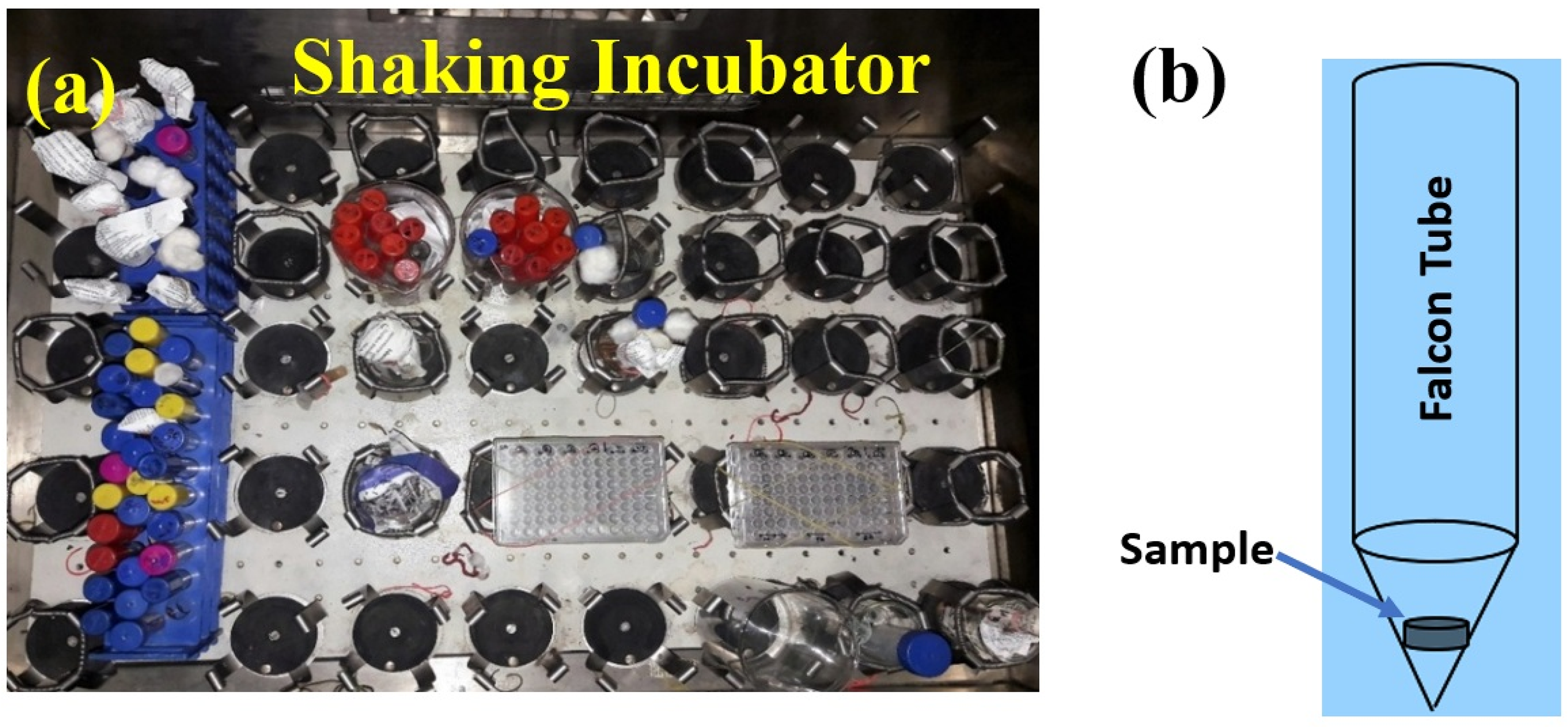
(A) Top view of the shaking incubator; (b) Positioning the samples in the falcon tube to be placed in the incubator.
Alizarin red assay
Initially, 100 mL of a 40 mM Alizarin Red solution was prepared. After the Alizarin Red S Assay, the ARS solubilised in the final sample solution was measured for absorbance using a UV spectrophotometer. These absorbance values were used to determine the Calcium Phosphate content in the textured surfaces of the different sample groups. Before measuring the absorbance of the ARS standards, they were checked for their absorbance maxima using a UV spectrophotometer. The absorbance maximum was observed at 436 nm. The amount of calcium phosphate deposited is directly proportional to the Alizarin Red dye bound to the sample surface. Moreover, the higher the absorbance value, the higher the concentration of the Alizarin Red Solution will be. Hence, to measure the exact calcium phosphate content, an ARS standard was prepared.
Results and discussion
Surface structure of laser-textured specimens
SEM images of the laser-textured specimens at different magnifications are shown in Figure 2(a-c). The non-textured surface of the samples shows a relatively smooth morphology with irregular microasperities. At higher magnification (500x), the surface appears flatter, with only limited irregularities. The smooth surfaces in non-textured regions will not support stronger cell attachment and bone integration. The groove-textured surface at lower magnification (50x shown in Figure 2(a)) reveals parallel, uniform microgrooves across the surface. At intermediate (250x) magnification, the grooves show clear edges and regular spacing, indicating controlled texturing. Higher magnification (500 x) highlights the detailed morphology of the grooves; due to localised laser ablation and rapid solidification, a few resolidified ridges are observed along the edges. These microgrooves can serve as attachment channels for cells and promote osteoblast growth, which is beneficial for improved bone implant integration.

Scanning electron micrographs of textured specimens at different magnifications (a) Groove pattern (b) Crisscross pattern (c) Wavy pattern.
The crisscross pattern consists of intersection grooves, which form an interconnected grid-like structure (Shown in Figure 2(b)). Intermediate magnification reveals deeper micro pockets formed at the intersection areas. Such interconnected regions will provide multiple anchoring sites for improved cell attachment and enhanced interlocking between the implant and bone tissue. The SEM images in Figure 2(c) show a wavy pattern with sinusoidal structures across the surface, which are clearly visible at lower magnification. The intermediate magnification reveals a smooth transition of peaks and valleys, while the 500x magnification highlights the surface irregularities along the wave path. This wavy pattern enhances cell proliferation and tissue integration.
Surface roughness analysis of textured specimens
The 3D topography and 2D roughness profiles obtained for different surface texture conditions are shown in Figure 3 (a-d). The untextured base metal surface (Figure 3(a)) exhibits a uniform isotropic surface with a low roughness of 7 ± 1 µm. The 2D profile shows random, low-amplitude fluctuations; this smooth surface reduces the number of active sites for protein adsorption, which may not be suitable for osseointegration in an implant. The groove texture (Figure 3(b)) shows an increase in roughness to 26.3 ± 1 µm, with periodic linear tracks clearly visible in both 3D and 2D profiles. These anisotropic structures provide alignment sites for proteins and cells and facilitate directional tissue growth. A slightly higher roughness of 28.3 ± 1 µm is obtained for the crisscross pattern (Figure 3(c)), with a network of interconnected peaks and valleys that form a grid-like structure in 3D topography. At the same time, the 2D profile reveals irregular, deeper valleys compared to the groove texture. This multidirectionally interconnected binding site enhances protein trapping efficiency and helps uniform protein adsorption. 27 The wavy pattern (Figure 3(d)) confirms the highest roughness of 46.43 ± 2 µm among all textures. The 3D graph shows periodic, smooth undulations, while the 2D plot shows regular peaks and valleys with large amplitudes. This continuous wave-like topography provides sufficient surface area for protein interactions and retention, leading to improved bioactivity.
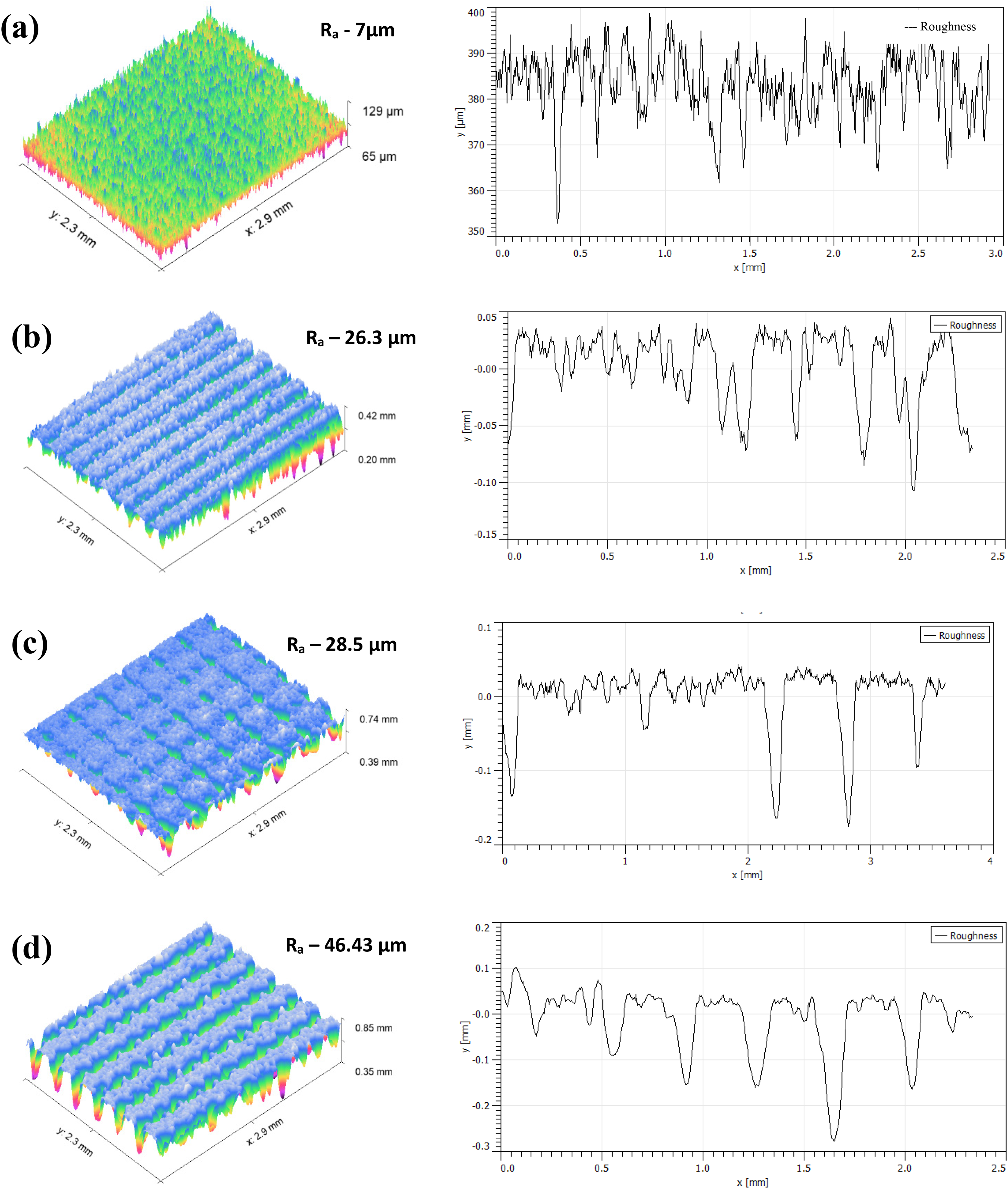
3D and 2D roughness profile obtained for (a) base metal (b) groove pattern (c) crisscross pattern (d) wavy pattern.
Wettability analysis of textured and untextured surfaces
Contact angle measurements were conducted after the laser-textured samples were stabilized under ambient laboratory conditions. Immediate post-processing wettability evolution was not investigated in the present work. The contact angle (CA) of textured and untextured surfaces (Figure 4) clearly shows the influence of surface smoothness on the wettability behaviour of Ti6Al4 V alloy samples. The untextured base-metal surface (Figure 4(a)) facilitates spreading of the liquid droplet and results in a low contact angle of 44.8° ± 5°, confirming its hydrophilic nature. On the linear groove surface, the CA increases to 76.75° ± 7°, indicating a more hydrophobic nature that partially limits liquid spreading (Figure 4(b)). In the crisscross pattern, the CA further increases to 119.9°± 8° (Figure 4(c)). The intersection structure formed in the crisscross pattern enhances the roughness and allows air pockets under the droplet, which is consistent with the Cassie Baxter wetting regime. The wavy surface exhibits a higher CA of 151.35 ° ± 10°, confirming its superhydrophobic nature, which drastically reduces the solid-liquid contact area and increases air entrapment (Figure 4(d)). This superhydrophobic surface supports better control of protein orientation and adsorption kinetics. 28 Figure 4 (e) shows the variation of CA for the different textures with an error bar. The variation of CA with surface roughness confirms (Figure 4(f)) the positive correlation between Ra and hydrophobicity. As Ra increases, the transition from hydrophilic to superhydrophobic becomes evident. The enhanced hydrophobicity observed in the crisscross and wavy textured surfaces can be explained through the Cassie–Baxter wetting regime, where trapped air pockets within the micro-textured grooves reduce the effective solid–liquid contact area. In this state, the liquid droplet partially rests on entrapped air rather than fully wetting the surface, resulting in higher apparent contact angles and superhydrophobic behavior. However, the biological implications of this wetting state differ from conventional smooth hydrophobic surfaces. In laser-textured biomaterials, hierarchical micro/nano-scale roughness can simultaneously promote localized protein anchoring and physical entrapment despite the reduced wetted contact area. The textured grooves and interconnected surface features provide favorable microenvironments for protein retention and stabilization, which subsequently influence cell attachment and mineralization behavior. Therefore, the improved biological response observed in the wavy textured surface is attributed not only to surface wettability but also to the combined effect of hierarchical roughness, air entrapment characteristics, and enhanced protein–surface interactions.28–31
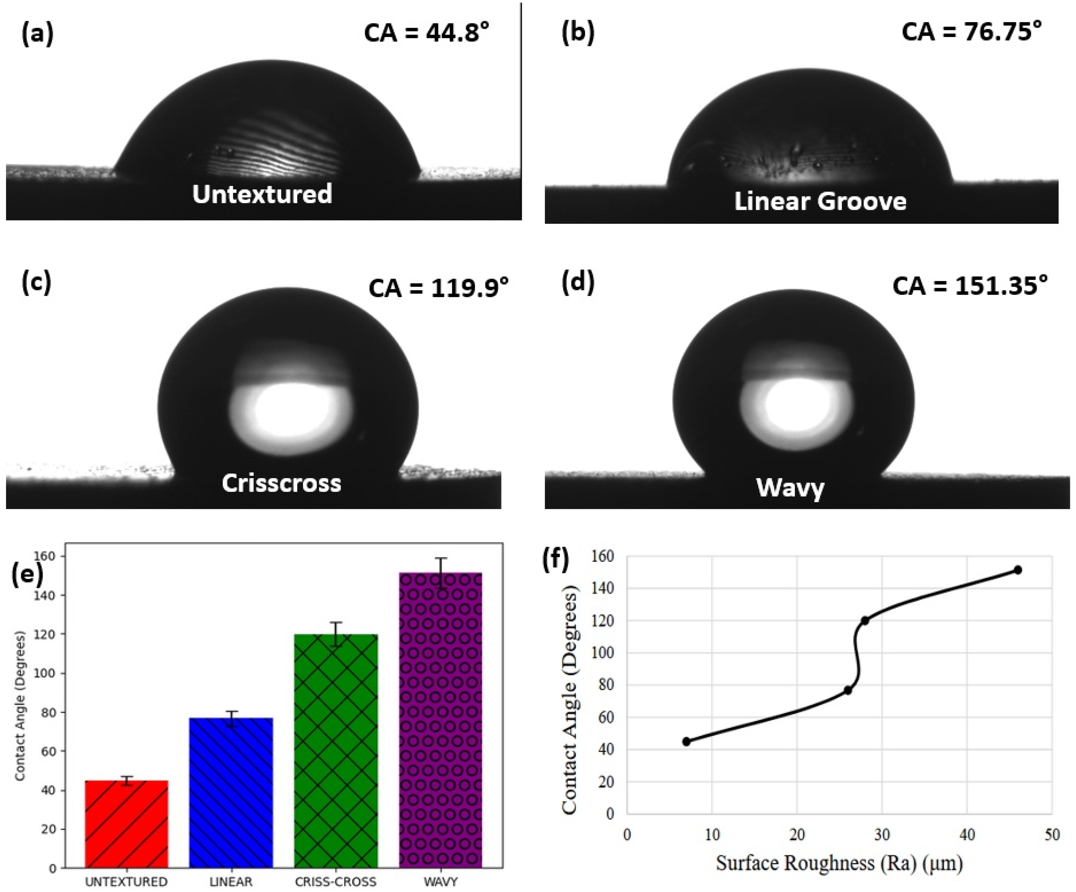
Contact angle in different surface conditions (a) Base metal (b) Linear groove (c) Crisscross (d) Wavy (e) Variation of CA (f) CA vs roughness.
Protein adsorption assay
The Lowry Assay was performed, and the samples’ absorbance was measured using a microplate reader. However, to correlate absorbance with the amount of protein remaining unabsorbed in DMEM containing 10% FBS, Bovine Serum Albumin solutions with different concentrations were prepared for absorbance measurements.
The standard calibration curve (Figure 5(a)) proves a solid linear relationship between absorbance and protein concentration, with a regression equation of y = 0.0038x + 0.0911 and a high coefficient of determination (R2 = 0.9941). This high linearity demonstrates the reliability and accuracy of the spectrophotometric technique used to quantify protein adsorption. The data points show nominal experimental deviation, validating the use of this calibration curve for determining unknown protein concentrations on laser-textured sample surfaces.
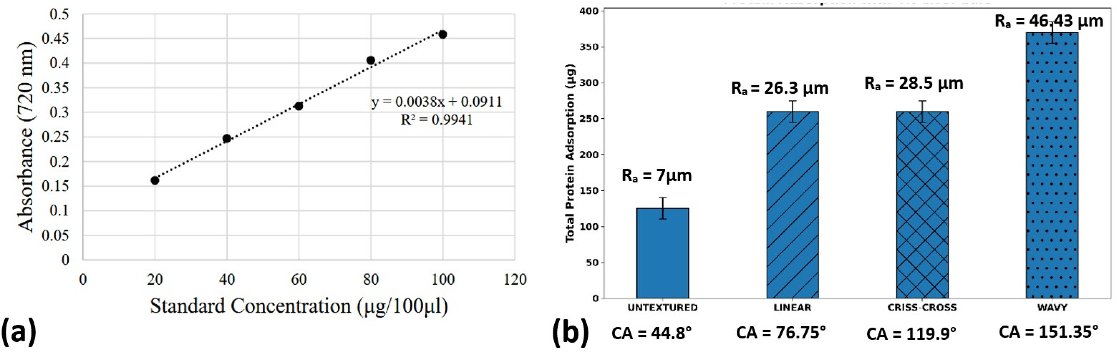
(a) Standard curve of absorbance vs. Protein concentration (b) Total protein adsorption in different surface texture conditions.
The protein adsorption results (Figure 5(b)) disclose a substantial influence of laser-pulsed surface texturing on protein binding behaviour. The untextured polished sample exhibits the lowest protein adsorption (138.33 ± 7 µg), agreeing with a quite smooth surface with inadequate active sites for protein attachment. Upon laser texturing, all different rough surfaces show a noticeable increase in protein adsorption, confirming that surface texturing improves bio-functional performance.
Among the laser-textured samples, the linear groove pattern (average Ra ≈ 26.3 µm) shows a substantial increase in adsorption (266.08 ± 8 µg), attributed to the formation of aligned microgrooves that promote protein anchoring over an enlarged surface area and directional wettability. The criss-cross pattern (average Ra ≈ 28.5 µm) exhibits slightly lower adsorption (248.46 ± 8 µg) compared to the linear pattern, despite having similar roughness. This may be due to the intersecting geometry, which could form localised trapping regions but also restrict uniform protein dispersion.
The wavy pattern (average Ra ≈ 46.43 µm) exhibits the highest protein adsorption (383.26 ± 10 µg), indicating that improved surface roughness and intricate topographic texture drastically increase protein interaction. The undulating structure likely provides a combination of a higher surface area, enhanced capillary effects, and favourable microenvironments for protein preservation. A strong correlation is also observed between contact angle (CA) and protein adsorption. The untextured surface, with a lower contact angle (44.8°), confirms reduced adsorption, whereas the wavy textured surface, with a higher contact angle (151.35°), shows maximum adsorption. This suggests that surface texturing not only alters roughness but also varies wettability characteristics, inducing protein–surface interactions. The increase in hydrophobicity, combined with micro and nanoscale roughness, likely promotes stronger protein adhesion and enhanced physical entrapment.29–31
Mineralisation Assay
The Alizarin Red Assay was performed, and the samples’ absorbance was measured. However, to correlate Absorbance with Calcium concentration at the sample surface, standard Alizarin Red solutions (ARS) with different concentrations were prepared by serial dilution. The standard calibration curve (Absorbance at 436 nm vs ARS concentration in mg/mL) is shown in Figure 6(a). After measuring the absorbance for triplicate samples from the four groups, the average calcium concentration for each group, in micrograms, was calculated using the standard curve.
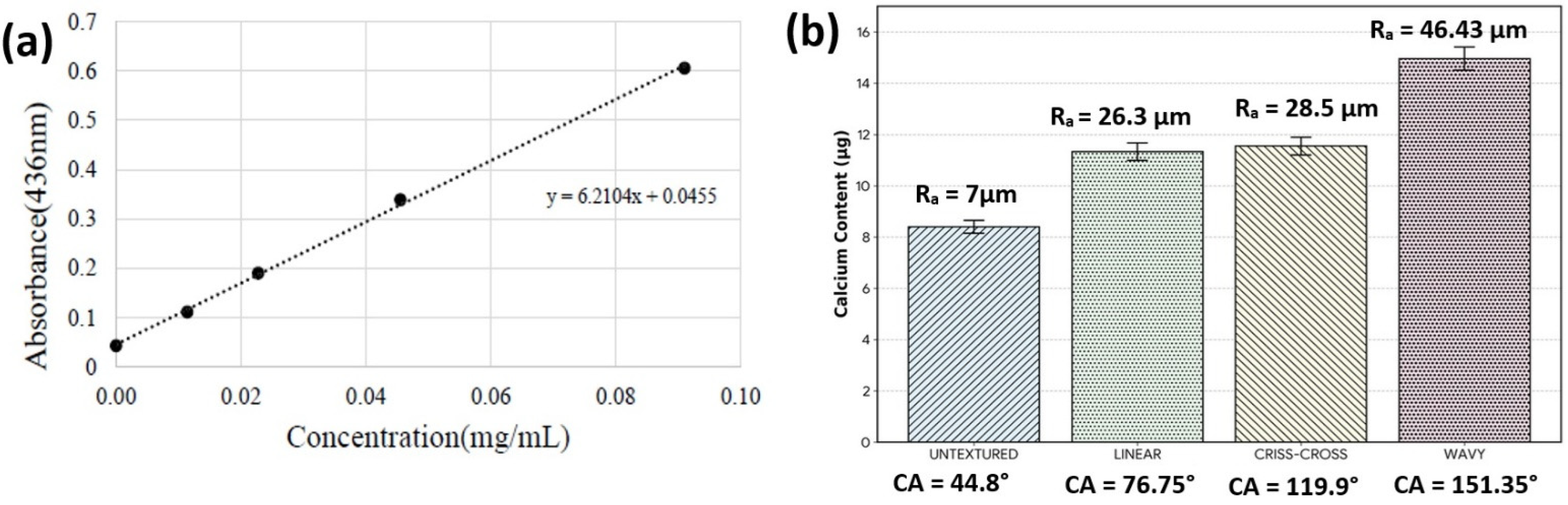
(a) Standard curve of absorbance Vs. Calcium concentration (b) Amount of Calcium mineralized in different surface textured samples.
A solid linear relationship is observed, as specified by the regression equation (y = 6.2104x + 0.0455). This linearity ensures the reliability and accuracy of the spectrophotometric method used for calcium quantification. The high sensitivity of the slope suggests that even small differences in calcium concentration can be effectively detected, making the method suitable for evaluating mineralisation on laser-textured surfaces. 32
Figure 6(b) shows the amount of calcium deposited on different surface-textured samples. A clear growing trend in calcium content is observed from untextured (8.4 ± 1 µg) to wavy textured samples (15 ± 2 µg). This shows that surface texturing significantly increases calcium mineralisation. The untextured base sample shows the lowest calcium content, with a smooth surface, which reduces nucleation sites for calcium deposition. In contrast, the laser-textured sample surfaces exhibit higher calcium adhesion due to increased surface roughness and altered wettability features. 33
The linear and criss-cross texture patterns exhibit moderate calcium deposition (11–12 µg), which may be linked to their structured grooves, which provide additional surface area and anchoring sites for mineral growth. However, the wavy texture pattern shows the highest calcium content (15 µg), suggesting more bioactivity. This complex topography, with higher hydrophobicity, likely enhances protein adsorption and ion interactions, thereby promoting the nucleation and growth of calcium-rich phases. 34
Conclusion
In this work, the effect of laser surface texturing on the biofunctional behaviour of Ti6Al4 V alloy was thoroughly explored through analyses of surface roughness, wettability, protein adsorption, and calcium mineralisation. The results evidently prove that surface structure plays a significant role in governing the biological response of the material.
Among the several textures, the wavy pattern exhibited the highest average surface roughness (Ra = 46.43 µm), thereby increasing the accessible surface area and providing advantageous microscale properties for biological interactions. Contact angle measurements showed that the moderately hydrophilic behaviour of the untextured surface transformed into a superhydrophobic (CA 151.35°) state in the wavy pattern. Because of its high contact angle, the hierarchical roughness of the wavy texture facilitated effective protein adsorption, indicating that surface structure can overcome wettability constraints in influencing biomolecular interactions.
The wavy pattern performed better in protein adsorption tests, which further confirmed that textured surfaces had higher protein attachment than the untextured sample. This increased protein adsorption (383.26 µg) is important because it is a prerequisite for cell joining and tissue formation. Calcium deposition studies confirmed higher mineralisation in the wavy-textured samples, supporting this trend and demonstrating the material's significant potential to promote bone joining and apatite formation.
Overall, the wavy texture is the most effective of the other designs due to the combination of increased roughness, and better biological interactions. This surface's greater calcium mineralisation and protein adsorption strongly imply that it is suitable for enhancing osseointegration in orthopaedic and dental implant applications.
Footnotes
Acknowledgment
The authors would like to acknowledge the CeNTAB (Centre for Nanotechnology & Advanced Biomaterials) at the SASTRA University for providing the testing facilities.
Ethical approval and informed consent statements
This article does not contain any studies with human or animal participants.
Author contribution(s)
Funding
The authors received no financial support for the research, authorship, and/or publication of this article.
Declaration of Conflicting Interests
The authors declared no potential conflicts of interest with respect to the research, authorship, and/or publication of this article.
Data availability statement
All data generated or analyzed during this study are included in this published article.