
Abstract
Objective:
This study was undertaken to establish an animal model of extracorporeal membrane oxygenation in rabbits.
Methods:
Ten New Zealand white rabbits weighing 2573±330 g were used in this study. Extracorporeal membrane oxygenation was established in these animals through cannulation of the right carotid artery and jugular vein for arterial perfusion and venous return. The components of the perfusion circuit were specially designed. Arterial blood pressure was measured with a blood pressure meter through cannulation of the right femoral artery. The heart rate and blood gas parameters were also monitored by electrocardiography and a blood gas analyzer (Radiometer ABL800, Bronshøj, Denmark), respectively.
Results:
The rabbit model of extracorporeal membrane oxygenation was established successfully. The hemodynamic and blood gas parameters were changed within an acceptable range during the extracorporeal membrane oxygenation process. The specially designed miniature membrane oxygenator was sufficient to meet the extracorporeal membrane oxygenation needs in this animal model.
Conclusion:
The rabbit model of extracorporeal membrane oxygenation established through right carotid artery and jugular vein cannulation is feasible, easily operated and economical. It is an ideal model for further research of the pathophysiology and organ protection offered through the application of extracorporeal membrane oxygenation.
Introduction
Extracorporeal membrane oxygenation (ECMO) is a kind of extracorporeal life support technique, which is the extension of the cardiopulmonary bypass technique and can be transferred to the intensive care unit. Today, it is commonly used to treat patients with end-stage lung, pulmonary vascular and/or heart disease.1,2 However, some of these patients ultimately need to have surgery or organ transplantation for long-term survival. The shortage of transplantable organs has reached critical proportions.3-5 As neocortical criteria of death has been accepted, it is hoped that there would be sufficient public support to expand organ resource,3,6 but brain death is also associated with a number of events that may complicate the management of multiple-organ donors and can lead to progressive organ deterioration. This deterioration limits the number of organs available for transplantation and may contribute to the postoperative morbidity and mortality associated with organ transplantation. Mechanisms contributing to organ deterioration include hemodynamic instability, endocrine failure, the inflammatory response, arrhythmias, hypothermia and coagulopathy. 7 In these patients, we conceive establishing ECMO before brain death to avoid organ deterioration. To test this assumption, an animal model and experimentation is needed.
The purpose of this study was to establish an economic, feasible and reliable animal model of ECMO in rabbits and also evaluate the effectiveness of this model from the viewpoint of the hemodynamics and blood gas state.
Materials and methods
Animal care
All animals received humane care in compliance with the ‘Principles of laboratory animal care’ formulated by the National Society for Medical Research and the ‘Guide for the care and use of laboratory animal resources’ published by the US National Institute of Health (NIH publication No. 85-23, revised 1996). The following experimental protocol was approved by the local Ethical Committee of Zhongshan Hospital, Fudan University, Shanghai.
Extracorporeal membrane oxygenation circuit
The ECMO circuit consisted of a non-pulsatile miniaturized Maquet Twin roller pump (Maquet Co. Ltd., Bremerhaven, Germany) (Figure 1A), a membrane oxygenator (Xijing Medical Co. Ltd., XIAN, China) (Figure 1B), a heat exchanger (Xijing Medical Co. Ltd.) (Figure 1C), tubing (Xiangsheng Medical Co. Ltd., Shanghai, China) (Figure 1D), and a specially designed cannula (Figure 1E). This system, in contrast to circuits for cardiopulmonary bypass, had no reservoir unit, therefore, blood-air contact was minimal. ECMO was established at a flow rate of 180-360 mL/min. Venous return was drained exclusively by the roller pump. The membrane oxygenator (oxygenation area 0.17m2) used during this procedure had been specifically conceived for this experimentation. The entire circuit priming volume was 25mL, of which the membrane oxygenator constituted 15 mL (Figure 2). Priming was carried out with synthetic colloid (10 mL) and Ringer’s solution (15 mL), without blood. The optimal flow rate was progressively reached and adjusted to a level that would maintain the desired arterial pressure. Gas flow (100% O2) was initiated at around 80-100 mL/min and adjusted in terms of sweep rate and FiO2 to maintain blood gasses in the physiological range. After 60 minutes, the animals were weaned from ECMO and the remaining priming volume re-infused.
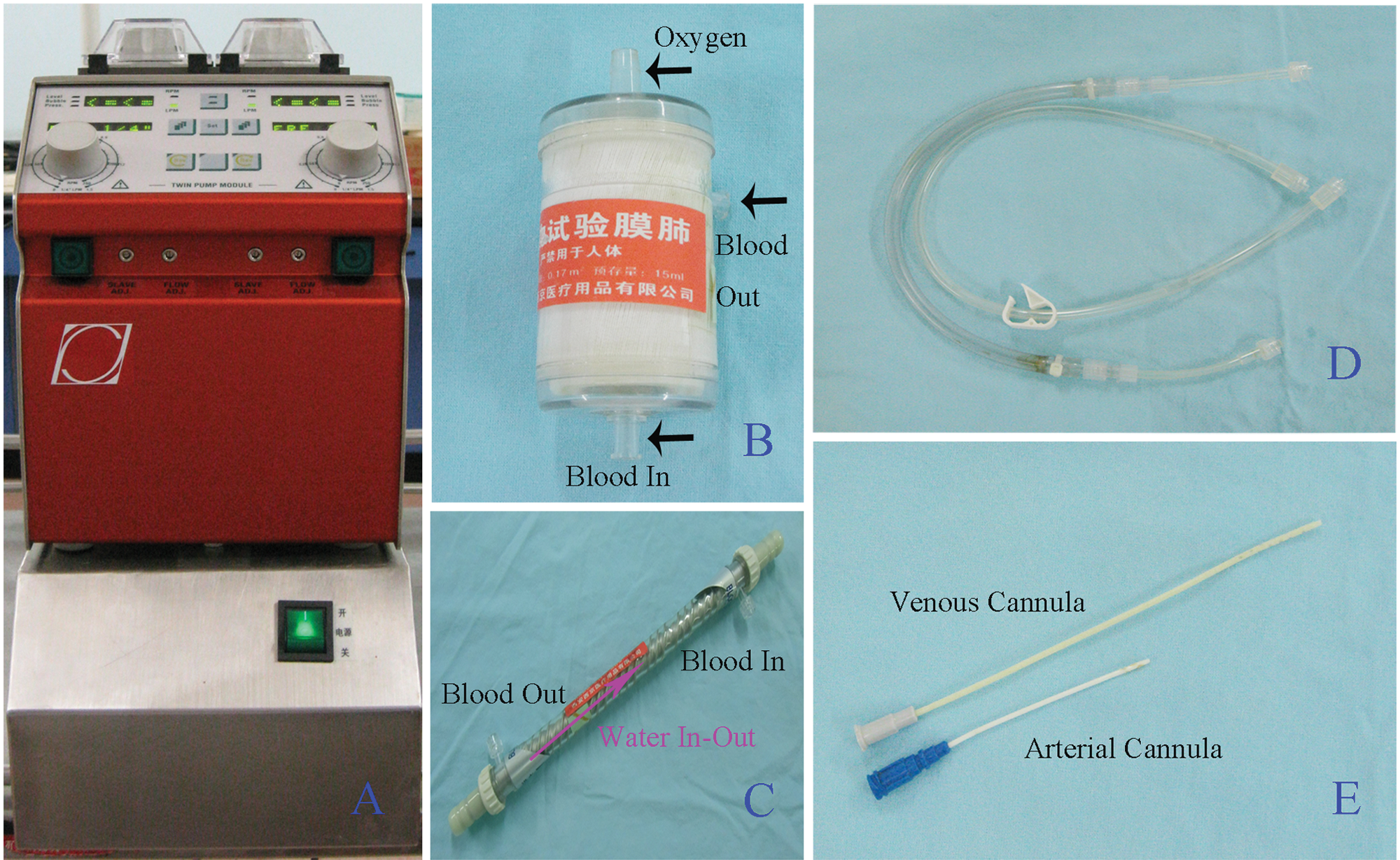
A: roller pump; B: membrane oxygenator; C: heat exchanger; D: conduit; E: cannula.
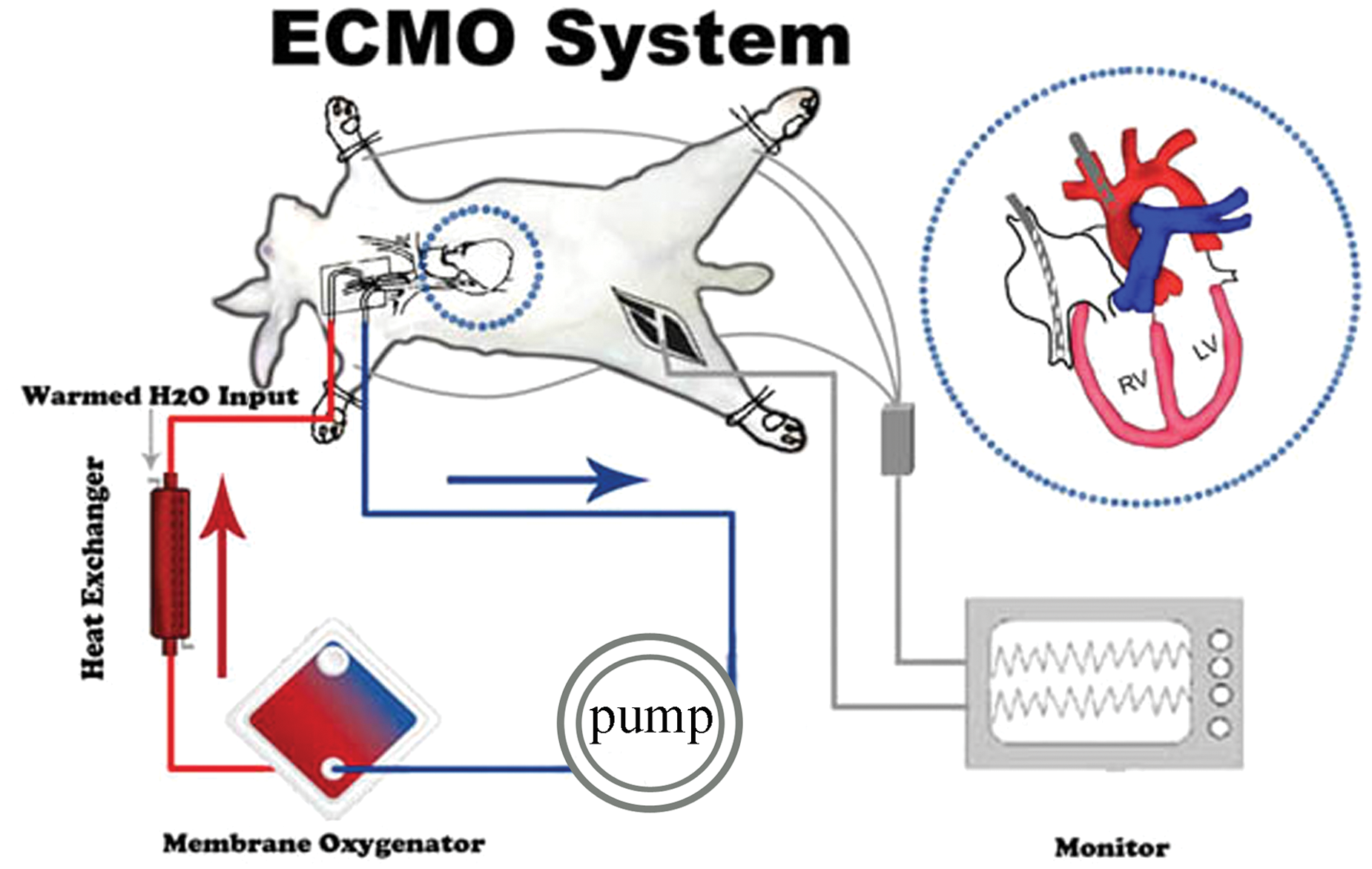
Overview of the ECMO circuit connected to the rabbit. The ECMO circuit comprised a roller pump, a membrane oxygenator, a heat exchanger and a cannula.
Surgical procedure
Ten New Zealand white rabbits (2573±330g) were used for all the experiments. They were anesthetized with an ear vein injection of amobarbital (5%) at a dosage of 1 mL/kg, providing 60 min of stable anesthesia while maintaining spontaneous ventilation during the entire operative procedure. Additional dosages were given if necessary. Ringer’s lactate solution was used as the maintenance fluid. An electrocardiogram (ECG) was carried out before the operation. For continuous arterial pressure monitoring, a 22-gauge Teflon catheter was inserted in the right femoral artery. Femoral arterial blood gas measurements were also performed at 0 min, 30 min and 60 min. The central temperature was monitored with a rectal probe and maintained at 36.8±1.2oC with the help of the heat exchanger and a heating lamp. The right carotid artery and jugular vein were exposed and cannulated with 18-gauge and 16-gauge specially designed Teflon heparinized catheters, respectively, while heparin was administered (125 units/kg).
Statistical analysis
Data were collected and entered into a dedicated Microsoft Excel table. Continuous variables were presented as mean±SD. Comparisons between different time points were made using the Student’s t-test, Pearson’s X2 test, or the non-parametric equivalent Mann-Whitney test. The significance level was set at p<0.05. Statistical analysis was performed using SPSS 14.0 software (SPSS Inc, Chicago, IL, USA).
Results
Ten New Zealand white rabbits were successfully established on extracorporeal membrane oxygenation without failure and all rabbits survived from the ECMO procedure.
Table 1 shows the hemodynamic data for the ECMO. The systolic arterial pressure, diastolic arterial pressure and mean arterial pressure were significantly decreased 30 min after ECMO compared with pre-operation (T30 min v T0 min, T60 min v T0 min, p-value <0.05), but remained stable during ECMO and 60 min after weaning from ECMO.
Arterial pressure over the study period (n=10)
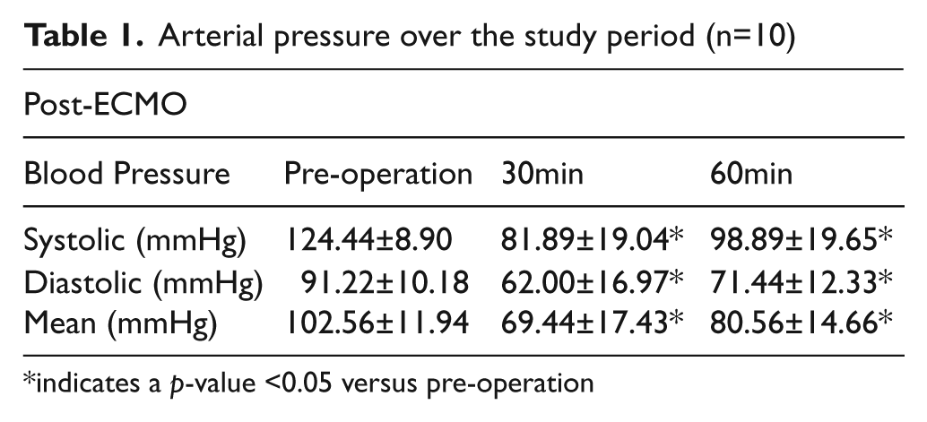
indicates a p-value <0.05 versus pre-operation
Table 2 summarizes the blood gas and biochemical parameters of the ECMO procedure. The PaO2 and PaCO2 (mmHg) were stable during ECMO, showing an excellent performance by the membrane oxygenator. Base excess and pH also remained stable throughout the experiment. Compared with pre-ECMO, the hematocrit (Hct) and hemoglobin (Hb) decreased significantly (T30 min v T0 min, T60 min v T0 min, p-value <0.05), but remained stable after T30.
Blood gas analysis results throughout the study period (n=10)
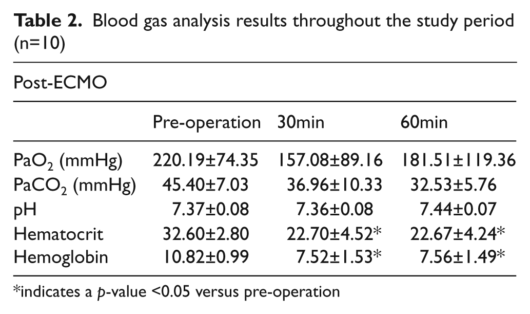
indicates a p-value <0.05 versus pre-operation
Discussion
In our study, we have demonstrated that our model of ECMO in rabbits was easily established and allowed excellent postoperative survival. In previous studies, large animal models of ECMO in dogs, sheep1,8 and animal models of cardiopulmonary bypass in dogs, cows, sheep and pigs were commonly used.9-14 There were also several studies that reported the establishment of an animal model of cardiopulmonary bypass in rats.9,10,15-17 In our opinion, large animal models of ECMO or cardiopulmonary bypass cost too much and there was less homology among individuals; small animal models, such as rats, increase the difficulty of the operation and usually end in failure. Meanwhile, the majority of cardiopulmonary bypass studies for small animals described in the literature require high priming volumes and the sacrifice of additional animals to achieve an acceptable hematocrit during the cardiopulmonary bypass period.9,10,18 Unlike these animals, the rabbit cerebrovascular anatomy is comparable to that of humans and many physiologic parameters of rabbits are virtually identical to human values. 18 Therefore, we think that rabbits may be ideal animals for us to establish ECMO. The heparin dosage was administered empirically (125units/kg). We did not evaluate anticoagulation activity during the whole procedure; however, neither thrombosis nor hemorrhage occurred in the rabbits with ECMO.
Satisfactory venous drainage is an important factor for the successful establishment of ECMO. Previous studies with an animal model of cardiopulmonary bypass reported that venous blood drainage by gravity was sufficient to allow adequate perfusion.9,10 In this rabbit ECMO model, venous drainage was achieved through active roller-head assist. A specially designed venous cannula with multiple side orifices in the forepart was placed in the mid-right atrium, allowing a perfusion flow rate of 180-360 mL/min.
Blood gas and hemodynamic analyses show that these parameters are fluctuating in an acceptable range. Compared with pre-ECMO, hematocrit and Hb decreased significantly, but remained stable after time point T30, mainly because of dilution of the blood, laboratory sampling and the surgical operation. To get satisfactory blood gas parameters, in our clinical experience, adding some sodium bicarbonate and potassium chloride routinely is necessary during bypass. Our current model of ECMO is associated with excellent success in rabbits. This model will allow for further physiological research8,19 and evaluation of the possible organ protective effect of ECMO.
Conclusion
This rabbit model of ECMO, established successfully via the right carotid and jugular vessel, is feasible, easily operated, safe and reliable. It is an ideal model for further pathophysiology and organ-protective research.
Footnotes
Funding
This research received no specific grant from any funding agency in the public, commercial, or not-for-profit sectors.
Statement of Responsibility
There are no conflicts of interest among the authors. The authors had full access to the data and take responsibility for its integrity. All authors have read and agree to the manuscript as written.