






Abstract
Introduction:
The substantial interpatient variability in heparin requirement has led to the use of a heparin dose response (HDR) technique. The accuracy of Hepcon-based heparin administration in achieving a target activated clotting time (ACT) using an HDR slope remains controversial.
Methods:
We prospectively studied 86 adult patients scheduled for cardiac surgery requiring cardiopulmonary bypass. The total dose of calculated heparin required for patient and pump priming was administered simultaneously to achieve a target ACT of 450 s for HDR on the Hepcon HMS system. Blood samples were obtained after the induction of anesthesia, at 3 min after heparin administration and after the initiation of CPB to measure kaolin ACT, HDR slope, whole-blood heparin concentration based on the HDR slope and anti-Xa heparin concentration, antithrombin and complete blood count.
Results:
The target ACT of 450 s was not achieved in 68.6% of patients. Compared with patients who achieved the target ACT, those who failed to achieve their target ACT had a significantly higher platelet count at baseline. Correlation between the HDR slope and heparin sensitivity was poor. Projected heparin concentration and anti-Xa heparin concentration are not interchangeable based on the Bland–Altman analysis.
Conclusion:
It can be hypothesized that the wide discrepancy in HDR slope versus heparin sensitivity may be explained by an inaccurate prediction of the plasma heparin level and/or the change in HDR of individual patients, depending on in vivo factors such as extravascular sequestration of heparin, decreased intrinsic antithrombin activity level and platelet count and/or activity.
Keywords
Introduction
Heparin is the most common drug to safely anticoagulate patients prior to the initiation of cardiopulmonary bypass (CPB). Inadequate anticoagulation with heparin in the setting of exposure of blood to foreign surfaces is known to lead to the generation of thrombin, 1 triggering a consumptive coagulopathy and several proinflammatory reactions.2,3
Because responses to a fixed-dose heparin bolus vary substantially among patients, Bull et al. attempted to develop the heparin dose response (HDR) technique, which has been used to optimize the dosing of heparin based on activated clotting time (ACT), most widely used to monitor anticoagulation during CPB. 4 The HDR slope is generated from a minimum of two ACTs (baseline ACT without heparin and in vitro ACT with a known concentration of heparin) and reflects individual variability in heparin responsiveness.
One of the automated HDR tests, the Hepcon HMS Plus system (Medtronic, Minneapolis, MN, USA) is theoretically advantageous for the early detection of heparin resistance to avoid both the delayed start of CPB and inadequate anticoagulation. Past studies showed that the use of Hepcon HMS was associated with higher total heparin and lower protamine doses in comparison with the traditional fixed-dose heparin bolus and is considered useful for providing patient-specific whole-blood heparin concentration measurements using an HDR slope and heparin/protamine titration on an individual basis.5,6 In our previous study, in vitro10% and 20% hemodilution and in vivo hemodilution with 500 ml intravenous fluid significantly increased the HDR slope and reduced the requirement for heparin. 7 It is estimated that the HDR slope in individual patients might be changed by complex and variable pharmacokinetics of heparin affected by hemodilution, antithrombin (AT) activity and platelet count and or activity as an operation progresses. The aim of this study was to evaluate the accuracy of Hepcon-based heparin administration at projected heparin concentrations to achieve a target ACT using the HDR slope in a comparison with heparin sensitivity by measuring anti-Xa heparin concentration during cardiac surgery with CPB.
Patients and methods
Ethics committee approval
Approval for this study (No. 2775) was provided by the ethics committee of Tokyo Women’s Medical University (Shunichi Miyazaki, Chair of Tokyo Women’s Medical University). Written informed consent was obtained from each patient enrolled in the study.
Patient selection
We prospectively studied 86 adult patients scheduled for elective cardiac surgery requiring CPB. Exclusion criteria included a history of any known coagulopathies, liver dysfunction, re-operations, preoperative abnormal coagulation profiles (international normalized ratio ⩾1.3, activated partial thromboplastin time >33 s) and exposure to anticoagulants and/or antiplatelets, which should be interrupted during the perioperative period. We included preoperative heparin therapy, defined as intravenous heparin that was not discontinued earlier than 24 h before surgery.
Cardiopulmonary bypass
All patients underwent normothermic CPB using a membrane oxygenator and biocompatible circuits (Capiox, RX-15 or 25; Terumo Corporation, Tokyo, Japan). Depending on the size of the patient, the extracorporeal circuit was primed with 550 mL of sodium bicarbonate, 30 g of mannitol and 5 mg of betamethasone per kilogram of body weight when the body surface area was <2.08 m2 or 800 mL of bicarbonate solution when the body surface area was ⩾2.08 m2.
Anticoagulation management
We used the Hepcon HMS for all patients, according to the manufacturer’s protocol, and the total dose of calculated porcine heparin (LEO Pharma, Ballerup, Denmark) for the estimation of patient blood volume and the pump priming volume was administered simultaneously to achieve a target ACT of 450 s for the HDR slope on the Hepcon HMS. Pump priming volumes were set at 1000 mL or 800 mL, based on the body surface area setting criterion of 2.08 m2. Because the calculation of blood volume of a patient based on the body surface area is approximate, a 3000 to 5000 IU loading dose of heparin was added to the CPB pump prime. Tranexamic acid at a dose of 1 g was routinely infused intravenously before heparin administration and blood sampling for measurement. No further continuous infusion was given.
Blood data collection
Blood samples were drawn via an indwelling arterial catheter after discarding approximately six dead-space volumes of the catheter and were immediately collected into a 3-mL Monoject syringe (Shearwood Medical, St. Louis, MO, USA) containing 3.2% sodium citrate and EDTA dipotassium salt dihydrate tubes (Venoject II, Terumo Corporation, Tokyo, Japan). Samples were obtained at the following times: (1) at baseline after the induction of anesthesia; (2) at 3 min after heparin administration; and (3) after the initiation of CPB.
At baseline, the measurement of kaolin ACT and HDR slope and the calculation of the projected heparin concentration from the HDR slope to reach a target ACT of 450 s were performed using HDR cartridges. HDR slope refers to the heparin dose response curve that is generated from three ACTs: baseline ACT without heparin and in vitro ACT with known concentrations of 1.5 and 2.5 U/mL heparin. In addition, plasma AT activity and complete blood count (CBC) were measured at baseline. Plasma AT activity was measured with a functional assay, using a chromogenic substrate specific for factor Xa, with bovine factor Xa added using a commercially available automated coagulation laboratory (ACL TOP 500CTS, Instrumentation Laboratory, Bedford, MA, USA) and CBCs were determined using the LH 780 (Beckman, Brea, CA, USA). Kaolin ACT using the Hepcon HMS Plus device was measured 3 min after heparin administration. The anti-Xa heparin concentration was evaluated using an automated chromogenic assay (two-stage assay) after initiation of CPB. In this assay, heparin is analyzed as a heparin–AT complex. After the addition of purified human AT to the plasma sample, an excess of factor Xa was then added to the sample and neutralized by the heparin–AT complex. The quantity of released chromophore cleaved by residual factor Xa is inversely proportional to the concentration of heparin (lower limit of detection by chromogenic assay: 0.1 U/mL).
Heparin sensitivity (s/unit/mL) was derived from the difference in ACT between baseline and at 3 min after heparin administration divided by the anti-Xa heparin concentration. The heparin sensitivity index (HSI; s/unit/kg) was calculated by the change in ACT between before and after heparin administration divided by the heparin dose per kilogram of body weight.
Statistical analysis
Data are presented as mean ± standard deviation (SD). Pearson’s correlation coefficients were determined for the HDR slope and heparin sensitivity and the projected heparin concentration using the Hepcon HMS Plus device and anti-Xa heparin concentration measured after the initiation of CPB. To assess the agreement of results for heparin concentration values, we used the Bland–Altman test, 9 setting a sensitivity limit of 0.7 IU/mL based on the fact that the cartridges for heparin/protamine titration method allows detection of heparin concentration by only 0.7 U/ml increments. 10 Differences less than 1.4 U/ml ( or ±0.7 U/ml) would be acceptable in clinical interpretation, considering the high heparin concentration usually attained during CPB (3~4 U/ml). Bland–Altman plots were used to show the limits of agreement, given as mean±2SD. The criterion for rejection of the null hypothesis was p<0.05. All statistical analyses were performed using IBM SPSS version 22.0 (IBM Corporation, Somers, NY, USA).
Results
The demographic data of 86 patients (49 males and 37 females) from cardiac surgery were as follows: age, 69.4±13.4 years; height, 158.3±8.6 cm; body weight, 58.2±12.6 kg. The initial dose of heparin administered was 13,453.5±4793.8 U (233.8±73 U/kg), resulting in an ACT of 425.2±81.2 s at 3 min after heparin administration and the heparin dose added to the CPB pump prime was 3142.9±1145.5 U (54.3±15.4 U/kg). Fourteen patients (16.3%) were required to be administered more than the standard dose of heparin (300 U/kg). The target ACT of 450 s was not achieved in 68.6% of patients after administration of the calculated heparin bolus dose although the HSI of those patients was above 1.0 (s/U/kg; Table 1). Compared with patients who achieved target ACT, those who failed to achieve their target ACT had a significantly higher platelet count at baseline although age, preoperative heparin treatment and AT activity did not differ between the two groups (Table 1). Correlation between the HDR slope and heparin sensitivity was poor (r=0.259, p=0.025; Figure 1). There was wide variation in anti-Xa heparin concentration among patients after administering the estimated heparin dose to reach target ACT (mean±SD 2.9±0.85 U/mL, median 2.9, interquartile range 1.2; Figure 2). There was a correlation between the projected heparin concentration and the anti-Xa heparin concentration after the initiation of CPB (r=0.625, p<0.001; Figure 3). According to Bland–Altman analysis, projected whole heparin concentration and anti-Xa heparin concentration after the initiation of CPB are not interchangeable based on the 95% limits of agreement (−1.174 to 1.713 U/mL), despite acceptable bias between the two methods (0.269 U/mL; Figure 3).
Comparisons of demographics, coagulation parameters, required heparin dose, HDR and HSI between the patients who did and did not achieve the target ACT.

ACT: activated clotting time; HDR: heparin dose response; HSI: heparin sensitivity index.
Data are shown as mean±standard deviation.
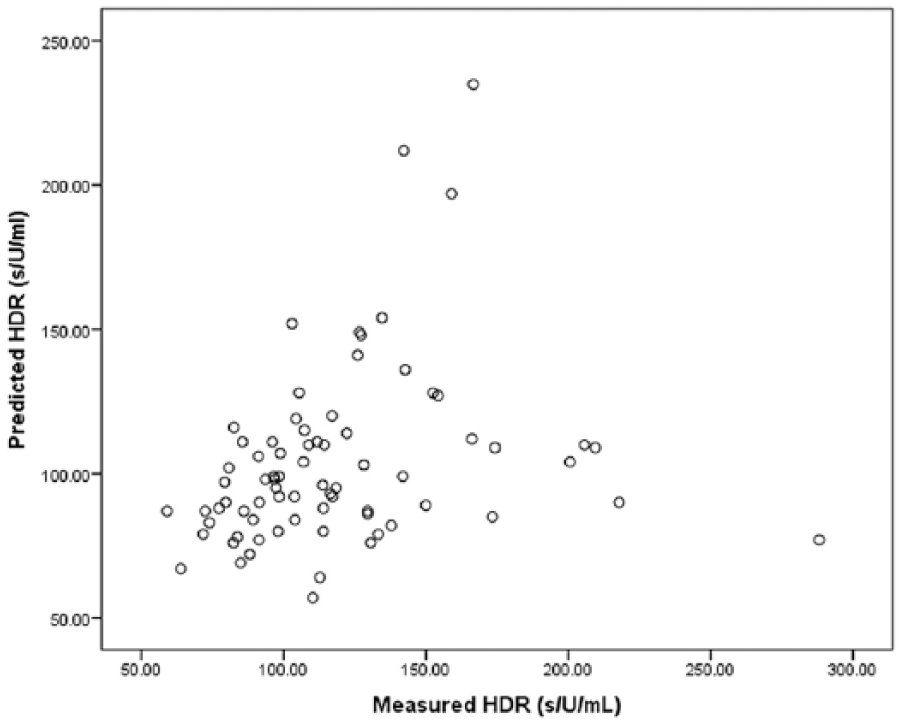
Correlation between the HDR slope and the calculated HDR (r=0.259, p=0.025).

Anti-Xa heparin concentration versus kaolin ACT (r=0.147, p=0.203).

Correlation plots and Bland–Altman plots showing correlation and agreement between plasma anti-Xa heparin concentration and predicted heparin concentration with the Hepcon HMS device after the initiation of cardiopulmonary bypass. In the Bland–Altman analysis, gray bold lines depict bias and dotted lines depict 95% prediction intervals.
Discussion
In this study, a high percentage of patients did not achieve the target ACT shortly after heparin administration. Although a calculated heparin dose derived from the HDR slope using the Hepcon HMS was administered to achieve the target ACT, these patients might not be regarded as heparin resistant, considering their HSI was above 1.0 s/units/kg. 8 Heparin resistance during cardiac surgery is defined as the failure of a standard dose of heparin (300–400 U/kg) to achieve the desired ACT (450–480 s). 10 The administered mean initial heparin dose for the patients who failed to reach target values was 209.5 U/kg, which was low in comparison with the standard dose, resulting in a mean ACT of 361.1 s. The HDR slope failed to consistently predict the heparin bolus dose, as our study demonstrated poor correlations of the HDR slope with heparin sensitivity, consistent with a past finding. 11 Garvin et al. 11 reported a non-linear relationship between measured HDR using the measured post-heparinization ACT and the measured heparin level and calculated HDR derived from the target ACT and the target heparin level. In this study, the HDR slope, as a result of calculation by the Hepcon HMS, was compared with an heparin-sensitivity ex vivo test of anticoagulation response to anti-Xa heparin concentration. Potential explanations for this discrepancy include a failure to reach the projected heparin concentration after administration of the required heparin dose based on calculated HDR and/or a failure of the in vitro calculated HDR to predict heparin’s true in vivo effect.
The results of Bland–Altman analysis in this study showed a lack of agreement between whole-blood projected heparin concentration calculated from the HDR slope and plasma anti-Xa levels after the initiation of CPB. We found that the anti-Xa heparin concentration tended to be lower than its projected heparin concentration calculated by the Hepcon HMS and the difference between the two became greater as mean heparin values increased. Raymond et al. 12 also found greater variation between anti-Xa heparin concentration and whole-blood heparin concentration measured by an automated protamine titration method, as the anti-Xa heparin concentration exceeded 3.4 U/mL in adult cardiac patients. This study compared projected whole-blood heparin concentration based on HDR with anti-Xa heparin concentration. The loss in agreement between the two as the mean heparin concentration increased is likely related to the past finding 13 that the HDR curve may not be linear at higher heparin concentrations. A concentration of heparin greater than 4.1 U/mL failed to produce statistically significant augmentation of ACT.
Previous studies of on-site whole heparin monitoring devices using the principle of heparin/protamine titration to quantitatively determine heparin level had varied results, with some studies 14 showing good correlation between whole-blood heparin concentration and laboratory plasma anti-Xa levels, whereas others15,16 showed unsatisfactory agreement between the two methods during CPB. Factors leading to a discrepancy of heparin concentration between the two methods include estimated patient blood volume based on Hepcon HMS, Allen’s formula, 17 a limited fidelity in reporting whole-blood concentrations with discrete categories rather than a continuous variable11,15,16 and inherent variability of the anti-Xa assay,18,19 among others. In particular, the severity of heart failure, type of cardiac diseases, 20 transfusion intervention and hemodilution on CPB 21 may be a substantial source of variation in blood volume estimates during cardiac surgery. This study was designed to compare the projected heparin level calculated from the HDR slope and a laboratory-based anti-Xa chromogenic assay to evaluate whether the projected heparin level could be reached after the administration of a calculated heparin dose. The whole-blood heparin concentrations demonstrated in this study were at slightly higher levels than those of plasma anti-Xa levels and did not predict the plasma heparin level accurately.
Unfractionated heparin response is complicated by the variability in heparin’s anticoagulant effect. 10 In this study, there was no significant relationship between ACT and anti-Xa heparin concentration, in agreement with the past finding that measurements of ACT were not correlated with the concentration of circulating heparin, 22 especially under conditions of deep hypothermia, hemodilution and variable platelet count and/or activity during CPB.23-25 Although there is wide variation in the response of the ACT to heparin among individual patients, the response of each individual should be relatively constant, which is the basis for using HDR. However, it is probable that several factors influence the HDR slope in vivo other than ex vivo factors and/or the progress of the procedure after anesthetics induction.
First, heparin binds non-specifically to various plasma proteins, including fibrinogen, factor VIII and histidine-rich glycoprotein, as well as macrophages and endothelial cells in circulation and is entrapped in the extravascular space. 26
Second, hemodilution reduces AT activity, which is the mediator of the anticoagulant effect of heparin. In our previous study, 7 AT activity was significantly decreased in vivo after hemodilution with 500 mL of intravenous fluid and 10% and 20% hemodilution in vitro. In addition, hemodilution during CPB is known to cause a decrease in AT activity level to approximately 50% of baseline values. 27 The ACT test is a measure of the intrinsic pathway of coagulation that detects the presence of fibrin formation. AT is a serine protease inhibitor that reversibly binds thrombin, neutralizing the effect of thrombin on fibrinogen. 28
Third, stimulated platelets release platelet factor 4 (PF4) which shows very tight binding to heparin and neutralizes its anticoagulant activity. 29 The release of PF4 during CPB is related to the amount of activated platelets. 30 In this study, the patients who failed to achieve their target ACT had a significantly higher number of circulating platelets after the induction of anesthesia compared with patients who achieved target ACT, which might be related to the greater amount of PF4 released to antagonize the anticoagulant effect of heparin.
In the present study, it was observed that HDR on individual patients might be changed, depending on a number of in vivo factors, such as extravascular sequestration of heparin, decreased intrinsic AT activity level and platelet count and/or activity. This observation was supported by the results of other investigators 22 whereby the ACT per unit of heparin did not remain constant during CPB and the ACT could not be employed to regulate quantitatively the heparin and protamine doses.
This study has a limitation. Anti-Xa heparin concentrations were measured after the administration of the required heparin dose for both patient and pump priming based on the Hepcon HMS in addition to a 3000 to 5000 IU loading dose of heparin in the pump for safety. However, Bland–Altman analysis indicated that anti-Xa heparin concentrations tend to be lower than projected heparin concentrations after the initiation of CPB.
Conclusions
The HDR calculated by the Hepcon HMS system correlates poorly with heparin sensitivity, resulting in a high percentage of failure to achieve target ACT. The discrepancy between the HDR slope and heparin sensitivity may result from failure to reach the projected heparin concentration after administration of the required heparin dose based on calculated HDR and/or inaccuracy in calculating the heparin loading dose to reach the target ACT, as heparin sensitivity might be changed, depending on in vivo factors and/or the operation progress after the induction of anesthesia.
Footnotes
Declaration of Conflicting Interests
The authors declared no potential conflicts of interest with respect to the research, authorship, and/or publication of this article.
Funding
The authors received no financial support for the research, authorship, and/or publication of this article.