
Abstract
Objectives:
The study aimed to determine the role of inward rectifier potassium channel 2.1 protein and connexin 40 expressions in regulating the duration of repolarization and conduction velocity of right atrial myocardium in rats following hypothermic ischemia-reperfusion.
Methods:
The Langendorff isolated rat cardiac perfusion models were divided into control (C) and hypothermic ischemia-reperfusion groups, with 8 models in group C and 16 models in group ischemia-reperfusion. Depending on the incidence of atrial arrhythmia after reperfusion, the models in group ischemia-reperfusion were further divided into reperfusion non-atrial arrhythmia or reperfusion atrial arrhythmia subgroup. Right atrial monophasic action potential duration at 50% and 90% of repolarization after 30 minutes of continuous perfusion in group C and group ischemia-reperfusion (T0), 105 minutes of continuous perfusion in group C or after 15 minutes of reperfusion in group ischemia-reperfusion (T1) and 120 minutes of continuous perfusion in group C or 30 minutes of reperfusion in group ischemia-reperfusion (T2) were recorded. Right atrial conduction velocity and effective refractory period were recorded at T2. Then, the expressions of inward rectifier potassium channel 2.1 protein and connexin 40 in the right atrial myocardium were detected.
Results:
Monophasic action potential duration at 50% and 90% were higher at T1 and T2 than those at T0 in subgroup reperfusion atrial arrhythmia (p < 0.05); monophasic action potential duration at 50% in subgroup reperfusion atrial arrhythmia were larger than group C and subgroup reperfusion non-atrial arrhythmia at T1 and T2 (p < 0.05); monophasic action potential duration at 90% in subgroup reperfusion atrial arrhythmia were larger than group C and subgroup reperfusion non-atrial arrhythmia at T1 and T2 (p < 0.05); effective refractory period in subgroup reperfusion atrial arrhythmia was greater than that in group C and subgroup reperfusion non-atrial arrhythmia, and the conduction velocity and the expressions of inward rectifier potassium channel 2.1 protein and connexin 40 were significantly lower than group C and subgroup reperfusion non-atrial arrhythmia (p < 0.05).
Conclusions:
The prolonged duration of repolarization and a decrease in conduction velocity of the atrial myocardium occur in rats after hypothermic ischemia-reperfusion. These observed effects may be related to the downregulated expressions of connexin 40 and inward rectifier potassium channel 2.1.
Introduction
Atrial arrhythmia is a common type of reperfusion arrhythmia (RA), among which the incidence of atrial fibrillation is the highest. 1 Hemodynamic disorders caused by RA is closely related to the prognosis of patients. 2 At present, research on reperfusion atrial arrhythmia (R-AA) is mainly focused on local ischemic myocardium.2,3 Previous studies in our group have revealed that prolonged duration of repolarization and decreased conduction velocity (CV) form the electrophysiological basis of ventricular arrhythmias induced by hypothermic global ischemia-reperfusion (IR),4,5 but that in atrial myocardium was not observed. Currently, changes in the duration of repolarization and CV in the atrial myocardium of hypothermic global IR atrial arrhythmia have not been reported.
Inward rectifier potassium current (Ik1) is the key current of action potential repolarization in atrial myocytes. The inward rectifier characteristics of Ik1 play an important role in maintaining a long plateau and phase-3 repolarization of cardiac myocytes. If activity of Ik1 decreases, the duration of repolarization of the action potential is prolonged. The main component of Ik1 is the inward rectifier potassium channel 2.1 protein (Kir2.1). 6 When the expression of Kir2.1 decreases, the duration of action potential repolarization is prolonged to increase the risk of atrial arrhythmia. 7 In addition, the expression of gap junction proteins, mediating direct intercellular communication, plays an important part in the incidence and maintenance of arrhythmia. Sun et al. 8 showed that connexin 40 (Cx40) is the most specific gap junction protein in the atrium and cardiac conduction system. Qiu et al. 9 reported that Cx40 can be downregulated depending on the physiological state of heart, and that humoral factors can disturb the intercellular communication function of the myocardium. Consequently, these changes can slow down the CV of the atrial myocardium and increase the probability of cardiac potential decoupling. It is not known whether the expressions of Cx40 and Kir2.1 are related to the changes in either repolarization or conductive function of the atrial myocardium in rats with atrial arrhythmia after hypothermic global IR.
This study aimed to observe changes in the duration of repolarization, CV and expressions of Kir2.1 and Cx40 in the right atrium of R-AAs. We were planed to establish the Langendorff isolated rat cardiac perfusion models and explore the role of the expressions of Kir2.1 and Cx40 in the changes of atrial repolarization and conductive function with R-AAs, thus to identify the potential underlying mechanisms of the occurrence and maintenance of R-AAs.
Materials and methods
Animal research approvals
The study was approved by the Ethics Committee of Guizhou Medical University (Animal Ethics No. 1900702). All animal protocols conformed to the Guidelines on the Care and Use of Laboratory Animals issued by the Chinese Council on Animal Research and the Guidelines of Animal Care.
Establishment of Langendorff isolated rat cardiac perfusion models
Healthy adult male SD rats weighing 310-390 g were provided by the Animal Experimental Center of Guizhou Medical University. Anti-coagulation was performed by intraperitoneal injection of 3% heparin (3 125 u/kg, Jiangsu Wanbang Biochemical Medicine Co., Ltd., Jiangsu, China) in rats. About 10 minutes after administration, intraperitoneal injection of 10% chloral hydrate (300 mg/kg, Shanghai Shangbao Biotechnology Co., Ltd., Shanghai, China) was used for anesthesia. After the anesthesia had taken effect, the heart was removed quickly and pruned in 4°C K-H solution at pH 7.4 [ingredients: 120 mmol/L NaCl, 4.50 mmol/L KCl, 1.25 mmol/L CaCl2, 1.20 mmol/L MgCl2.6H2O, 1.20 mmol/L KH2PO4, 20 mmol/L NaHCO3, 10 mmol/L C6H12O6]. The aorta was then intubated and connected to a Langendorff perfusion device (Shanghai Alcott Biotechnology Co., Ltd., Shanghai, China). The whole process was completed in 2 minutes.
Isolated hearts were perfused with K-H solution saturated with 95% O2% and 5% CO2 at constant temperature (37°C), constant pressure (8.65 KPa), and non-cyclic retrograde perfusion. Restoration of a normal rhythm within 3 minutes of continuous perfusion, and an HR at the end of 3 minutes of continuous perfusion of more than 180 beats per minute indicated the successful establishment of the isolated rat cardiac perfusion models.
Study groups and treatment
Twenty-four Langendorff isolated rat cardiac perfusion models were successfully established and randomly divided into the control group (group C) and hypothermic ischemia-reperfusion group (group IR), with 8 models in group C and 16 models in group IR. The occurrence of atrial arrhythmia after reperfusion was used to further divide the rats into a reperfusion non-atrial arrhythmia (R-NAA) and R-AA subgroups. In group C, the heart was continuously perfused with K-H solution at 37°C for 120 minutes. In group IR, the heart was continuously perfused with K-H solution at 37°C for 30 minutes and then stopped. Cardiac arrest was induced for 60 minutes with the injection of Thomas solution (20 mL/kg) at 4°C. Perfusion of Thomas solution at 4°C (10 mL/kg) was performed again at 30 minutes of cardiac arrest, and the heart was resuscitated by the reperfusion of the K-H solution at 37°C for 30 minutes after 60 minutes of cardiac arrest. During the whole cardiac arrest, Thomas solution was maintained at 4°C to protect the heart. The perfusion process of the two groups of rats is shown in Figure 1.
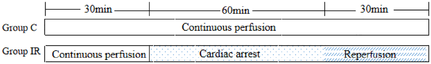
Perfusion treatment flow chart of two groups of rats.
Monitoring of electrophysiological indicators
After 30 minutes of continuous perfusion, a self-made composite electrode was slowly placed on the anterior wall of the right atrium.10,11 The electrode wire was connected with the signal input line of the BL-420 F Biological Function Experimental System (Chengdu Tai League Software Co., Ltd., Chengdu, China). Right atrial monophasic action potential (MAPs) at 30 minutes of continuous perfusion in group C and group IR (T0), at 105 minutes of continuous perfusion in group C, or 15 minutes of reperfusion in group IR (T1) and 120 minutes of continuous perfusion in group C or 30 minutes of reperfusion in group IR (T2), were recorded, and MAPDs at 50% and 90% of repolarization (MAPD50 and MAPD90) were measured. The incidence of atrial arrhythmia was observed and recorded after 90 minutes of continuous perfusion in group C or immediately after reperfusion in group IR. At T2, a compound recording electrode and a stimulation electrode could be placed on the surface of the free anterior wall of the right atrium. A reference electrode was placed at the root of the aorta, and the stimulation electrode wire was connected to the DF-5A cardiac stimulator (Suzhou Dongfang Electronic instrument Factory, Suzhou, China). The recording electrode wire was connected to the signal input line of the BL-420 F biological function experimental system. Using a programmed electrical stimulation technique, 8 pacemaker stimulus waves (S1) were successively released, and an early beat stimulus wave (S2) (S1 S1 = 400 ms) was detected. The S1 S2 interval was reduced by 20 ms at each time from 400 ms until S1 S2 is decreased to 110 ms. Subsequently, the S1S2 interval was reduced by 10 ms at a time. If S2 could induce an action potential, the S1 S2 interval would continue to be reduced by 10 ms. Conversely, if S2 could not evoke the action potential, the S1 S2 could increase by 10 ms, and then it was reverse scanned within −1 ms until S2 failed to evoke the action potential again. At this time, the S1 S2 interval is the atrial effective refractory period (ERP). The distance between the recording electrode and the stimulating electrode was fixed at 3 mm. After 8 pacemaker stimulus waves (S1) were continuously released using the programmed electrical stimulation technique, an early beat stimulus wave (S2) (S1S1 = 400 ms) was released. The trial CV was measured and calculated. The CV is equal to the distance between electrodes divided by the excitation time. The activation time is defined as the time interval from the start of the stimulation wave to the peak of MAP. The expressions of Kir2.1 and Cx40 were detected immediately after the experiment.
Western blotting
The expressions of Kir2.1 and Cx40 proteins in the atrial myocardium of rats in each group were detected by Western blotting. After adding a specific proportion of protein lysate and protease inhibitor, the atrial myocardium tissue was homogenized at 4°C and centrifuged for 15 minutes at 12, 000 rpm. The BCA colorimetric assay was used to quantify sample proteins, which were then diluted to the same concentration by PBS. Protein samples were boiled at 100°C for 5 min and then separated by SDS-PAGE using a 10% separating gel and 5% loading gel. Electrophoresis was carried out in an electrophoretic buffer and the target bands transferred to a PVDF membrane by semi-dry transfer (10 V, 15 min). The membrane was blocked with 5% BSA and incubated in TBST for 2 hours. It was then washed with TBST and incubated overnight with Kir2.1 goat anti-rabbit first antibody (1:500, ABcam, UK) and Cx40 goat anti-rabbit first antibody (1:2000, ABcam, UK, at 4°C. The next day, after washing with TBST, the membrane was incubated at room temperature for 1 hour with rabbit anti-goat second antibod (1:12,000, Beijing Boosen Biotechnology Co., Ltd., Beijing, China) labeled with horseradish peroxidase. After washing with TBST, ECL hypersensitive reagent was added to the membrane before visualizing using an automatic gel imaging analysis system (ZF-158, American Bio-rad Company). Image J software was used to measure the band intensity of each group. GAPDH protein levels were measured as an internal loading control.
Statistical analysis
Statistical analysis was performed using SPSS 22.0 Software (IBM Inc., Chicago, IL, USA). The data are presented as the mean and standard deviation (x̅ ± s). Comparisons within each group were performed through repeated measures analysis of variance. A t-test was used for comparison between groups, and the χ2 test was used to compare enumeration data. A p-value threshold of < 0.05 was used to determine statistical significance.
Results
A case of atrial premature beats occurred in group C after continuous perfusion for 90 minutes. Three cases of atrial fibrillation, and two cases of atrial premature beats occurred in group IR after reperfusion. In total, there were 11 cases observed in subgroup R-NAA and 5 cases in subgroup R-AA.
The changes of MAPD50 and MAPD90
From the results summarized in Table 1, MAPD50 was higher at T1 and T2 than that at T0 in subgroups R-NAA and R-AA (subgroup R-NAA, p = 0.000 and p = 0.003; subgroup R-AA, p = 0.003 and p = 0.011). MAPD50 in subgroups R-NAA and R-AA was larger than that in group C at T1 (p = 0.020 and p = 0.001, respectively); MAPD50 in subgroup R-AA was larger than that in group C at T2 (p = 0.018); MAPD50 in subgroup R-AA was larger than that in subgroup R-NAA at T1 and T2 (p = 0.008 and p = 0.049, respectively); MAPD90 was higher at T1 and T2 than that at T0 in subgroups R-NAA and R-AA (subgroup R-NAA, p = 0.001 and p = 0.018; subgroup R-AA, p = 0.000 and p = 0.000). MAPD90 in subgroup R-NAA was larger than that in group C at T1 and T2 (p = 0.001 and p = 0.014, respectively); MAPD90 in subgroup R-AA was larger than that in group C and subgroup R-NAA at T1 and T2 (subgroup R-AA vs group C, p = 0.000 and p = 0.000; subgroup R-AA vs subgroup R-NAA, p = 0.001 and p = 0.005);
Effects of hypothermic ischemia-reperfusion on MAPD50 and MAPD90 in the isolated rat atrial myocardium (x ± s).
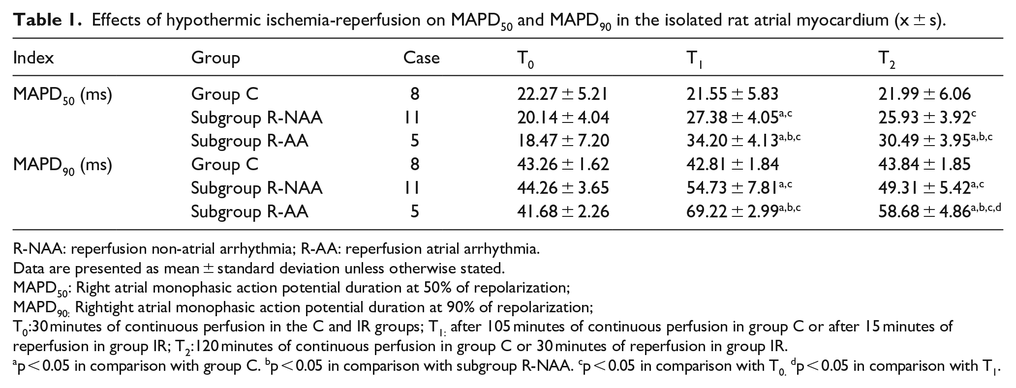
R-NAA: reperfusion non-atrial arrhythmia; R-AA: reperfusion atrial arrhythmia.
Data are presented as mean ± standard deviation unless otherwise stated.
MAPD50: Right atrial monophasic action potential duration at 50% of repolarization;
MAPD90: Rightight atrial monophasic action potential duration at 90% of repolarization;
T0:30 minutes of continuous perfusion in the C and IR groups; T1: after 105 minutes of continuous perfusion in group C or after 15 minutes of reperfusion in group IR; T2:120 minutes of continuous perfusion in group C or 30 minutes of reperfusion in group IR.
p < 0.05 in comparison with group C. bp < 0.05 in comparison with subgroup R-NAA. cp < 0.05 in comparison with T0. dp < 0.05 in comparison with T1.
Changes in CV and ERP
From the results shown in Table 2, the CV in subgroup R-AA was lower than that in group C and subgroup R-NAA (p = 0.003 and p = 0.033, respectively). ERP in subgroup R-NAA was greater than that in group C (p = 0.007). ERP in subgroup R-AA was greater than that in group C and subgroup R-NAA (p = 0.000 and p = 0.004, respectively).
Effects of hypothermic ischemia-reperfusion on CV and ERP in the isolated rat atrial myocardium at T2(x̅ ± s).
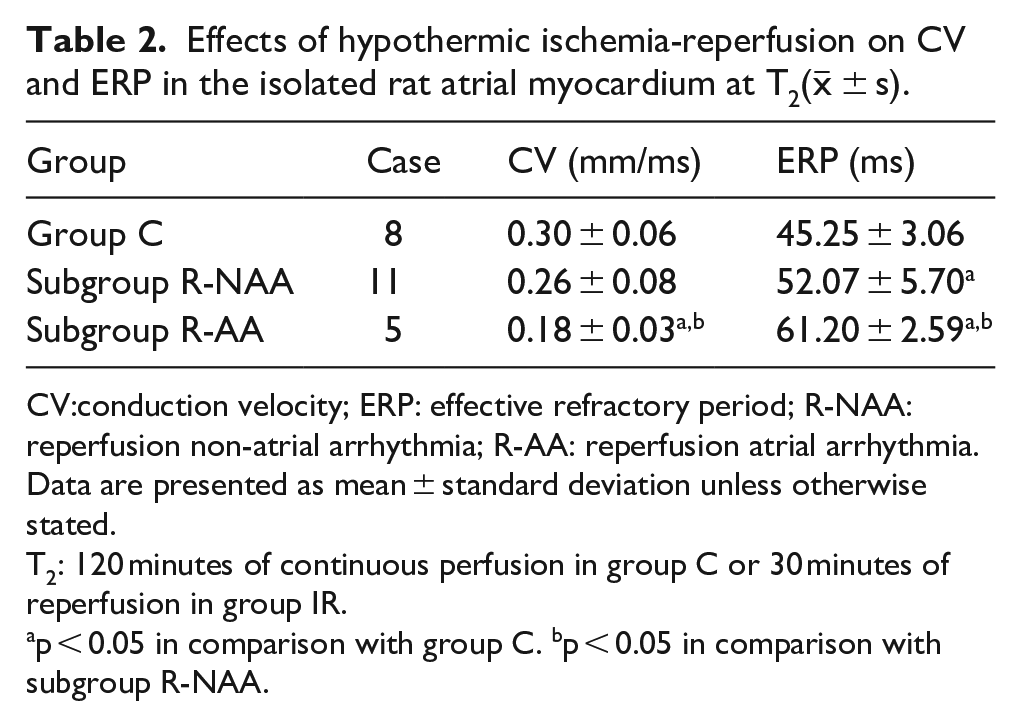
CV:conduction velocity; ERP: effective refractory period; R-NAA: reperfusion non-atrial arrhythmia; R-AA: reperfusion atrial arrhythmia.
Data are presented as mean ± standard deviation unless otherwise stated.
T2: 120 minutes of continuous perfusion in group C or 30 minutes of reperfusion in group IR.
p < 0.05 in comparison with group C. bp < 0.05 in comparison with subgroup R-NAA.
Expression of Kir2.1 protein by Western blotting analysis
Western blotting results showed that the relative expressions of Kir2.1 protein in the C, subgroups R-NAA and R-AA were 1.68 ± 0.25, 1.18 ± 0.26 and 0.79 ± 0.23, respectively. The expressions of Kir2.1 in subgroups R-NAA and R-AA were significantly lower than that in group C (p = 0.001 and p = 0.000, respectively). The expression of Kir2.1 in subgroup R-AA was significantly lower than that in subgroup R-NAA (p = 0.006) (Figure 2).
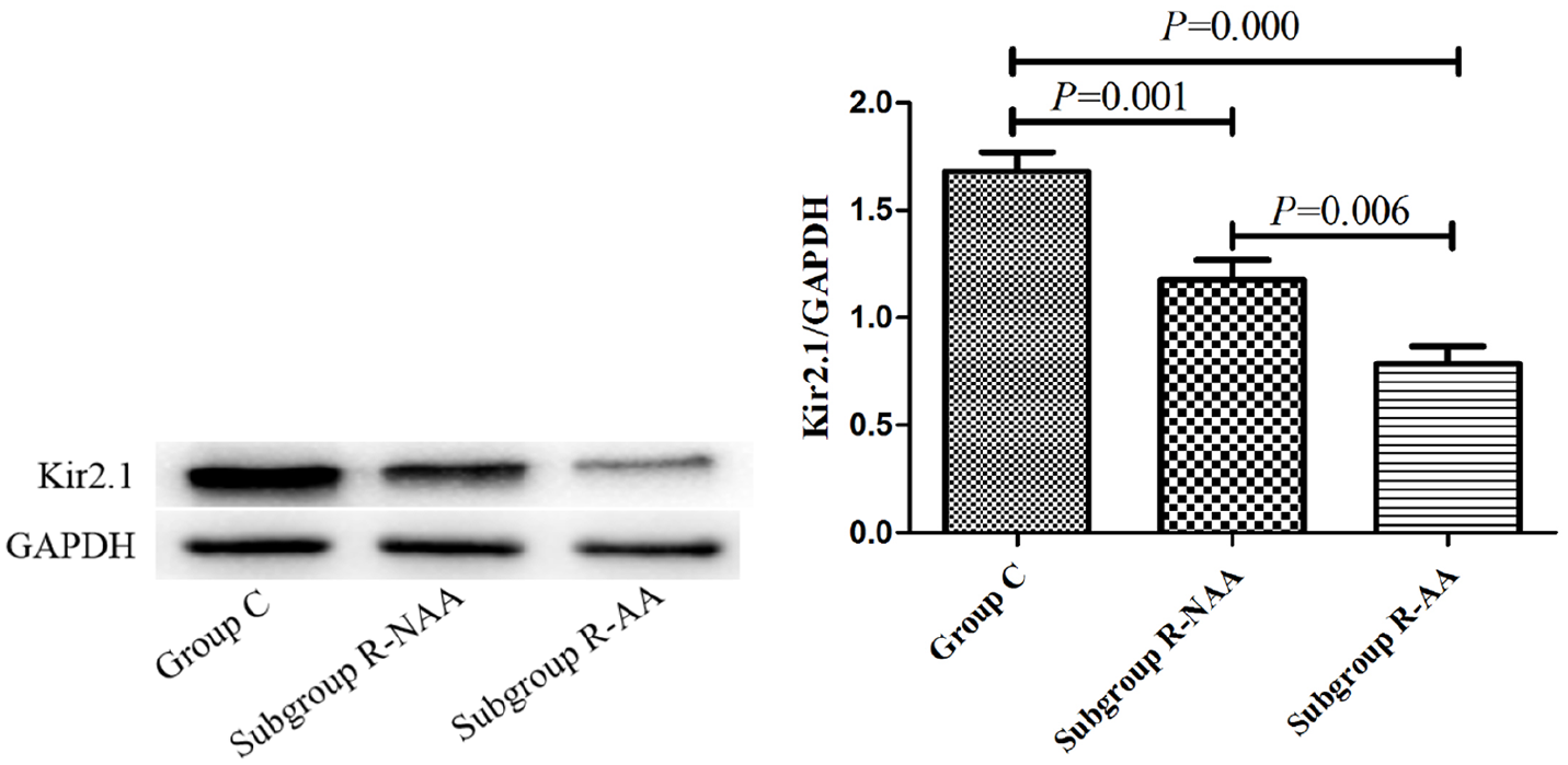
Effects of hypothermic ischemia-reperfusion on the expression of Kir2.1 protein in the isolated rat atrial myocardium.
Expression of Cx40 protein by Western blotting analysis
Western blotting analysis results showed that the relative expressions of Cx40 protein in group C, subgroups R-NAA and R-AA were 0.95 ± 0.26, 0.74 ± 0.23 and 0.44 ± 0.24, respectively. The expression of Cx40 protein in subgroup R-AA was significantly lower than those in group C and subgroup R-NAA (p = 0.001 and p = 0.021, respectively). (Figure 3).
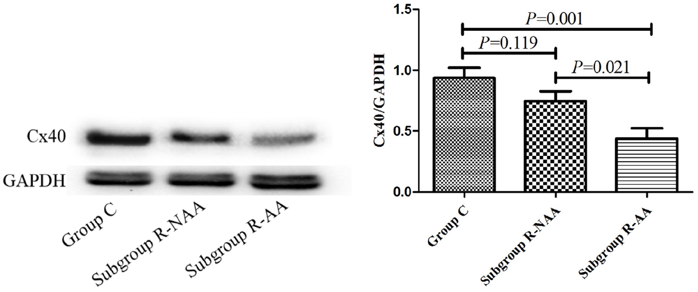
Effects of hypothermic ischemia-reperfusion on Cx40 expression in the isolated rat atrial myocardium.
Discussion
Pathological changes associated with atrial arrhythmia mainly include atrial electrical and structural remodeling. The early stage of atrial arrhythmia is manifested as atrial electrical remodeling, including changes in electrophysiology and ion channels. As R-AA mainly occurs in the early stage, and this study explored the electrophysiological mechanisms of R-AA from the perspective of electrical remodeling. R-AA is one of the characteristic manifestations of myocardial IR injury. Previous experimental studies have confirmed that decreased CV, prolonged duration of repolarization, over-activation of L-type Ca2+ current (ICaL), early post-depolarization, oxidative stress8,12–14 are common causes of atrial arrhythmia. Although the molecular mechanisms of atrial arrhythmia have been extensively investigated, there is limited information about the occurrence of R-AA after hypothermic global ischemia reperfusion.
Some studies have reported that hypothermic IR is one of the causes of increased susceptibility to atrial arrhythmia. Hypothermic IR can damage the mitochondrial and ion channel activity of atrial myocardium, increasing the risk of atrial arrhythmia. 15 Atrial electrical remodeling is a key link in the occurrence and maintenance of atrial arrhythmia. Therefore, accurate monitoring of electrophysiological changes of atrial electrical remodeling and in-depth study of its mechanisms are of major interest toward establishing an effective mechanism for basic treatment. Previous studies in our group revealed that prolonged duration of repolarization and decreased CV of MAP form the electrophysiological basis of ventricular arrhythmias induced by hypothermic global ischemia reperfusion.4,5 However, the changes in repolarization duration and CV of atrial myocardium with R-AAs were not observed. Kir2.1 and Cx40 proteins are closely related to atrial repolarization duration and CV, respectively. When the expressions of Kir2.1 and Cx40 were downregulated, the repolarization duration of atrial myocytes was prolonged, and the intercellular CV decreased.7,16 Detection of Kir2.1 and Cx40 may be particularly important in predicting the duration of atrial repolarization and CV.
This study observed the changes in the duration of atrial repolarization, CV and the expressions of Kir2.1 and Cx40 in a rat model with hypothermic global IR atrial arrhythmias. Overall, this approach aimed to explore the molecular mechanism of atrial repolarization and conduction changes of atrial myocardium with R-AAs.
In this study, according to whether atrial arrhythmia occurred after reperfusion, group IR was further subdivided into subgroups R-NAA and R-AA to more accurately reveal the electrophysiological changes of reperfused atrial arrhythmias. The results showed the incidence of atrial fibrillation was higher than other types of atrial arrhythmias in group IR, which was in agreement with the previous findings of J Alexandre et al. 1 B Maesen and colleagues17–19 found that the incidence of atrial fibrillation was higher in perioperative arrhythmias. This was as high as 20-40% after coronary artery bypass grafting (CABG), and the postoperative incidence of CABG and mitral valve surgery was as high as 64%. RA was shown to mainly occur during reperfusion within 15-30 minutes. 20 In the current study, we used ischemia for 60 minutes and reperfusion for 30 minutes as the model construction standard.
The expressions of Kir2.1 and Cx40 were regulated by many factors including hormones, body fluids and signaling molecules. In this study, the Langendorff cardiac perfusion models were used to eliminate the effects of hormones, body fluids and other factors on the experimental results. Thus, this model can be used to accurately evaluate the effects of hypothermic IR on atrial electrophysiology.
MAPD reflected the repolarization of action potential of cardiomyocytes during phases 0-3. Among these, MAPD50 and MAPD90 were related to phases 2 and 3, respectively.21,5 Kir2.1 protein is encoded by the KCNJ2 gene and is closely related to the duration of atrial repolarization. 22 The study of Lammers et al. 23 revealed that expression of kir2.1 is inversely correlated with the duration of repolarization: the downregulation of kir2.1 protein expression prolonged the repolarization duration. Our results suggest that the duration of atrial repolarization in subgroup R-AA was significantly longer than that in group C and subgroup R-NAA. At the same time, Western blotting indicated the expression of Kir2.1 in subgroup R-NAA was significantly lower, which may be related to the denaturation and remodeling of Kir2.1 in the atrial myocardium after hypothermic IR. These findings agree well with that of previously reported by Xia et al., 24 suggesting that downregulation of Kir2.1 may be related to the prolongation of atrial repolarization duration in rats with atrial arrhythmia.
Conductivity is an important physiological characteristic of myocardial tissue, which is closely related to other physiological characteristics such as excitability. ERP is an index of myocardial excitability. The atrial myocardium during the phase of ERP is unable to respond to any stimulation, which causes the atrial conduction to be blocked. When the myocardium is in the relative refractory period, depolarization and CV slow down, and so prolongation of ERP indicates decreased CV. CV is an important index for evaluating myocardial conduction, which indicates the decrease of conductive function. 25 Therefore, ERP and CV were included in this study to evaluate the conductive function of the atrial myocardium. In terms of the determinants of CV, gap junctions play a more significant role compared to ion channel currents. Gap junction proteins are differentially expressed in different parts of the heart. Cardiac myocytes express three gap junction proteins, namely Cx40, Cx43, and Cx45. Cx40 is encoded by GJA5 gene and specifically distributed in atrial myocytes. The protein is mainly expressed in the intercalated disk of the myocardium and is the key protein of atrial electro-activation conduction. Abnormal changes in Cx40 can significantly delay the CV of the atrial myocardium with several consequences including conduction block, reentry conduction, and increased heterogeneity of myocardial electrical conduction. These changes may act to promote the generation of atrial arrhythmia.26,27 Other studies show that the electro-physiological changes in Cx40 knockout mice are mainly manifested in the prolongation of the P wave, P-Q, and QRS intervals and the slowdown of atrial CV, thus increasing the susceptibility to atrial arrhythmia.28–31 Kanthan et al. 32 further confirmed that when human Cx40 mutated, the coupling ability of myocardial electricity decreased. Also, myocardial conduction was delayed and the dispersion of the action potential duration enlarged, and the conduction of myocardial anisotropy was not uniform. In this study, the CV and the expressions of Cx40 protein in subgroup R-AA were lower than those in group C, and the ERP was longer. These findings agree with those previously reported by Kanthan et al. 32 in rabbits. Therefore, the decreased CV in the atrial myocardium of R-AA may be related to the downregulation of Cx40 protein expression.
Conclusion
In summary, the downregulation of Kir2.1 and Cx40 may be related to the prolonged duration of repolarization and decreased CV in the atrial myocardium after hypothermic IR.
Limitations
First, this study did not observe the changes in atrial repolarization duration and CV after hypothermic IR in isolated heart after Kir2.1 and Cx40 protein overexpression or inhibition, which is the deficiency of this study and needs further study. In addition, the study missed differentially expressed genes between the groups with their fold changes.
Footnotes
Data Availability
All data that support the findings of this study are available from the first author upon request.
Declaration of Conflicting Interests
The author(s) declared no potential conflicts of interest with respect to the research, authorship, and/or publication of this article.
Funding
The author(s) received no financial support for the research, authorship, and/or publication of this article.