






Abstract
Middle aortic syndrome (MAS) is a rare disease characterized by distal thoracic aorta or abdominal aorta coarctation, and thoraco-abdominal aortic bypass grafting is an effective treatment for this condition. However, significant trauma is associated with the conventional surgical approach. We report a 26-year-old woman with MAS who presented with hypertension and needed thoraco-abdominal bypass grafting. In this operation, we adopted the endoscopic technique to improve the conventional surgical approach (reduce the incision). This case report shows that it is safe and feasible to use an endoscopic technique to reduce the trauma during this kind of operation, and provides a reference for similar treatments.
Introduction
Middle aortic syndrome (MAS) is a rare disease that is characterized by distal thoracic aorta or abdominal aorta coarctation. 1 The etiologies include abnormal development, neurofibromatosis, Takayasu arteritis and so on. 2 Patients with this condition may present hypertension and stenosis distal hypoperfusion. Timely treatment can effectively alleviate these symptoms and improve patient’s quality of life.
There are several treatment methods, that can be divided into surgical or endovascular treatment. 3 The surgical treatments include patch aortoplasty, aortoaortic bypass, and interposition aortic graft. Endovascular treatments include balloon angioplasty and stent grafting. The appropriate treatment method can be selected according to the degree and location of the coarctation. Some types of MAS force us to choose the surgical treatment; however, we often are not willing to accept the huge surgical trauma that is associated with this method. In this article, we report a case of a young female with MAS who was successfully treated with thoraco-abdominal aortic bypass grafting assisted by endoscope, and propose a method that reduces surgical trauma.
Case report
The patient is a 27-year-old female, who presented to our hospital with headache that intensified with movement or mood fluctuation. Her highest blood pressure was 178/116 mmHg and could be controlled with antihypertensive treatment. We found that the blood pressure in both of her legs was lower (left upper extremity: 119/88 mmHg; right upper extremity: 127/90 mmHg; left lower extremity: 118/84 mmHg; right lower extremity: 113/85 mmHg). CT angiography (Figure 1) showed occlusion of the part of abdominal aorta below the renal artery, and the length of the occlusion was about 30 mm. The main and branches of the superior mesenteric artery were obviously tortuous and dilated. One of the branches was connected with the lower abdominal aorta. After discussion, we decided to treat the patient with thoraco-abdominal aortic bypass grafting assisted by endoscope.

The CT angiography of the patient before operation, the green arrow pointed to the lesion site of abdominal aorta. Dilated superior mesenteric artery and its branches can be observed.
First, the patient was placed in left lateral decubitus position under general anesthesia, and thoracoscopy was used to expose the target vessels. We planned to anastomose the blood vessels through the thoracoscopic incision under direct vision, but this was difficult to perform. Therefore, we decided to extend the small incision (Figure 2). After prolonging the incision (5–12 cm), heparin was given (1 mL/kg) to prevent clotting, and the target blood vessels were partially clamped. Then the 12 mm GORE-TEX artificial vascular graft was anastomosed end-to-side to the thoracic aortic, and the procedure required 30 min. After hemostasis, the distal end of the vascular graft was ligated, and a 2 cm incision was made on the diaphragm to penetrate the vascular graft into the abdominal cavity. The chest tube was retained, and the incision of chest wall was sutured.
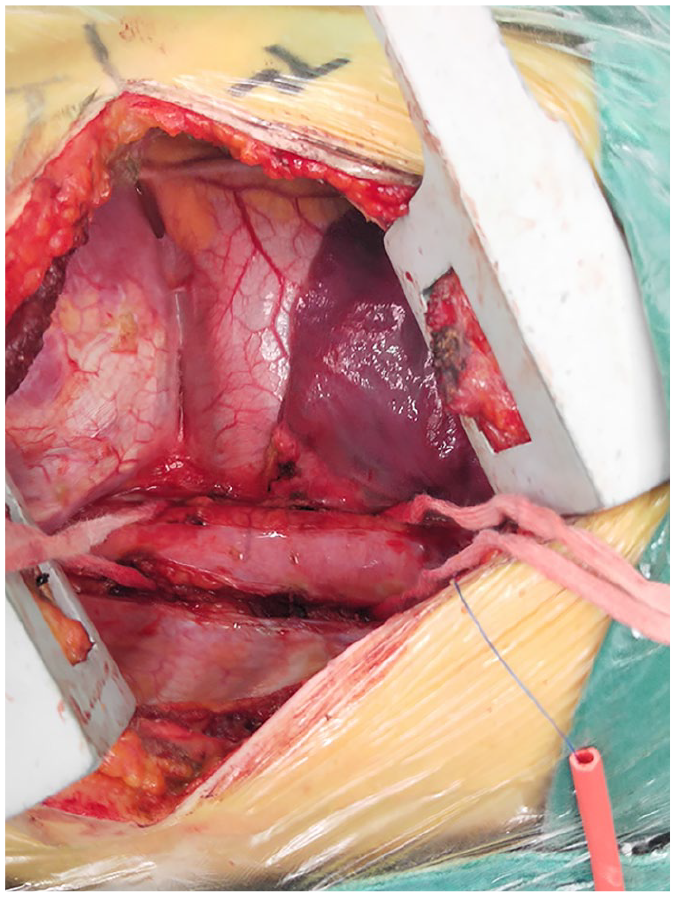
Showing the incision of the chest wall and the prepared thoracic aorta.
The patient’s position was changed from side to supine for the next part of the operation. After laparoscopic enterolysis, the tunnel of the artificial vessel was established along the lesser curvature of the stomach, the posterior wall of the stomach, the back of the transverse colon, and the medial edge of the descending colon. The end of the artificial vessel was placed near the target abdominal aorta, and this portion of the abdominal aorta was exposed and partially clamped. Then, the distal end of the artificial vessel was anastomosed with the target abdominal aorta under direct vision through a small incision (Figure 3), and the air of the artificial vessel is discharged. The abdominal incision was sutured routinely. The operation was successfully completed, and the patient returned to the ward safely.

Anastomose the artificial blood vessel with the abdominal aorta through the laparoscopic incision.
Postoperative, the blood pressure measurement of the patient showed: arms 101/75 mmHg, legs 128/86 mmHg. The result of the CT angiography showed that the artificial vessel bypass was unobstructed, and the superior mesenteric artery dilation obviously improved (Figure 4). The blood pressure returned to normal after the operation, and the patient was advised to walk on the second day after the operation.

The CT angiography after operation, the artificial vessels are marked with green arrows. The dilation of superior mesenteric artery was improved obviously.
Discussion
According to the 1990 American College of Rheumato-logy Classification Criteria for Takayasu arteritis, the cause of MAS in this patient was Takayasu arteritis. As mentioned in the introduction, there are many treatment options for MAS, 4 and main reasons for choosing this method were as follows. First, the degree of MAS was occlusion rather than narrowing, so the endovascular treatment could not be used. Second, the CT angiography demonstrated that the collateral circulation in the abdominal cavity was abundant. To reduce the handling of abdominal visceral organs and collateral circulation, we choose to build a bypass between the thoracic and abdominal aorta rather than the upper and lower part of the occlusion.
The form of thoraco-abdominal aortic bypass grafting was not single. In the surgical approach, we chose a divided incision rather than a thoraco-abdominal combined incision. We also tried to reduce surgical trauma with assistance from endoscopy, on the premise of preparing for open surgery. Although we extend the incision of the chest in the first part of the procedure, we successfully completed the abdominal aortic anastomosis through the small incision. In terms of the artificial vessel tunnel, we chose to fix the artificial vessel in the abdominal cavity to avoid possible renal artery compression that is caused by the retroperitoneal route.
The most dangerous complication of this treatment is downstream ischemia caused by the clamp during anastomosis. 5 In this operation, we should pay attention to this complication because of the limited incision. Protection of the distal blood supply was reflected in this operation: the thoracic aorta and abdominal aorta clamping was partial, 6 we tried to shorten blocking time as much as possible, and prepared the patient for cardiopulmonary bypass. The thoracic aorta and abdominal aorta clamping time during this operation were 32 minutes and 30 minutes respectively, which are within the safe time limit. If the anastomosis preoperative prediction is complex and the clamping time may be prolonged, partial cardiopulmonary bypass can be employed to keep perfusion of the lower extremities.7,8 The operation was successful, and the patient returned to the ward safely. However, we cannot relax our vigilance. This kind of patient may present graft narrowing with thrombus and neointimal formation, graft kinking, anastomotic dehiscence, infection, and development of false aneurysms. 5 Surgeons should ensure long-term follow-up of such patients. 6
Conclusion
Cases of MAS are rare, and fewer cases have used endoscopy to reduce the surgical trauma of thoraco-abdominal aortic bypass grafting. This case report provides a strategy for reducing surgical trauma for similar diseases and shows that it is feasible to use the endoscopic technique to reduce surgical trauma during this kind of operation.
Footnotes
Declaration of conflicting interests
The author(s) declared no potential conflicts of interest with respect to the research, authorship, and/or publication of this article.
Funding
The author(s) disclosed receipt of the following financial support for the research, authorship, and/or publication of this article: National Natural Science Foundation of China (No.81660074).
