






Abstract
Background:
The aim of this study was to evaluate the ipsilateral lower extremity (ILE) outcomes of patients who underwent bedside angiography via the distal perfusion catheter while on femoral veno-arterial extracorporeal membrane oxygenation (VA ECMO).
Methods:
This is a retrospective analysis of all patients placed on VA ECMO at a single center from January 2017 to December 2019 who underwent bedside angiography via the distal perfusion catheter.
Results:
Twenty-four patients underwent bedside angiography via the distal perfusion catheter after being placed on VA ECMO. A vasodilator was directly administered in three patients for suspected spasm. One patient had distal thrombus and underwent thrombectomy and fasciotomy. One patient had a dislodged catheter and underwent thrombectomy, fasciotomy, and replacement of the catheter. One patient had severe ILE ischemia, however was not intervened upon due to critical acuity. Finally, one patient had inadvertent placement in the saphenous vein and had a new catheter placed in the SFA. No patients underwent amputation. Ultimately, 21 patients (87.5%) had no ILE compromise at the end their ECMO course. Survival to decannulation was 66.7% (n = 16).
Conclusions:
Bedside angiography of the distal perfusion catheter is feasible and can be a useful adjunct in informing the need for further intervention to the ILE.
Classifications:
extracorporeal membrane oxygenation, ischemia
Keywords
Introduction
For patients in acute cardiogenic shock, veno-arterial extracorporeal membrane oxygenation (VA ECMO) can be useful as a bridge to recovery, durable mechanical circulatory support, or transplantation. A feared complication with the use of peripheral VA ECMO with common femoral artery cannulation is ipsilateral lower extremity (ILE) ischemia. In order to mitigate the risk of this complication, often a distal perfusion catheter is placed in the superficial femoral artery (SFA) of the ILE, usually under ultrasound guidance. 1 However, at time of VA ECMO initiation, these patients may have a high pressor and inotrope requirement, which can lead to vasoconstriction and vasospasm of peripheral arteries. This, in concert with the non-pulsatile flow from VA ECMO, can make it difficult to palpate a pulse or obtain a doppler signal in the ILE. We report a series of patients who underwent bedside angiography of the distal perfusion catheter to confirm placement and adequate blood flow to the ILE.
Patients and methods
Study design and patients
With Institutional Review Board approval (HP-00088089), a retrospective observational study of patients who were placed on VA ECMO from January 2017 to December 2019 was performed. The need for informed consent was waived. All 293 patients placed on VA ECMO during this time period had a distal perfusion catheter placed at the time of cannulation. Of those, 24 patients had an absence of palpable pulse or doppler signals and therefore underwent bedside angiography via the distal perfusion catheter while on VA ECMO support. A manual review of patient charts was performed to obtain demographics, ECMO variables, angiographic variables, and patient outcomes.
Technique
Patients underwent a percutaneous bedside cannulation for VA ECMO under ultrasound guidance. A 5000 unit heparin bolus was administered prior to cannulation with a goal PTT of 60–80 seconds (1.5–2 times the upper normal limit) once on VA ECMO. Arterial cannula size was determined by aiming for a goal index of 2.2 L/min/m2 solely based on predicted extracorporeal flows. A 6 Fr wire-reinforced sheath (Teleflex, Morrisville, NC, USA) was placed in the superficial femoral artery as a distal perfusion catheter as previously described. 2 This was preferentially placed prior to the insertion of the arterial cannuIa, with the exception of emergent cannulation and extracorporeal cardiopulmonary resuscitation (ECPR). If placed after insertion of the arterial cannula, a percutaneous approach was still used with the ultrasound probe being placed just inferior to the arterial cannula insertion site to avoid acoustic shadowing. In the absence of a palpable pulse or doppler signals of the ILE, patients underwent bedside angiography via the distal perfusion catheter to confirm placement in the SFA and ILE perfusion. Iohexol contrast at a concentration of 300 mg/mL (10–30 mL) was injected via a 3-way stopcock in the distal perfusion catheter after which the stopcock was opened to allow blood flow from the arterial cannula to the distal perfusion catheter. Immediately following this, a plain static film of the leg was taken using a portable x-ray machine in order to visualize lower extremity arterial vasculature. If the trifurcation below the knee to the anterior tibial, posterior tibial, and peroneal artery were visualized the image was considered adequate. If not the angiogram was repeated and more proximal films were taken to evaluate for abnormalities.
Outcomes
The primary outcome was ILE ischemia requiring thrombectomy, fasciotomy, or amputation. Survival to decannulation was also reported.
Statistical analysis
Continuous variables are reported as median and interquartile range (IQR) as they were non-parametric and were compared using a Mann–Whitney U-test. Categorical variables are presented as a number and percentage and were compared using a Fisher exact test. All data analysis was performed using R version 3.4.0 software.
Results
Patient characteristics
The baseline demographics of the 24 patients who underwent bedside angiography of their ILE via the distal perfusion catheter are reported in Table 1. Of note, only five patients (20.8%) had a pre-existing peripheral vascular disease.
Patient demographics.

BMI: body mass index; CAD: coronary artery disease.
Of the 24 patients, the majority were placed on VA ECMO for massive pulmonary embolism (n = 7, 29.2%), cardiogenic shock (n = 4, 16.7%), post-cardiotomy shock (n = 4, 16.7%), or refractory ventricular fibrillation/ventricular tachycardia (n = 4, 16.7%). Ten patients (41.7%) were placed on ECMO during active cardiopulmonary resuscitation. Most patients had either a 17 Fr (n = 8, 33.3%) or a 19 Fr (n = 12, 50%) arterial cannula. Only eight patients (33.3%) had the distal perfusion catheter placed before the arterial cannula, with the other 16 having the distal perfusion catheter placed immediately after institution of ECMO flows (Table 2).
Extracorporeal membrane oxygenation characteristics.

CFA: common femoral artery; ECMO: extracorporeal membrane oxygenation; ECPR: extracorporeal cardiopulmonary resuscitation; PE; pulmonary embolism; VF; ventricular fibrillation; VT: ventricular tachycardia.
Bedside angiography was performed a median of 136 (75–260) minutes after ECMO cannulation. Twenty-three patients (95.8%) were on continuous vasoactive medications at the time of the bedside angiography, with the majority being on two (n = 11, 45.8%) or three (n = 11, 45.8%) agents. Patients who had a bedside angiography were on more vasopressor agents than patients who did not have a bedside angiography (2 agents (2–3) vs 1 agent (0–1), p < 0.0001). Seventeen patients (70.8%) had well visualized flow of the anterior tibial, posterior tibial, and the peroneal arteries (Figure 1). Three patients (12.5%) had diffuse, smooth tapering of vessels, concerning for spasm (Figure 2(a)). Two patients had an abrupt cut-off of vessels, concerning for thrombus (Figure 2(b)). Finally, two patients had a catheter not in the SFA, one which had become dislodged (Figure 2(c)) and the other which was inadvertently placed in the saphenous vein (Figure 2(d) and Table 3). There was no difference in arterial cannula size between the seven patients with abnormalities on angiogram compared to the 17 patients with well-visualized flow (17 Fr (16–19) vs 19 Fr (17–19), p = 0.17).

These two normal bedside angiographies show the patency of the anterior tibial, posterior tibial, and peroneal arteries.
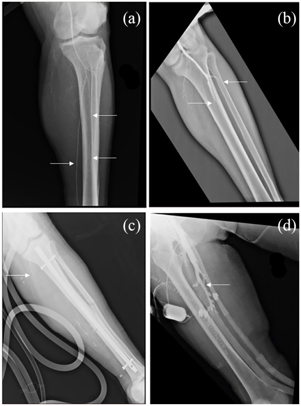
Patients with abnormal bedside angiography: (a) patient with diffuse tapering of vessels (arrows) concerning for spasm, (b) patient with abrupt cut-off of vessels (arrows) concerning for thrombus, (c) patient with dislodged distal perfusion catheter. Minimal contrast is seen in the vasculature of the leg (arrow), and (d) patient with distal perfusion catheter inadvertently placed in the saphenous vein (arrow).
Bedside angiography characteristics.

SFA: superficial femoral artery.
Defined as epinephrine, norepinephrine, or vasopressin.
Outcomes
The three patients (12.5%) with diffuse tapering of vessels were thought to have spasm and a vasodilatory agent was administered directly into the distal perfusion catheter with improvement of ILE pallor and doppler signals. The patient with inadvertent placement in the saphenous vein had another catheter placed in the SFA at bedside and had no ILE complication. The patient with a dislodged distal perfusion catheter underwent open thrombectomy, four compartment fasciotomy, and replacement of the catheter in the SFA. Of the two patients with an abrupt cutoff of vessels, one underwent lytic and vasodilator administration directly via the distal perfusion catheter, however ultimately required a thrombectomy and fasciotomy. The other was unlikely to survive without ECMO support given severe multi-organ failure, and the family requested transfer to another center prior to intervention. Ultimately, 21 patients (87.5%) had no compromise of their ILE while on ECMO support and 16 patients (66.7%) survived to decannulation (Table 4).
Outcomes and interventions.

DPC: distal perfusion cannula; ICU: intensive care unit; ILE: ipsilateral lower extremity; SFA: superficial femoral artery.
Patients who underwent bedside angiography had a higher rate of ILE ischemia than those who did not (12.5% vs 0.7%, p = 0.004). Patients who underwent bedside angiography compared to those who did not had a similar survival-to-decannulation (66.7% vs 69.1%, p = 0.82), survival-to-discharge from the intensive care unit (58.3% vs 61.3%, p = 1), and survival-to-discharge from the hospital (54.2% vs 59.1%, p = 0.67).
Discussion
VA ECMO is a potentially life-saving modality of support for patients with cardiac or cardiopulmonary failure. Cannulation can be performed using an open technique, however percutaneous access with ultrasound guidance can be rapidly performed at the bedside and has become the preferred method of many centers, including our own. 3 ILE ischemia is a potential complication of common femoral arterial cannulation, and the use of a distal perfusion catheter placed in the SFA has been associated with its prevention.1,2,4,5 However, placement of distal perfusion catheter does not prevent ILE ischemia in all patients. 2 Patients with pre-existing vascular disease, hypercoagulable disorders, or states of low flow, either systemically or to their extremities, such as with high dose vasopressors, are at increased risk for ILE complications.5,6
While placement of the distal perfusion catheter prior to the insertion of the arterial cannula is preferred in percutaneous cannulation, there are several clinical scenarios that do not allow for this. In emergent cannulations or an ECPR setting, institution of systemic circulation takes priority. In these patients, who are also often on high doses of vasopressors, there can be reduced flow to the ILE, which may lead to thrombus formation, as well vasoconstriction and vasospasm of their peripheral vasculature. 7 This can increase the difficulty of placement of a distal perfusion catheter, and in the absence of a palpable pulse or doppler signal, it is imperative to ensure proper placement and perfusion to the ILE. Correct placement in the setting of thrombus has the potential for distal embolization, while incorrect placement can lead to ILE ischemia, hematoma formation, and thigh compartment syndrome. 8 In addition, even a correctly placed distal perfusion catheter can become dislodged from the superficial femoral artery in the setting of an expanding groin hematoma, as was seen in one of our patients.
Formal angiography or open placement can confirm placement of the distal perfusion cannula in the SFA, and ILE perfusion, however these may require fluoroscopic capabilities, and potentially add the morbidity of a groin incision, respectively. Previously described bedside methods to confirm placement in the SFA are with micro-bubble administration via the distal perfusion cannula visualized under ultrasound, 9 and the visualization of a guidewire passed through the distal perfusion cannula under ultrasound. 10 Another method to assess blood flow is via color-flow doppler ultrasonography down the ILE. 11 However, these methods are suited to confirm placement in the arterial system and assessing perfusion in all three major arteries of the leg with ultrasonography requires a higher level of user skill. Direct monitoring of the distal perfusion flow is also possible, however the distal perfusion flow can be variable depending on arterial cannula size, ECMO flow rate, and the arterial blood pressure at the infusion site. 12 A patient’s hemodynamics can be quite dynamic over the course of an ECMO run, and as the heart recovers and coaxial pressure waves pass by the return cannula, the distal perfusion flow may decrease substantially. In addition, there is a paucity of data of the minimal acceptable flow for adequate ILE perfusion, and in our experience we have found that in the setting of misplaced or dislodged catheters, there is still flow. The use of near infrared spectroscopy to monitor distal tissue perfusion to the ipsilateral lower extremity has been reported by several centers.13–15 Low regional oxygen saturation, drops from baseline values, or large differences compared to the contralateral lower extremity may serve as an early indication that further interrogation or intervention is needed.
The 24 patients included here were of severe acuity, and in 66% systemic circulation needed to be established prior to the insertion of a distal perfusion catheter. This included both patients who underwent thrombectomy and fasciotomy, as they received ECPR. Bedside angiography confirmed correct placement of the distal perfusion catheter in 22 patients, a dislodged catheter in one patient, and incorrect placement in one patients which was rapidly corrected. It also allowed for identification of patients with compromised perfusion to their ILE, due to suspected thrombus, and subsequent expedient intervention.
We do not routinely perform confirmatory bedside angiography in all patients who are placed on VA ECMO. Generally, in patients with palpable pulses or a doppler signal after placement of a distal perfusion catheter, no further confirmation is done. These patients represented a subset of extremely critically ill patients at the time of cannulation, and the 12.5% rate of profound ILE ischemia is higher than seen in our overall VA ECMO patient population. The overall rate of ILE ischemia was 1.7% (5/293). There were only two patients who experience ILE ischemia of the 269 patients placed on VA ECMO who did not undergo bedside angiogram. Neither underwent bedside angiogram as in one patient the distal perfusion catheter was unable to be placed at bedside after ECPR and required open placement, and the other patient clearly had a dislodged distal perfusion catheter requiring operative intervention.
While broader experience with larger cohorts of patients will be required to more comprehensively assess the applicability of this procedure, bedside angiography can serve as a useful adjunct to other methods of distal perfusion assessment, such as ultrasound, doppler, and near infrared spectroscopy. It can be done rapidly and has become our preferred method for confirmation of distal perfusion catheter placement when there is question of placement or ILE perfusion.
Footnotes
Declaration of conflicting interests
The author(s) declared no potential conflicts of interest with respect to the research, authorship, and/or publication of this article.
Funding
The author(s) received no financial support for the research, authorship, and/or publication of this article.
