
Abstract
Introduction
In children with respiratory failure, veno-venous extracorporeal membrane oxygenation (v-v ECMO) is commonly utilized when traditional ventilatory support fails. 1 Conventionally, this requires the placement of two large venous cannulas; one for inflow and one for outflow. The Avalon Elite bi-caval dual lumen catheter (AEC) (Maquet Cardiopulmonary GmbH, Germany) provides the opportunity to use one venous cannula as both the inflow and outflow with its dual lumen configuration.1,2 In order for the ECMO support to be effective, the cannula must be precisely placed with the tip in the inferior vena cava (IVC) and the outflow infusion port directed toward the tricuspid valve. 2 Moreover, cannula dislodgement is common, and sometimes repositioning is difficult with classic maneuvers.2–4
Case report
A 2-year-old male presented with acute hypoxemic respiratory failure secondary to severe acute respiratory distress syndrome. He required emergent placement on v-v ECMO utilizing a 20 Fr AEC through the right internal jugular vein. The cannula was sutured to the skin to stabilize its positioning. On ECMO day 9, the cannula had migrated deep into the IVC. It was successfully repositioned by pulling it to a higher position. On ECMO day 42, he developed hypoxemia. Transthoracic echocardiography (TTE) showed ECMO cannula migration into the middle hepatic vein. Attempts at repositioning the cannula with classic manual manipulation and TTE guidance failed. After thorough disinfection of the catheter, the skin at the insertion site, and the inflow tubing close to the cannula, a five Fr sheath was inserted in the inflow tubing (Figure 1(a)). The AEC tip was retracted to the right atrium. A 0.035″ J-tip wire was advanced into the sheath, which led to a small amount of air being introduced to the circuit, presumably through the valve of the sheath from the negative pressure in the inflow tubing. This problem was mitigated by brief (less than 10 s) ECMO pauses during subsequent manipulations. A combination of a four Fr Angled Glide catheter (Terumo, Japan) and 0.035″ J-tip wire was inserted inside the sheath (Figure 1(a)) and used to access the IVC under fluoroscopy guidance. The cannula was slid over the catheter and wire into the IVC (Figure 1(b)). The wire and catheter were removed. The v-v ECMO inflow tubing was clamped proximal and distal to the sheath insertion site and cut at the sheath insertion site (Figure 1(c)). Ends were reconnected using a sterile ¼” to ¼” straight connector, and the connector was secured using plastic zip ties (Figure 1(d)). This required less than 10 s of ECMO circulation pause. TTE confirmed appropriate positioning of the cannula with improvement in the patient’s oxygen saturation. Muscle relaxants were administered during the cannula adjustment. The patient was undergoing bacteremia treatment with ceftaroline, and no additional antibiotics were adminstered. On ECMO day 49, the cannula became dislodged again and the same technique was utilized to achieve successful repositioning. With the last repositioning, no air was introduced into the circuit as the ECMO was briefly paused during manipulations, which was well tolerated. (a) The five Fr sheath (one arrow) is directly inserted in the inflow ECMO tube. The Angled Glide catheter (two arrows) and the J-tip wire (3 arrows) are inserted inside the sheath. (b) The cannula (one arrow) was successfully slid over the wire and catheter (two arrows) in the inferior vena cava. (c) Clamps are positioned proximal and distal to the sheath in preparation for clamping and cutting the inflow tube at the sheath insertion site. (d) The connector (black arrow) is secured in position using plastic zip ties (white arrows).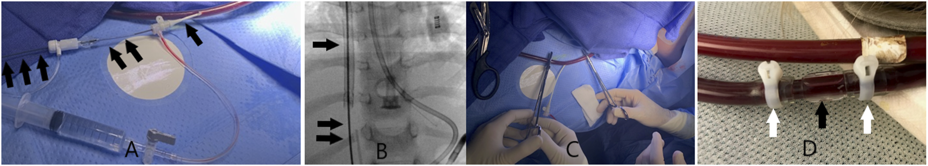
The patient passed away on ECMO day # 67. With no additional cannula repositioning since the last adjustment.
Discussion
Avalon Elite bi-caval dual lumen cannulas provide the opportunity to utilize v-v ECMO through a single cannula, however, cannula migration with associated v-v ECMO dysfunction is not uncommon.
2
Manual repositioning with bedside TTE guidance is usually attempted. If it fails, different techniques can be utilized (snaring the catheter or accessing the distal end of the catheter with “mother-and-child” technique). Those techniques are performed in the cardiac catheterization lab requiring transporting a sick patient, and additional femoral venous access needs to be obtained.2–4 Kalbhenn and colleagues described a bedside technique where the inflow tube is cut and a “Y-connector with a valve” is connected to allow insertion of a sheath through the valve to reposition the AEC.
5
This technique requires pausing ECMO circulation to place the Y-connector and to remove the Y-connector.
5
Our technique is simpler. Directly accessing the inflow tube precludes the need for a Y-connector and reduces the time off ECMO. When compared to other reported methods, our technique has advantages, including not obtaining additional venous access and the ability to be performed at the bedside avoiding the hassle of transporting a sick patient with a bulky ECMO machine.3,4 Although different catheters could potentially be used to access the IVC, the authors are more familiar with the Angled Glide catheter and commonly use it for different interventions. This catheter is hydrophilically coated, allowing smooth tracking. Its unique distal curve (Figure 2), torquability, and softness allow access to many structures with relative ease.6,7 Image of an Angled Glide catheter.
Conclusion
When AECs migrate out of appropriate position, attempts must be made to reposition to maximize efficacy of therapy. Our technique can be utilized to reposition a migrated cannula without requiring patient transportation, additional venous access, or extended ECMO circulation pauses. Caution should be exercised to avoid air introduction during sheath placement and wire and catheter manipulations.
Footnotes
Declaration of conflicting interests
The author(s) declared no potential conflicts of interest with respect to the research, authorship, and/or publication of this article.
Funding
The author(s) received no financial support for the research, authorship, and/or publication of this article.