






Abstract
Emergencies during phlebology procedures are rare. Nonetheless, a phlebologist needs to be prepared to manage such a situation (like syncope due to anaphylaxis) if it occurs, to distinguish a true emergency from a more benign scenario (like vasovagal syncope), and even to manage an emergency unrelated causally to the procedure itself (like a heart dysrhythmia which happens to occur during sclerotherapy). The focus of such preparations should be actions and information which affect patient outcomes. Physician mental and even medical team rehearsal of such scenarios can improve phlebologist and team responses. This article discusses the differential diagnosis and management of the patient with syncope in a phlebology practice, with emphasis on anaphylaxis.
Clinical scenario
A 55-year-old woman returns for sclerotherapy of remaining varicose veins after technically successful thermal ablation of her great saphenous vein. She is otherwise healthy, has no known allergies, takes no regular medicines, and has no acute complaints. During sclerotherapy, she complains of nausea and lightheadedness and then faints.
Introduction
Key causes of syncope for phlebologists.
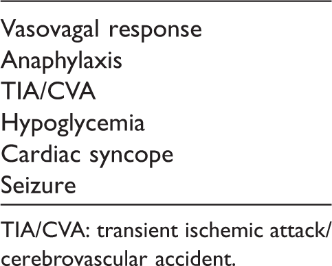
TIA/CVA: transient ischemic attack/cerebrovascular accident.
The final common pathway of syncope is a hypoperfused brain. In this scenario, strongly suggestive of vasovagal response, a reasonable early step in a responsive patient is to put head down to increase cerebral perfusion. A quick return to normal level of consciousness with rapid improvement in initial symptoms suggests vasovagal reaction.
If altered mental status is persistent, then resuscitation measures should be initiated. Unresponsiveness should prompt advanced cardiac life support protocols such as airway and pulse checks. Continued altered mental status in a responsive patient merits a search for anaphylaxis and an intravenous (IV) line. A phlebologist can access any vein in an urgent situation, including a saphenous vein, and ultrasound guidance skills can be useful. Seizure or postictal state is also possible. Additional monitoring equipment, such as blood pressure, pulse, telemetry and pulse oximetery, can be used if available.
Anaphylaxis
Anaphylaxis is a severe, potentially life-threatening, allergic response to an environmental exposure. 1 The key pathophysiological step is basophil and mast cell release of chemical mediators producing symptoms. These chemical mediators include histamine, leukotrienes, and cytokines, and their release is mediated through immunoglobulin E. In the past, much ink was spilled attempting to distinguish anaphylaxis from anaphylactoid reaction, but this distinction is not clinically significant.
By consensus opinion, anaphylaxis is considered highly likely if one of the following three criteria or met. 1

Gastrointestinal and cardiovascular symptoms and signs are somewhat less common during anaphylaxis. 1 Symptoms such as nausea, vomiting, diarrhea, and crampy abdominal pain occur in around 50% of cases. Syncope, incontinence, dizziness, tachycardia, and hypotension also occur in around 50% of cases.
Prescribing instructions for polidocanol and sodium tetradecyl sulfate recommend that treating physicians be able to manage anaphylaxis if it occurs.2,3 Sodium tetracdecyl sulfate intructions 2 also recommend a test dose to check for allergy before a full sclerotherapy session. This author recommends against such a trial session, which can produce anaphylaxis with no benefit to the patient, and which does not guarantee there will not be anaphylaxis at a later session if negative.
Vasovagal response
A vasovagal response occurs after overstimulation of the parasympathetic versus the sympathetic nervous system. 4 It is the most common cause of simple loss of consciousness. Characteristic of vasovagal response is dysfunction of the autonomic nervous system, with parasympathetic activation resulting in an initial bradycardia, and loss of sympathetic stimulation resulting in initial hypotension. An environmental trigger, such as a needle stick, is a common cause.
Vasovagal reaction typically presents with a prodrome of nausea, pallor, and diaphoresis, although sudden loss of consciousness is also possible. 4 Other common symptoms include lightheadedness, feeling hot, and tinnitus. Lack of blood flow to the brain can result in confusion or even syncope. For a patient with the clear diagnosis of vasovagal reaction, the main issue is to avoid vasovagal triggers. Cardiology consultation can be considered if the patient has a history of recurrent vasovagal reactions, an unclear diagnosis, or a profession such as an airplane pilot in which further syncopal episodes could be dangerous.
Differential diagnosis
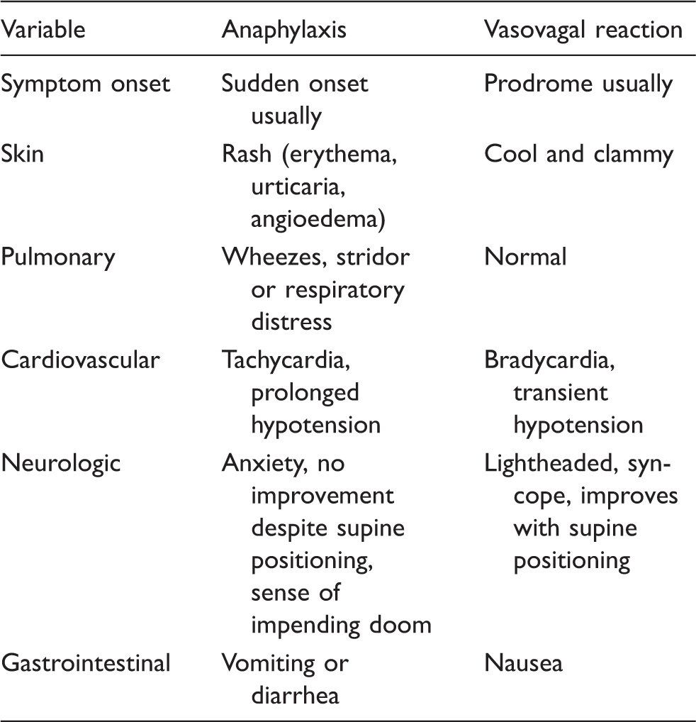
Differential findings in anaphylaxis versus vasovagal reaction.
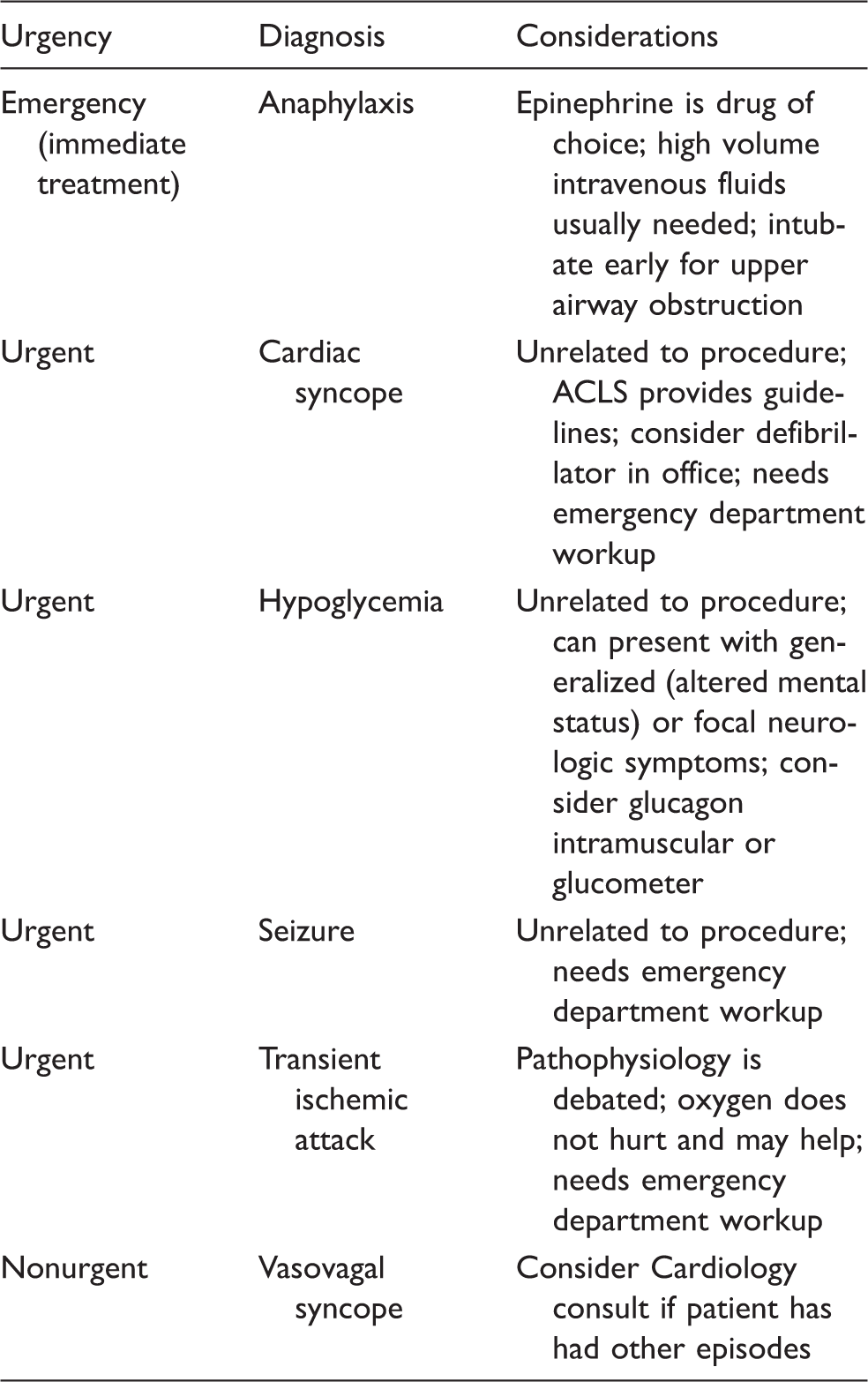
Differential diagnosis considerations.
Emergency equipment to consider for syncope.
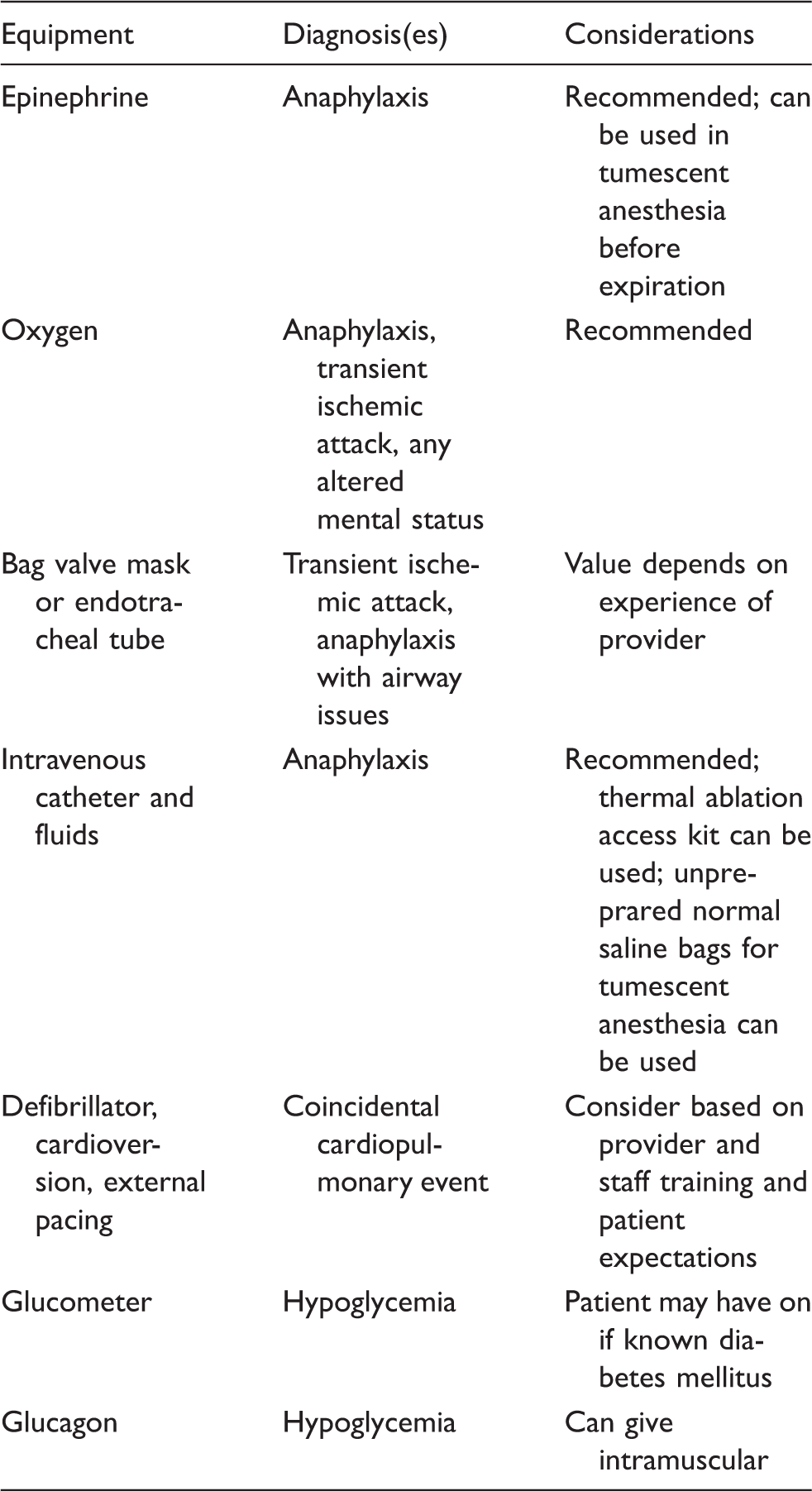
Provider experience and credentials may also impact how a physician manages an emergency. An experienced physician in airway management may elect to have advanced airway equipment and processes in place for the potential difficult airway in an anaphylaxis patient. Experienced airway managers know that the bag valve mask can help a patient through most airway emergencies even if endotracheal tubes are not available, although the occasional anaphylaxis upper airway obstruction can be an exception. Credentials such as advanced cardiac life support and/or basic life support can help providers and their assisting staff think through team emergency responses with extra knowledge.
Hypoglycemia can present with syncope or even with focal neurological signs. A glucometer is helpful to diagnose hypoglycemia if available. If the patient can tolerate oral fluids with minimal risk of aspiration, he/she patient can simply be given a sugar-containing beverage. If the patient cannot safely tolerate oral intake, he/she can be given glucagon intramuscular (IM), if available. The patient with known diabetes mellitus may have their own glucometer available.
Transient ischemic attacks and cerebrovascular attacks have been reported after venous stripping, phlebectomy, thermal ablation, foam sclerotherapy, and liquid sclerotherapy. 5 Various pathological mechanisms have been proposed for these rare events, including paradocal thrombus embolization, paradoxical gas embolism (for foam sclerotherapy), and enodthelin-mediated vasospasm (for foam or liquid sclerotherapy). A neurological exam is prudent to exclude any focal neurological finding suggesting such an event in a patient with alteration in mental status. High flow oxygen, if available, may help, and will not hurt, the patient. Transport to an emergency department (ED), or even possibly a hyperbaric chamber for a suspected gas embolism event, is recommended if such an even cannot be ruled out. Syncope plus a sudden severe headache suggests the possibility of subarachnoid hemorrhage, which would be an unfortunate coincidence to a phlebology procedure.
Cardiac syncope, such as heart dysrhythmia, myocardial ischemia, or congestive heart failure, is an important cause of syncope to be ruled out if the patient’s presentation is not consistent with other etiologies. In the scenario above, such a cause would be very unlikely. Still, if the loss of consciousness is without any prodrome, or if the duration lasts more than a few seconds, this diagnosis should be considered, since cardiac syncope is an important cause of future sudden death. 4 A history of dysrhythmias or heart disease is also suggestive. A pulse, blood pressure, stethoscope, and telemetry are useful to check for dysrhythmias. Concomitant chest pain or shortness of breath with syncope would indicate an ED workup.
Seizure is suggested by persistent confusion after syncope, or syncope plus other symptoms suggesting seizure, such as tongue biting or incontinence. A first seizure merits an urgent workup and a seizure in a known seizure disorder patient generally merits checking medication levels.
Management of anaphylaxis
Common errors in the management of anaphylaxis.
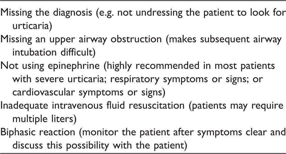
Epinephrine is the drug of choice for management of anaphylaxis. 6 Definite indications include upper airway obstruction, dyspnea, or hypotension. In adults 0.5 mg is given IM at the anterolateral thigh, where absorption is most rapid. A dose of 0.5 mg is 0.5 mL of 1:1000 (1 mg/mL) epinephrine. This epinephrine concentration is preferred to a dose of 5.0 mL of 1:10,000 (0.1 mg/kg) epinephrine. Self-injectable epinephrine delivers a dose of 0.3 mg. Epinephrine injections can be repeated up to every 5 min if clinically necessary. There are no absolute contraindications to epinephrine in a patient with anaphylaxis. Epinephrine can also be used in patients with severe urticaria if young and healthy. The alert patient should be warned that he/she may sense their heart racing or beating strongly.
IV access should be obtained. The need for multiple liters of IV fluid resuscitation should be anticipated. 6 Fluid resuscitation is clearly indicated in hypotension and recommended for tachycardia. Telemetry and pulse oximetry are useful for monitoring when available. Although IM epinephrine is preferred to IV epinephrine initially due to greater safety, IV epinephrine can be considered if the patient is unresponsive to IM epinephrine and IV fluids.
Other recommended medications include oxygen, antihistamines (H1- and H2-blockers), 7 and corticosteroids. 6 While antihistamines with or without corticosteroids are sufficient for mild cases of acute urticaria, they are not sufficient for a true anaphylaxis. Inhaled beta-agonists should be considered in the setting of wheezing. 6
After initial anaphylaxis treatment, some patients can experience a biphasic reaction, a return of moderate to severe anaphylactic symptoms. 1 This event has been reported 1–72 h after the initial symptoms. It is speculated that this occurs when the epinephrine wears off and also that corticosteroids may decrease this risk.
The patient should be observed for some period of time if anaphylaxis symptoms clear and sent to the ED if symptoms do not clear. Four to six hours of observation has been proposed 1 but is difficult logistically in a clinic setting. Patients can be observed in the ED if needed. Extra caution is advised in patients with initially severe anaphylaxis symptoms, with any symptoms of reactive airway disease, or with difficulty in accessing care. If discharged, consideration should be given to prescribing self-injectable epinephrine for a return of symptoms, and continuing antihistamines and corticosteroids for three days to minimize symptoms. Allergy consultation should be considered.
Conclusions
In any potential emergency, it is best to assume the worst and hope for the best. Assume an emergency condition until it is ruled out. In the clinical scenario above, anaphylaxis should be assumed until it can (or cannot be) ruled out. A patient with vasovagal reaction should rapidly return to baseline status. Prolonged altered mental status or hypotension requires urgent management. If an emergency cannot be ruled out, consult early and utilize ED if unsure. For an acute allergic reaction, the emergency physician is likely the initial specialist of choice. The ED can also monitor for allergic biphasic reaction.
Anaphylaxis management has a number of potential pitfalls, even after it is correctly diagnosed. Standard resuscitation measures may need to be rehearsed (at least mentally), since phlebology emergencies are rare. The airway needs particularly careful monitoring for upper airway obstruction symptoms and findings such as hoarseness, voice changes, and stridor. Epinephrine is the drug of choice for anaphylaxis. Patients should be warned about the possibility of a biphasic reaction.
Footnotes
Funding
This research received no specific grant from any funding agency in the public, commercial, or not-for-profit sectors.
Conflict of Interest
None declared.