
Abstract
Laser ablation involves the delivery of laser light through a glass fibre placed into the lumen of a refluxing vein. This energy is converted into heat inducing a permanent, non-thrombotic occlusion. This highly effective and safe approach has significant advantages over traditional surgical treatment and has driven the endovenous revolution in the management of varicose veins. This chapter will explore the mechanism of action, present the evidence of laser' clinical and cost effectiveness, and analyse specific and generic aspects of laser ablation technique.
Introduction
Laser ablation has driven the endovenous revolution in the management of varicose veins. This chapter will summarise the mechanism of action, provide evidence of laser’s clinical and cost effectiveness, and analyse specific and generic aspects of laser ablation technique, which may warrant further investigation.
Mechanism of action
The aim of this treatment is to induce a permanent, non-thrombotic occlusion of a refluxing vein using the intraluminal application of laser energy. The laser energy is converted into thermal energy, which induces mural inflammation and ultimately fibrosis with resultant vein obliteration. Laser is an acronym for light amplification by the stimulated emission of radiation. Lasers emit light of very specific characteristics. First, it is monochromatic or “one colour” which is determined by its wavelength. Endovenous laser ablation (EVLA) of varicose veins is commonly performed using a wavelength of between 810 nm and 1500 nm. Laser light is unique in that it is coherent (with all waves in phase, allowing an intense energy transfer) and collimated (delivered in a non-divergent beam rather than spreading out in many directions like the light from a light bulb).
Substances within the vein absorb the laser photons and are referred to as chromophores. Different chromophores will preferentially absorb different wavelengths. Water is a common chromophore in this application, absorbing photons of around 1470 nm. Absorption of photons induces molecular excitement in the chromophore and de-excitation produces thermal energy by photothermolysis. This thermal energy causes vein wall tissue destruction with associated permanent vein occlusion, which via a complex remodelling process gradually transforms into a fibrous scar.
There are different mechanisms of heat transfer at play during EVLA, and the relative contribution of each mechanism to vein occlusion may be academic; however, understanding of these mechanisms may facilitate their manipulation, in order to maximise efficiency and reduce side effects and minor complications.
The first chromophore encountered by the laser energy during EVLA is the blood within the vein. The blood absorbs a proportion of the energy, and is heated rapidly beyond its boiling point, transferring heat energy to the vein wall via convection and conduction. This is the predominant mechanism by which EVLA achieves vein occlusion.
The sudden and extreme transfer of energy at the interface between the fibre and the blood results in molecular ablation releasing carbon which then coats the fibre. This black carbonised layer has a very high absorption coefficient for all wavelengths of light and is thought to absorb around 45% of the emitted photons. This results in very rapid heating in excess of 1000°C. Direct conduction of this tip causes super-heating of the surrounding blood and acts as a black body radiator emitting different wavelengths of radiation into the blood and towards the vein wall.
The super-heated blood forms steam bubbles. The significance of these steam bubbles is unknown, but they have been observed to behave in a manner similar to a “heat pipe”, with the bubbles travelling down the lumen and condensing back into water, releasing additional energy. This may propagate the laser’s thermal impact.
The laser energy not absorbed by the carbonisation or the blood may be absorbed by chromophores within the vein wall, causing a direct cellular thermal injury. Evidence suggests that this mechanism is responsible for only a small proportion of the temperature profile.
Direct contact of the fibre with the vein wall with associated conduction of heat was initially thought to be the primary mechanism of action. This causes extreme localised injury and frequent perforations to the vein wall, which is thought to contribute to some of the side-effects of treatment such as pain and bruising. Therefore, direct contact of the fibre with the vein wall is considered by some to be detrimental. However, others see contact as beneficial, facilitating a reduction in the energy required for durable occlusion.
Inflammation is the final mechanism contributing to vein occlusion following EVLA. This explains why veins experimentally harvested immediately following early EVLA technology demonstrated mainly localised injuries from fibre contact, whereas veins from a delayed harvest showed significant changes throughout the entire circumference of the vein wall sections.
Comparative evidence
The safety and efficacy of EVLA are supported by the strongest evidence base of all of the minimally invasive techniques for the treatment of varicose veins, and thus EVLA represents the standard against which all other techniques should be compared. There have been a range of meta-analyses identifying a large number of comparative studies to date. One extracted data from 64 varying studies treating 12,320 limbs and found success rates (absence of a refluxing axial vein on ultrasound) following EVLA to be around 95% at 5 years and that was significantly higher than seen following surgical ligation and stripping (76%), foam sclerotherapy (74%) and older radiofrequency technology (80%). 1 Since this time, changes in radiofrequency technology have improved the efficacy; however, there are only a small number of studies to confirm this.
A health technology assessment study funded by the UK National Institute of Health Research performed a review of the available randomised controlled trials (RCTs) and found evidence that thermal ablation is associated with less recurrence than surgery or foam sclerotherapy and a faster recovery than surgery with low rates of significant adverse events. 2 This study went on to find foam sclerotherapy to be the most likely to be cost-effective, due to its significantly lower costs. However, another cost-effectiveness modelling study found that EVLA had the highest probability of being cost-effective, due to the low efficacy and presumed higher re-intervention rates associated with foam sclerotherapy. 3
The largest RCT comparing different treatments for varicose veins was recently published. This study randomised 785 patients in 11 centres to EVLA, surgical ligation and stripping or foam sclerotherapy. 4 EVLA was associated with superior quality of life (QoL) at 6 weeks when compared with surgery, reflecting the reduced postoperative morbidity associated with EVLA, whilst the improvement in venous symptoms was less following foam sclerotherapy than the other treatments. Foam sclerotherapy was associated with worse clinical severity scores and lower ablation rates (55% at 6 months). The associated detailed cost-effectiveness analysis estimated that EVLA had around an 80% probability of being cost-effective at conventional willingness to pay thresholds and was significantly superior to the other treatments. 5 Similar findings were seen in another detailed RCT comparing 280 patients randomised to surgery or EVLA 6 and this group found a difference in favour of EVLA in terms of clinical recurrence. 7
The UK National Institute of Health and Care Excellence performed a detailed review of RCT evidence of the clinical and cost-effectiveness of endothermal ablation and recommended that, in technically suitable patients, endothermal ablation should be considered as the first choice treatment in the UK NHS. 8 This review did not distinguish between EVLA and radiofrequency ablation but considered them as equivalent “endothermal” treatments. Studies comparing EVLA and modern RFA have concluded that short-term efficacy is equivalent, but EVLA is associated with more pain and bruising.9–12 The significance of these findings is unclear as no study has shown any significant benefit in QoL relating to the reported differences and the EVLA outcomes are inferior to those previously reported in the literature. In addition, the evidence to support the medium-term efficacy of radiofrequency ablation is limited.
EVLA parameters
There are multiple EVLA parameters which can be varied such as the laser power, the withdrawal speed of the fibre, the energy delivered, the wavelength of the laser and the design of the fibre delivering the laser energy. Alterations of these can and do affect occlusion rates, complications and post-procedural recovery. The number of variables and complexities make EVLA appear more daunting to the novice than other procedures which have been fully standardised. This should not be the case, however, and it is possible to have a standardised protocol which needs little or no adjustment for clinical practice providing excellent clinical results and high patient reported QoL and satisfaction. Many surgeons use the same power and pullback rates for every case with excellent results.
It can be challenging to disentangle the evidence regarding these parameters from commercial marketing, but this is attempted in the following sections. What is clear is that further evidence is needed to establish the optimal combination of these variables.
Power, pullback rate and energy
These parameters are closely linked. The power of the laser in Watts is the number of Joules of energy delivered in 1 s. The pullback rate therefore determines the number of Joules of energy delivered to each centimetre of vein at a given power.
The energy delivered per unit length is critical for durable vein occlusion. Too little energy and recanalisation will occur, too much and there is a risk of injury to the surrounding tissues. An early case series with an 810 nm laser and a bare fibre found that the mean energy density of treatment failures was 47 J/cm compared to 63 J/cm for successes and no failures were seen over 80 J/cm. 13 The authors then performed a prospective case series with a target of 80 J/cm, seeing a large increase in their success rates; however, failures were still seen at a mean of 98 J/cm. 14 Several other studies went on to note similar thresholds of recanalisation below 60–80 J/cm.15,16 Thus, with the original bare fibre and short wavelength lasers, 80 J/cm is the point at which recanalisation becomes uncommon, however it is still seen. So, how high must we go to achieve 100% occlusion rates? Some authors have experimented with variation of the energy depending upon the vein diameter and some purposefully calculate different pullback rates for different vein segments, explicitly or by “feel”. Whilst these “artisan” approaches seem attractive to the expert, there is little evidence that they make any difference and they certainly add to the complexity of the procedure. A more obvious approach is to simply increase the target energy delivery in all cases, but where would this start to become unsafe?
A detailed secondary analysis of 232 patients randomised to receive EVLA in RCTs with an 810 nm laser and a bare fibre found no evidence of any differences in pain, analgesia, recovery time, QoL or complications between energies of 44–158 J/cm. 17 This large safe operating range may not be true for longer wavelengths as an early case series using 1470 nm noted that great saphenous vein (GSV) treatment with more than 100 J/cm was associated with an increase in paraesthesia from 2.3% to 15.5%, although this did improve over time. 18
The way in which the energy is delivered can be varied. The power and pullback can be changed simultaneously whilst keeping the energy constant; however, there is clearly an optimum operating window in both of these parameters. Most procedures are performed using a power between 5 and 14 W. This leads to the question of whether there is any difference in outcomes between the “fast burn” (high power and pullback rate) or the “slow cook” (low power and pull back rate).
An early clinical study randomised patients to receive 810 nm EVLA using either 12 W pulsed or 14 W continuous power EVLA with a bare fibre. 19 In the 12 W pulsed group, the energy delivery was less reliable and clinical recurrence rates at five years were higher. An interesting study used mathematical modelling for each thermal transfer mechanism involved along with data from basic science experiments. 20 This found that vein wall temperatures were higher at higher powers even though the actual energy applied was the same. If this were true, it would mean that higher power setting will allow a slightly lower energy to be applied per unit length, whilst still achieving the same temperature at the vein wall. This would also reduce procedural times as pull back rates could be increased.
Wavelength
The wavelength or colour of the laser is currently fixed for each device. Its impact upon clinical results is contentious. The wavelength selects which chromophore absorbs the laser energy, with shorter wavelengths being absorbed by haemoglobin and longer wavelength lasers being absorbed by water. Commercial marketing claims that the latter primarily occurs within the vein wall cells, and that this “selective thermolysis” allows a reduction in the energy thus less vein wall perforations, leading to less pain, bruising and complications. However, as discussed previously, the direct absorption of laser energy by the cells is thought to be responsible for very little of the thermal damage.
Currently, level 1 evidence demonstrating improved outcomes for longer wavelengths is lacking as variation in fibre tip is often investigated simultaneously. A multicentre pseudorandomised trial (randomisation by alternation) allocated 180 patients to receive EVLA with either a 980 nm or 1500 nm laser. 21 Confounding factors included higher energy densities, younger patients and longer treated vein lengths in the 980 group, all of which are likely to have increased reported pain. The 1500 nm group required a shorter duration of analgesia (1.8 vs 2.9 days) and had 4% superior improvement in the disease-specific CIVIQ 2 QoL score, but a non-significant trend towards higher recanalisation rates.
An experimental mathematical modelling study found that the calculated temperature profiles at the vein wall are independent of wavelength. 22 Experimental in vitro models using thermocouples found similar temperature profiles for both 940 and 1470 nm lasers, appearing to support the theoretical model’s findings. 23 A second more detailed modelling study demonstrated that the scattered photons reaching the vein wall explain only a very small amount of the temperature profile when compared with the hot tip and super-heated blood. 20
Fibres
The delivery system for the laser energy into the lumen of the vein is the fibre tip. The first fibres were bare tipped and delivered a forward firing beam with little divergence, concentrating its energy over a small area in front of the fibre, perforating and cutting the vein whilst in contact. Three design modifications are in common clinical use and alter either the contact of the fibre with the vein wall and, or reduce the intensity of the beam by increasing the incident surface area by divergence. These aim to reduce energy requirements and reduce the number of perforations and consequently pain, bruising and complications.
The Tulip-Tip fibre (Figure 1, Tobrix Waalre, The Netherlands) is a bare fibre, but has springy “petals” around the tip acting to mechanically centralise the fibre within the vein lumen. This prevents contact of the bare tip with the vein wall and promotes an even distribution of energy application. A two centre RCT randomised 174 patients equally to a bare or Tulip-Tip fibre, using a 1470 nm laser with similar energy delivery.
24
Small benefits were noted in pain in the first week; and bruising and QoL at 2 weeks.
The tulip tip fibre. Images reproduced with kind permission of Tobrix.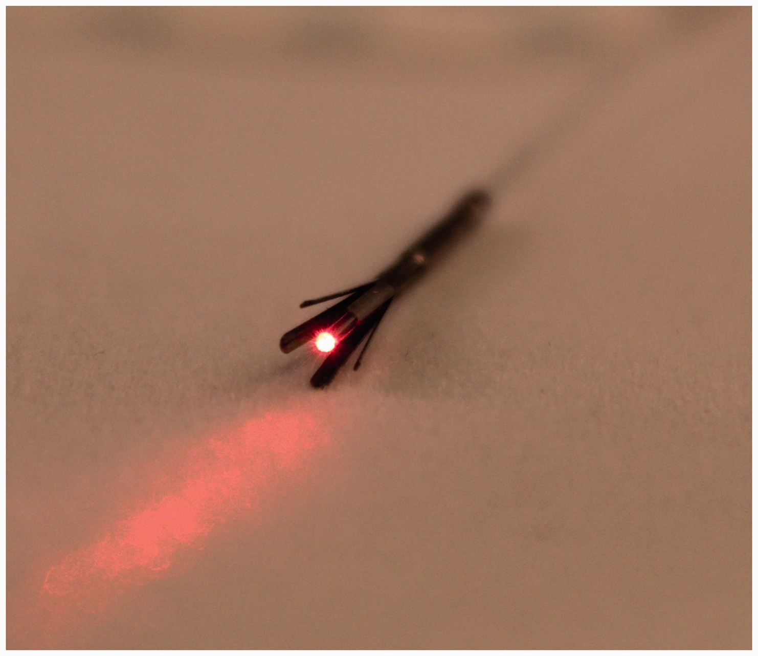
The jacket tip fibre (Figure 2, NeverTouch, Latham, NY, USA) has a curved glass weld at the fibre tip which is encased in a protective metal jacket. The glass weld significantly increases beam divergence, and the jacket centres the fibre within the vein lumen and reduces vein wall contact. An experimental science study on a human vein filled with porcine blood in warm saline; seems to support the claims of this technology demonstrating reduced vein perforations with jacket tip fibres.
25
Whilst this is highly encouraging data, there is currently no convincing comparative clinical evidence to support any benefit for jacket tip fibres.
The nevertouch fibre. Images reproduced with kind permission of Angiodynamics.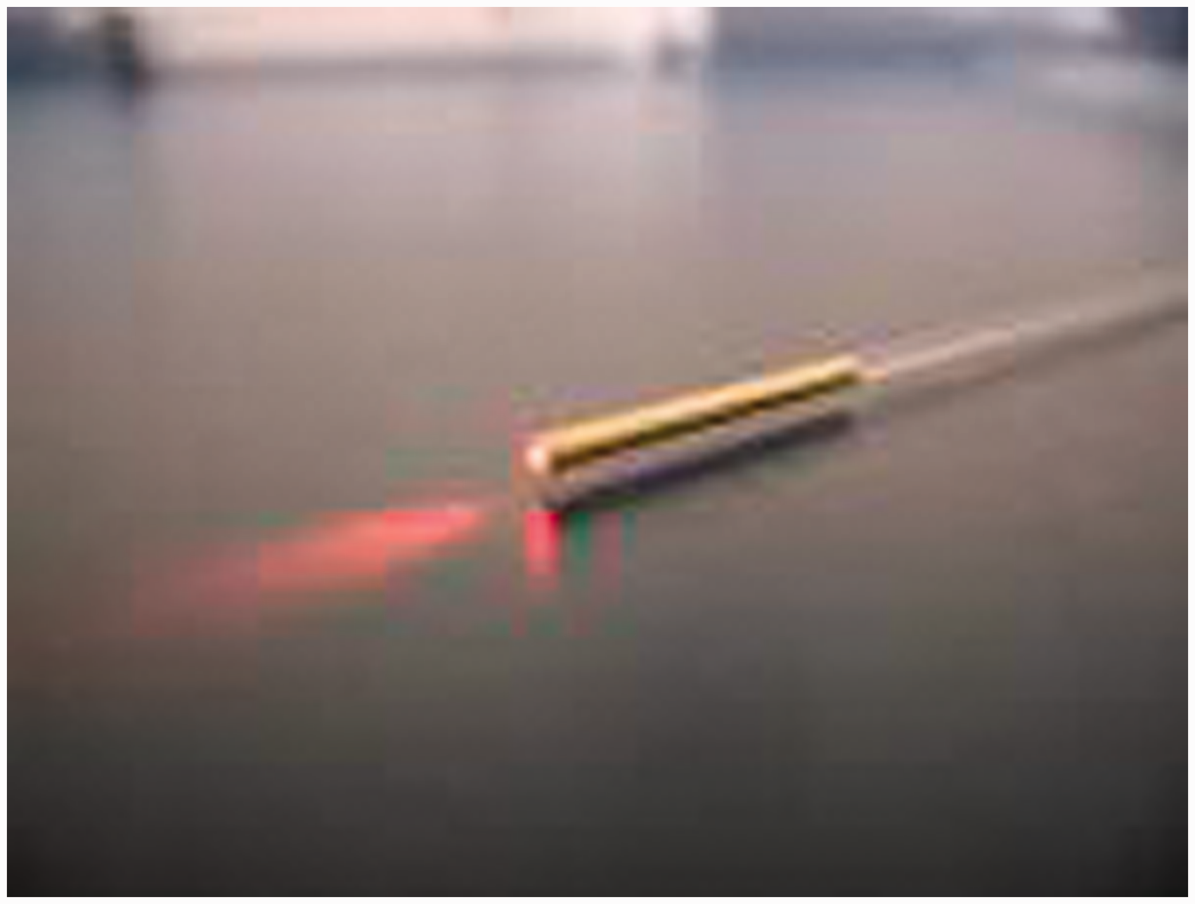
The radial firing fibre (Figure 3, Biolitec Jena, Germany) has a cone within the tip, which causes the beam to be emitted radially from the fibre in a 360° ring directly towards the vein wall. It is conceivable that this may increase the proportion of laser energy directly reaching the vein wall cells and therefore may result in differences in the temperature profile. Two retrospective “before and after” studies compare bare and radial fibres at 1470 nm. The first included 453 legs in 215 patients with a similar energy delivery and concomitant phlebectomy.
26
The radial fibre was associated with less analgesia requirement (0.9% vs 7.4%) and a lower proportion of patients with greater than 25% surface area bruising (1.9% vs 19.4%), although the significance of these findings is unknown as there was no QoL assessment and greater volumes of tumescent anaesthesia were infiltrated with the radial fibre. The second study treated 312 legs in 286 patients, but used lower power and less energy in the radial group (10 W and 57 J/cm vs 15 W and 79 J/cm).
27
Less analgesia was required in the radial fibre group who also had less bruising, but again no QoL data were reported. At 3 months, there was no difference in efficacy between the two groups despite the difference in energy delivery. No long-term follow-up is available.
The radial fibre. Images reproduced with kind permission of Biolitec GmbH.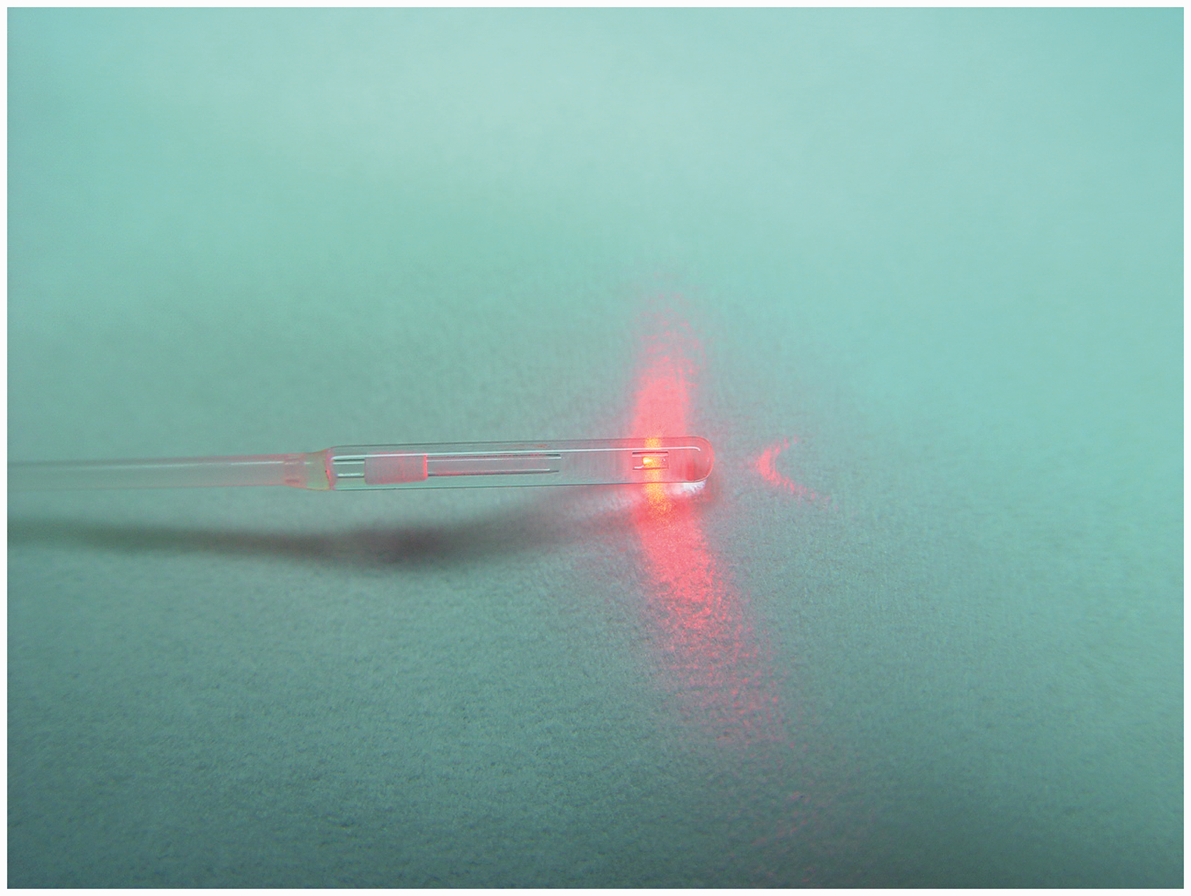
Finally, an RCT compared the bare fibre (980 nm) with a radial fibre (1470 nm). 28 The differences in the wavelength used are disappointing; however, the power and energy delivery were equivalent between the groups. Sixty patients were randomised and the radial 1470 nm group reported a lower duration of pain (2.2 vs 3.2 days) and analgesia (5.8 vs 7.1 days), and also less induration, bruising and paraesthesia. This led to a small difference in initial recovery times (1.6 vs 2.3 days). Clearly, this study alone is not able to ascribe these modest improvements to wavelength or the fibre in isolation.
Tumescent anaesthesia
The infiltration of perivenous tumescent anaesthesia has several valuable roles; it provides local anaesthia, compresses the vein around the fibre, provides hydro-dissection pushing nerves and other structures away from the laser fibre and finally acts as a heat sink limiting heat conduction. It is therefore unlikely that despite being the most uncomfortable part of the procedure, EVLA is ever likely to become “tumescentless”. Recent attention has focused on altering the pH and temperature of the tumescent solution in order to minimise the associated discomfort.29,30
Post-procedural management
There is considerable variability in the post-procedural management of patients following EVLA, and this promises to be a fruitful area for future research in order to guide practice standardisation. 31
The provision of post-procedural analgesia and its duration are highly variable. Approximately 40% of practitioners routinely prescribe post-procedural analgesia, usually in the form of non-steroidal anti-inflammatory agents.
Most practitioners following EVLA almost universally apply some form of post-procedural compression, but the method and duration of compression are highly variable. NICE recommends this as an area for future research. 8
Guidelines recommend a routine postoperative Duplex scan; 32 however, given the high occlusion rates and low complication rates this appears unnecessary. Clearly, a scan would be indicated where there seems to be a clinical requirement, e.g. residual veins or clinical evidence of a DVT.
Conclusion
The clinical and cost effectiveness of endovenous laser ablation is superior to surgery and sclerotherapy and NICE recommends it as first line treatment for symptomatic varicose veins. Future research should focus on maximising efficacy and patient acceptability.
Footnotes
Conflict of interest
DC and IC are involved in the development and delivery of educational and training events for a range of organisations including Angiodynamics at the Charing Cross vein symposium.
Funding
This research received no specific grant from any funding agency in the public, commercial, or not-for-profit sectors.