
Abstract
The pathophysiology of calf perforators is presented. Bidirectional flow within calf perforators with a prevailing inward, into deep veins oriented component arises during calf pump activity in varicose vein patients, as evidenced by venous pressure measurements, plethysmographic findings, duplex ultrasonography, and electromagnetic flow measurements. Reflux within calf perforators is an inward, not outward, flow; the opinion that the outward flow within calf perforators is a reflux is at odds with the reality. During calf muscle contraction, the pressure in the posterior tibial vein is higher than in the great saphenous vein; it induces the harmless outward flow within calf perforators, which runs further via great saphenous vein in the physiological direction toward the heart. Deep and superficial veins of the lower leg form conjoined vessels, as documented by nearly equal pressure curves registered simultaneously in the posterior tibial and great saphenous veins both in varicose vein patients and in healthy people. Calf perforators do not entail ambulatory venous hypertension or any other hemodynamic disorder, even if enlarged and incompetent; their ligation is needless. The diameter of calf perforators is influenced by the intensity of saphenous reflux; it enlarges with increasing intensity of saphenous reflux and diminishes after the abolition of reflux.
Introduction
Linton, 1 in 1938, and Cockett and Jones, 2 in 1953, emphasized the importance of incompetent lower leg perforators for the development of leg ulcers. Linton 1 drew his conclusions from 10 anatomical studies on bodies and from 50 operations. He reported on perforating veins connecting the posterior tibial, anterior tibial, peroneal, and popliteal veins with the greater and lesser saphenous veins, and proposed the ligation of these perforators. Cockett and Jones 2 argued on the basis of their anatomical studies on bodies (they did not specify the total number of studies, mentioned only two examinations) and of their 10 operated cases with a follow-up of several months that the perforating veins drained venous blood from the skin and subcutaneous tissue of the gaiter area selectively into the posterior tibial vein (PTV), and contested their connections with the great saphenous vein (GSV). The authors claimed that in healthy people these perforating veins were fitted with valves enabling only unidirectional flow from superficial into deep veins, and that they impeded the outward flow from the PTV into the GSV. In pathological situations, if these perforators enlarge and get incompetent, a considerable amount of blood is allegedly pressed out from deep into superficial veins during calf muscle contractions, which induces venous hypertension and leads to ulcer formation. Cockett and Jones called this condition “blow-out syndrome.” This opinion has since been widely accepted. As a consequence, perforating veins have been divided into competent ones found in healthy subjects and incompetent ones found in pathological situations. However, this conception was based on subjective speculations and was not substantiated by functional tests and hard facts. No evidence has so far been presented that, first, calf perforators in healthy persons are indeed fitted with competent valves and impede the outward flow from deep to superficial veins, and, second, that incompetent calf perforators really cause ambulatory venous hypertension.
Results of venous pressure and electromagnetic flow measurements, plethysmographic, phlebographic, and anatomical findings presented in this article rebut the theory of incompetent calf perforators and document that calf perforators do not cause hemodynamic disturbance, even if enlarged. Linton himself acknowledged ultimately that reality with his well-known saying:
“The perforator ligation plus compression is as effective as compression alone.”
Calf perforators behave as conjoined vessels both in varicose vein disease and in healthy people
Anatomical studies
There are anatomical studies pointing to the fact that calf perforators are either valveless or fitted only with valve remnants. McMullin et al. 3 cited Barber and Shatara 4 who performed meticulous dissections of the veins of cadaveric limbs; they found that perforating veins of less than 1 mm in diameter had no valves at all. Hadfield 5 dissected 25 cadaveric limbs and reported that smaller perforating veins had no valves at all and the larger ones had only rudimentary valve remnants.
Simultaneous pressure recordings in superficial and deep veins
When considering the competence or incompetence of calf perforators, it must be taken into account that there is a lot of perforators beneath the knee, and that they form a complex system. Consequently, it is necessary to study the competence or incompetence of the whole system, not only of single perforators. Nobody has as yet provided conclusive proof that calf perforating veins as a whole system enable exclusively unidirectional, inward flow from superficial veins into deep veins. Duplex ultrasonography with its key-hole view examining individual perforators is not the relevant method to solve this problem. Persuasive evidence concerning the competence or incompetence of the whole system of calf perforators can be gained by simultaneous pressure recordings in superficial and deep veins of the leg.
In primary varicose veins, simultaneous pressure recordings in the PTV and GSV showed that the pressure curves were nearly identical (Figure 1). Analysis of the peak pressures detected that during calf muscle contraction the pressure in the PTV was higher than in the GSV: the difference amounted to about 13 mm Hg and was not influenced by the presence or absence of saphenous reflux.
6
Consequently, the systolic pressure difference propels venous blood from the PTV into the GSV via calf perforators. Calf muscle contraction evokes a centripetal, toward the heart-directed flow; the main outflow channel is the deep venous axis (popliteal, femoral), but some blood escapes from the PTV via calf perforators into the GSV and streams further through the sapheno-femoral junction into the femoral vein; it can be registered as a flow wave in the thigh segment of the GSV. During calf muscle relaxation the pressure gradient turns round; the pressure in the GSV is higher than in the PTV. In this phase, the venous blood streams from the GSV into the PTV. Saphenous reflux enhances significantly the diastolic pressure gradient between the GSV and PTV (Figure 2). Arnoldi
7
found similar pressure differences between the PTV and GSV in varicose vein patients during calf muscle contractions and relaxations.
Simultaneous recordings of the mean pressure in the GSV (upper curve) and the PTV (lower curve) in a varicose vein patient after interruption of saphenous reflux showing physiological decrease in pressure in both veins. The curves are nearly identical in the quiet standing position, during ambulation, as well as during the recovery phase. This is the typical pressure pattern of conjoined vessels. Peak pressure differences between the PTV and GSV during calf pump activity in primary varicose veins. A = with reflux, B = after interruption of reflux. During contractions (left) the pressure in the PTV was 13 mm Hg higher than in the GSV; it induces the outward flow within calf perforators. Saphenous reflux did not influence the peak pressure difference. During calf muscle relaxations (right), the pressure in the GSV was higher than in the PTV. Saphenous reflux increased significantly the pressure difference.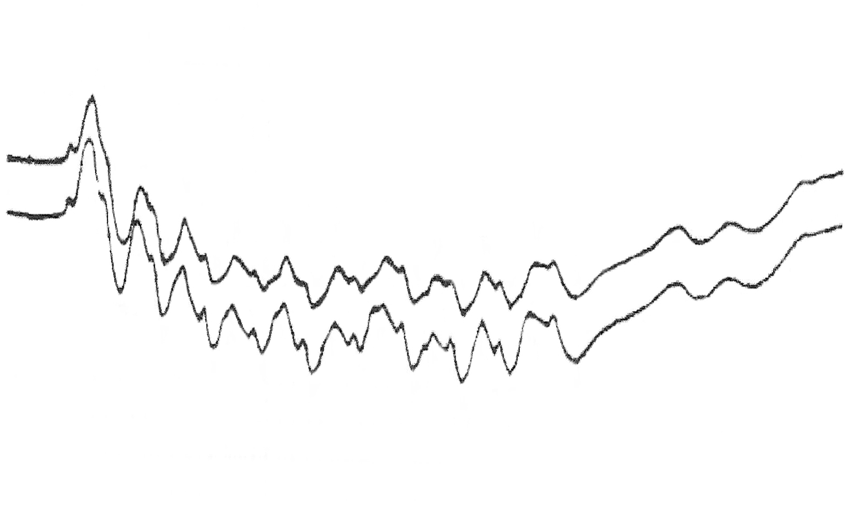
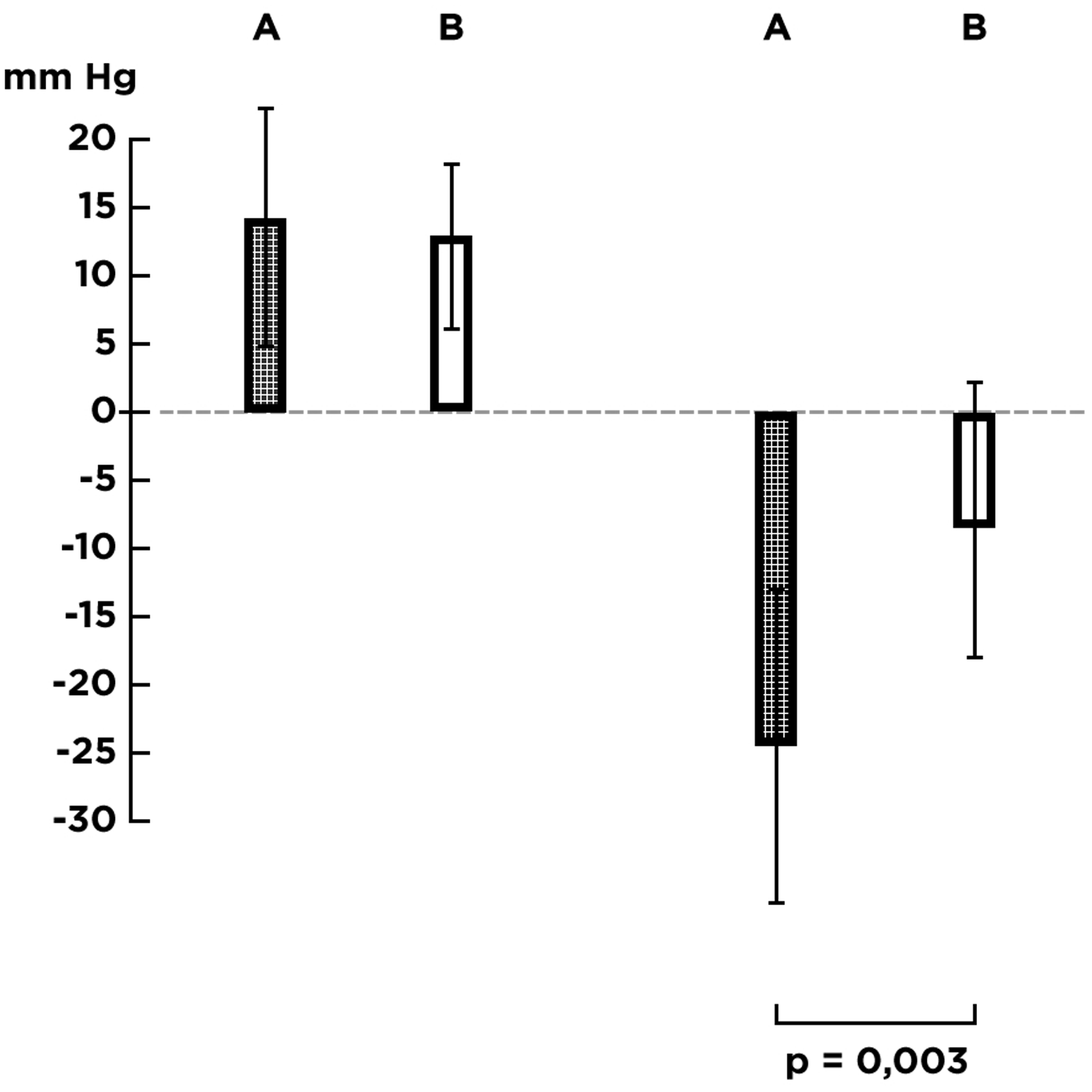
During calf pump activity, the flow within large incompetent calf perforators has a bidirectional pattern; the vector of the bidirectional flow is oriented clearly inward, not outward, as documented by electromagnetic flow registrations in incompetent perforators performed by Bjordal. 8 This finding is at odds with Cockett’s blow-out syndrome conception. The distinctly higher pressure difference occurring during calf muscle relaxation (about 25 mm Hg) in comparison with than one during calf muscle contraction (about 13 mm Hg) is in accordance with Bjordal’s findings. 6
Similar conditions exist also in healthy people. Arnoldi found that the systolic pressure in the PTV was about 41 mm Hg higher than in the GSV; during calf muscle relaxation, the pressure in the GSV was about 11 mm Hg higher than in the PTV.
9
Höjensgard and Stürup10 performed simultaneous pressure recordings in the PTV and GSV in healthy persons. During calf pump activity, both pressure curves were nearly identical documenting free pressure transmission within calf perforators in either direction. There was a steep increase in pressure during each calf muscle contraction in the PTV as well as in the GSV, and decrease in pressure during relaxation; this is the pressure pattern typical of conjoined vessels. The steep rise of pressure in the GSV is a persuasive proof that venous channels connecting PTV and GSV are either valveless or incompetent and do not hinder pressure transmission. Competent valves in calf perforators would have impeded the systolic pressure transmission and the rise of pressure in the GSV. Figure 3 illustrates schematically how the pressure curve in GSV would look like during calf pump activity if valves in the connecting channels between the PTV and GSV were competent: while the steep rise in pressure during each contraction is the characteristic feature documenting an incompetent valve, a competent valve precludes pressure transmission, which would have resulted in a staircase form of the pressure curve.
Schematic illustration of the different pressure curves in the GSV. Left: Pressure pattern in the presence of incompetent calf perforators with steep increase in pressure during each calf muscle contraction. Right: Pressure pattern in the presence of competent calf perforators. No increase in pressure during calf muscle contractions, staircase form of the pressure curve.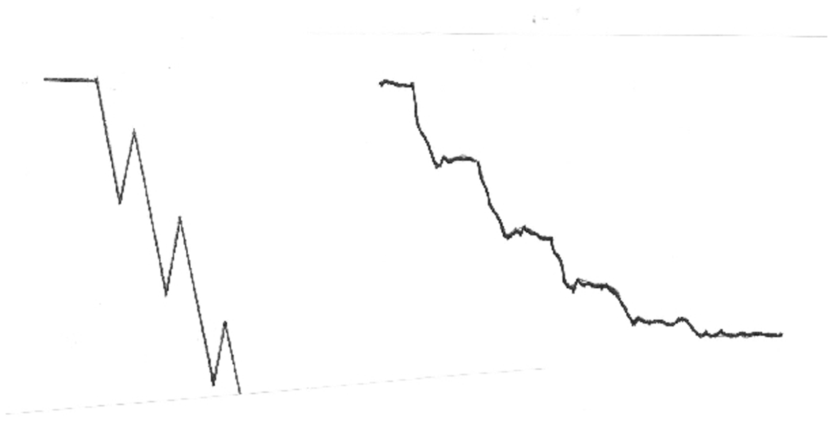
The increase in pressure in the GSV is caused by blood inflow. This can be documented by the pressure behaviour across the mitral valve in the heart. If the mitral valve is competent, there is a steep rise of systolic pressure in the left ventricle to about 120 mm Hg but no rise of pressure in the left atrium, where the pressure remains at the level of 10 mm Hg. Incompetent mitral valve enables escaping of blood from the left ventricle into the left atrium, which is accompanied by systolic rise of pressure in the left atrium. Thus, if there is no blood displacement, there is no increase in pressure in the compartment. The systolic outward flow through calf perforators in healthy people was revealed by Sarin et al. using duplex ultrasonography; 11 they stated that the outward flow should not be regarded as a pathological phenomenon but as a variance of normal.
Simultaneous pressure recordings in the PTV and GSV in healthy persons documented incompetence of calf perforators because of the steep rise of pressure in both veins during calf muscle contraction. Nevertheless, an exceptional event occurred during one muscle contraction in the picture presented by Höjensgard and Stürup: the pressure increased in the PTV but there was no increase in the GSV; in this exceptional case, the calf perforator(s) behaved as competent, whatever the reason for this event might be. The pressure pattern in this exceptional situation complied with that one across the competent mitral valve.
Valves in the veins of the lower extremity have one essential function – to hinder the centrifugal, pathological flow. Competent valves located in the deep veins of the lower leg and in the saphenous system preclude centrifugal (backward) flow during calf muscle relaxation and enable decrease in pressure in the veins below the knee from about 80 mm Hg (equivalent to the hydrostatic pressure during quiet standing) to about 25 mm Hg. On the other hand, there is no physiological reason for hindering the to and fro flow in communicating channels connecting parallel venous trunks of the lower leg. In the next chapter, it will be explained more comprehensively that the outward flow within calf perforators is no pathological “regurgitation” as in the case of mitral valve incompetence in the heart; the outward flow in calf perforators continues via GSV in the physiological centripetal direction toward the heart, without causing any hemodynamic harm. Moreover, competent valves in calf perforators would constitute a considerable disadvantage in cases of acute thrombotic occlusion of the femoral and/or popliteal veins; they would significantly compromise the venous return from the lower leg. Perforators enabling outward flow protect the leg from severe venous congestion in the setting of acute deep venous thrombosis.
Lower leg perforators ensure pressure equalization between deep and superficial veins of the lower leg, which is realized by flow in both directions. From this point of view, the term “competent calf perforator” is misleading; the division into “competent” and “incompetent” calf perforators is arbitrary, freely contrived, and unjustified. Lower leg perforators do not function in healthy persons as a competent, one-way system.
Because the pressure recordings in PTV and GSV showed unhindered pressure transmission in either direction within calf perforators and prompt systolic and diastolic pressure equalization both in varicose vein patients with large “incompetent” perforators and in healthy persons with “normal” perforators, we can conclude that deep and superficial veins of the lower leg form conjoined vessels
Physiological and pathological streaming within calf perforators
There is a common meaning that the inward flow within calf perforators is a physiological phenomenon, whereas the outward flow a pathological reflux. The term “reflux within incompetent calf perforators” (i.e. systolic outward streaming from deep into superficial veins), although generally used, is de facto a misnomer: in reality, the outward flow is no harmful reflux but a harmless systolic centripetal flow running in the physiological direction toward the heart, without causing any hemodynamic disturbance.
When we are speaking about reflux, we must bear in mind four substantial features of each reflux: (1) Pressure difference setting off the flow (2) Source and mouth of reflux (3) Reflux carrying incompetent conduit (4) Centrifugal direction of reflux, i.e. from the heart to the periphery.
Because there is no consensus as to the real substance and characteristic attributes of venous reflux, the definition of venous reflux is presented.
Venous reflux in the lower extremity is a diastolic, centrifugal, pathological flow of venous blood within incompetent venous channel(s) connecting both poles of the ambulatory pressure gradient that arises during the diastolic phase of the calf pump activity and amounts to 37.4 ± 6.4 mm Hg. The higher pole of the pressure gradient lies in the popliteal, femoral, or iliac vein, the lower pole in deep lower leg veins. Reflux takes place during relaxation of the calf musculature, exceeds the duration of the physiological centrifugal streaming lasting 200–300 milliseconds, and stops as soon as the ambulatory pressure gradient has been equalized. It can be also evoked by increased intra-abdominal pressure propagating into the iliac, femoral, and popliteal vein. Reflux interferes with physiological decrease in pressure arising during calf pump activity in deep and superficial veins of the lower leg and foot. It causes ambulatory venous hypertension, the degree of which depends on the intensity of the centrifugal flow expressed in ml/s.
When confronted with this definition, the outward flow within calf perforators contravenes the essential reflux characteristics; it is, in reality, the exact opposite of reflux. Venous reflux is a diastolic (i.e. occurring during calf muscle relaxation), centrifugal (i.e. directed away from the heart), pathological flow. The outward flow in calf perforators is a systolic (i.e. occurring during contraction of calf muscles), centripetal (i.e. streaming towards heart) flow evoked by the contraction of the “peripheral heart” and running via the GSV or other superficial veins in the physiological direction toward the heart. The outward flow in calf perforators does not interfere with the physiological decrease of venous pressure arising during calf pump activity and does not cause ambulatory venous hypertension or any other hemodynamic disorder. Thus, the genuine venous reflux within calf perforators is an inward, not outward, flow as documented by Bjordal’s findings. 8
The hemodynamic disorder in primary varicose veins is caused by saphenous reflux, which is set off by the ambulatory pressure gradient; it streams during the diastolic phase in the centrifugal direction through the incompetent GSV and inward into deep lower leg veins through the calf perforators. During calf muscle contractions, the venous blood streams toward the heart through two parallel channels: through the main channel, i.e. via popliteal and femoral vein, and through an additional channel, i.e. via calf perforators and GSV into the common femoral vein. The calf pump has a double-barreled outflow; the flow in both pathways is centripetal, it runs toward the heart. Regardless of whether the blood flows through the deep or superficial system, it causes no harm; it just returns to the heart, be it in normal people or in varicose vein patients with chronic venous insufficiency.
Large calf perforators do not cause ambulatory venous hypertension
The still prevailing opinion that incompetent calf perforators cause ambulatory venous hypertension was never evidenced by hard facts. On the contrary, the results of direct venous pressure measurements in incompetent calf perforators, in deep and superficial veins, as well as the plethysmographic findings evidenced unequivocally that incompetent calf perforators are innocent structures that do not produce any pathological hemodynamic disorder.
Bjordal carried out direct venous pressure measurements in incompetent calf perforators. Physiological decrease in ambulatory pressure came about during calf pump activity once the saphenous reflux was interrupted.
8
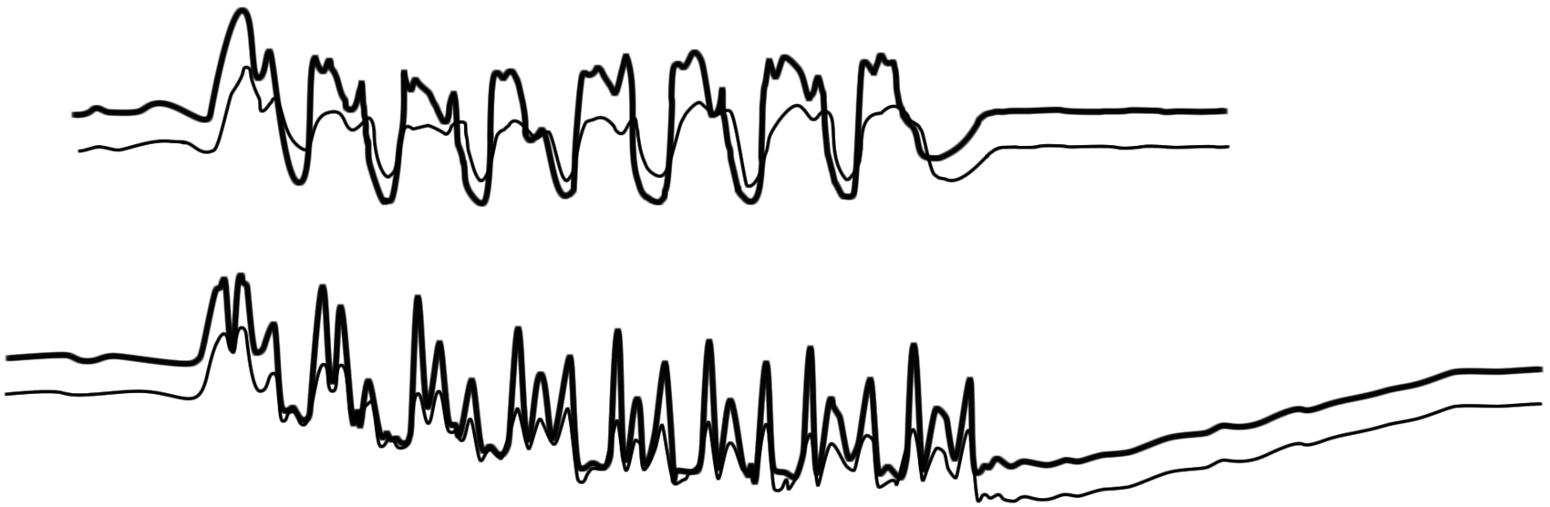
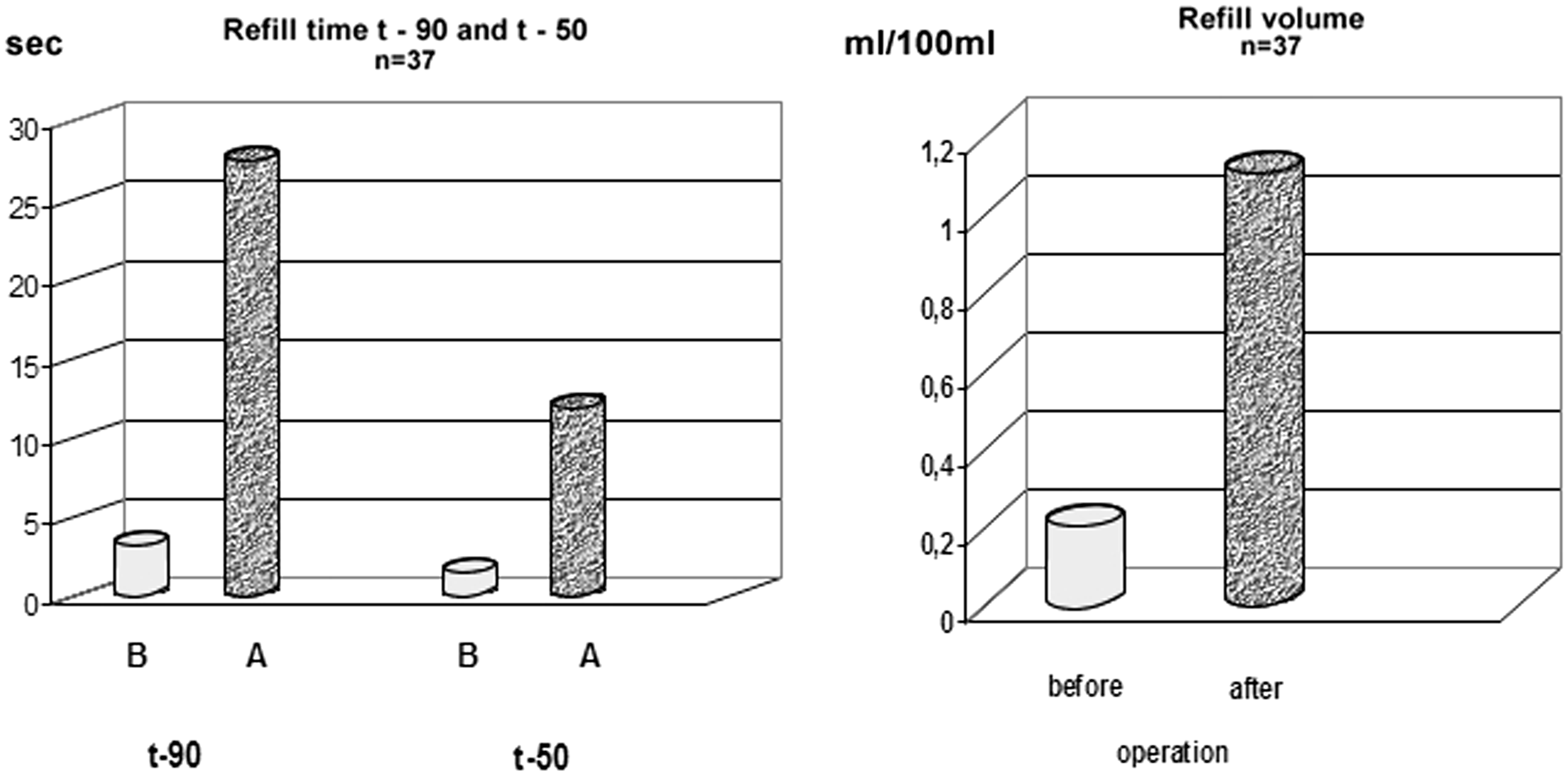
This is the direct proof that large incompetent calf perforators do not induce ambulatory venous hypertension. Simultaneous venous pressure recordings in the PTV and GSV in patients with severe saphenous reflux and large incompetent calf perforators detected very severe ambulatory venous hypertension, which was manifested by no decrease in pressure during calf pump activity. After compression of the incompetent GSV and interruption of saphenous reflux, a physiological decrease in pressure during calf pump activity was restored despite the presence of large incompetent calf perforators,12,13 as shown in Figures 4 and 5. The results of strain gauge plethysmography corroborated the findings gained by venous pressure measurements and confirmed the harmlessness of incompetent calf perforators. Patients with chronic venous insufficiency, strong saphenous reflux, and large incompetent calf perforators displayed very severe hemodynamic disturbance. Abolition of saphenous reflux at the sapheno-femoral junction eliminated the severe pre-treatment hemodynamic disturbance and restored normal plethysmographic values one week after the crossectomy in spite of the presence of large incompetent calf perforators
14
(Figure 6).
Venous pressure recordings in the GSV above the ankle in a patient with severe saphenous reflux, large incompetent calf perforators, and leg ulcer. Top: There was no decrease in venous pressure during calf pump activity documenting the gravest form of ambulatory venous hypertension. Bottom: The same patient as above. Pronounced decrease in pressure during calf pump activity after interruption of saphenous reflux in the thigh and prolonged return to the starting level during the recovery period document restoration of physiological pressure conditions in spite of the presence of large incompetent calf perforators. Simultaneous pressure recordings in the PTV (upper trace, bold) and GSV (lower trace). Above: No decrease in pressure in the PTV as well as in the GSV under the influence of the strong saphenous reflux. Below: After interruption of the saphenous reflux, the venous pressures in both veins returned to the physiological status in spite of large incompetent calf perforators. Impact of saphenous reflux on the hemodynamic disturbance evaluated by strain-gauge plethysmographic parameter refill volume and refill time. (B) before treatment, (A) one week after crossectomy. Abolition of saphenous reflux eliminated the gravest pre-treatment hemodynamic disorder and re-established physiological hemodynamic values in spite of large incompetent calf perforators.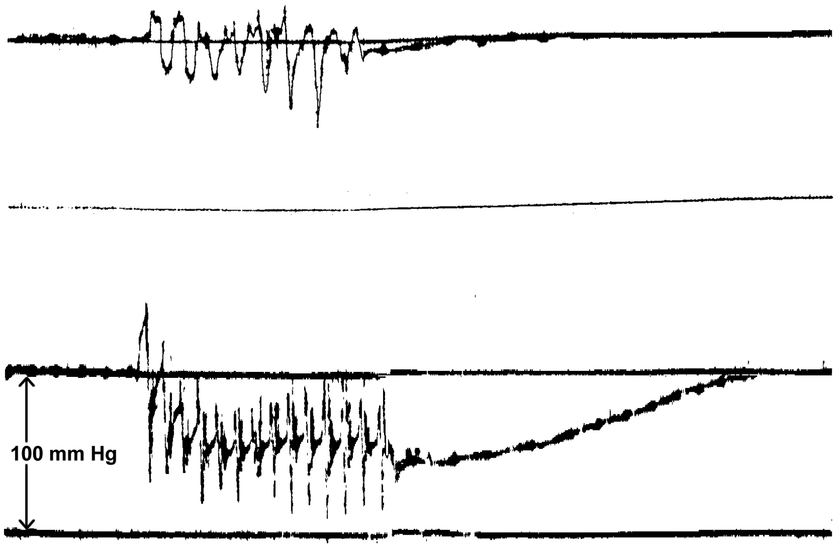
During calf pump activity, the high systolic pressure in the PTV and in the GSV disappears within a fraction of a second and is replaced by decrease in pressure well below the starting level during the ensuing muscle relaxation. The instantaneous short-term rise in pressure causes no harm either to the muscular or to the epifascial tissue.
The simple and well-known Perthes test documents that incompetent calf perforators in primary varicose veins do not produce ambulatory venous hypertension. When the refluxing flow in the incompetent saphenous vein in patients with severe form of chronic venous insufficiency is interrupted by compression and the patients activate the calf muscle pump (e.g. by walking, tiptoe movements or knee bending), the bulging varicose veins empty. According to the blow-out theory the varicose veins should yet more fill up.
The findings of venous pressure measurements and plethysmography unequivocally rebut the belief that incompetent calf perforators cause ambulatory venous hypertension and, therefore, promote formation of leg ulcers. Because of this belief many surgeons interrupted enlarged calf perforators either by the direct access or using subfascial endoscopic perforator surgery (SEPS). In addition to SEPS, abolition of saphenous reflux was routinely carried out but the good results were attributed solely to the SEPS effect, disregarding the fact that saphenous reflux is the determining pathological phenomenon inducing the hemodynamic disturbance. Consequently, SEPS was ironically renamed
Selective abolition of “incompetent” calf perforators does not improve the hemodynamic disorder
Burnand et al. 15 measured venous pressure in 20 patients with incompetent calf perforators before operation and 3 months after selective perforator ligation. The ambulatory venous pressure improved very little after surgery and remained far below normal. Akesson et al. 16 reported the results of selective interruption of incompetent calf perforators in 25 patients with chronic venous insufficiency (30 limbs treated). Deep venous insufficiency was seen on descending phlebography in all cases; in 12 limbs, mild reflux was limited to above the knee level, and in 18 limbs severe reflux extended below the knee level. The patients were examined by foot volumetry and venous pressure measurements. Twelve limbs with a clinical evidence of superficial incompetence underwent saphenous stripping and excision of varicosities. Three months later extensive subfascial ligation of incompetent perforating veins was performed on all 30 limbs. Venous pressure was measured initially and after each surgical procedure. Ambulatory venous pressure improved significantly after superficial vein surgery but did not change after ensuing subfascial ligation of incompetent perforators. Foot volumetric parameters behaved in a similar manner: they improved after superficial vein surgery but did not change after subsequent perforator ligation. In the trial by Scriven et al., 17 patients with combined deep and perforating vein incompetence but without saphenous reflux were evaluated using photoplethysmography. Before treatment, the findings demonstrated a global abnormality of venous function. The pathological findings persisted after perforating vein surgery. According to the study of Fitridge et al., 18 superficial vein surgery removed the hemodynamic disorder but no further improvement was achieved by additional perforator ligation.
The quoted papers evidenced that selective interruption of incompetent calf perforators did not improve ambulatory venous hypertension and/or venous hemodynamics both in primary varicose veins and in deep venous incompetence.
Thigh and lower leg perforators feature different hemodynamic behaviour
From the hemodynamic point of view, there is a substantial difference between the perforators located above and below the knee. Thigh perforators are connected to the higher pole of the ambulatory pressure gradient. 19 When incompetent, they enable pathological centrifugal flow resulting in ambulatory venous hypertension. In contrast to that, calf perforators emptying into the deep veins of the lower leg where the lower pole of the ambulatory pressure gradient is located cannot be the feeding source of centrifugal refluxing flow. They drain the saphenous reflux into deep calf veins and are also called “terminal perforators” or “re-entry points.”
The sapheno-femoral junction and the sapheno-popliteal junction are de facto thigh perforators. In healthy persons, the terminal valves as well as the other valves in the GSV and small saphenous vein are competent and hinder the outward flow through the sapheno-femoral and sapheno-popliteal junction as well as the centrifugal flow through the GSV and small saphenous vein. During calf muscle contraction, the pressure increases also in the popliteal vein, although distinctly less than in the PTV. The systolic escape of blood through the sapheno-popliteal junction is blocked in healthy people by competent valves. When the sapheno-popliteal junction becomes incompetent, the systolic blood escapes from the popliteal vein into the small saphenous vein and continues toward the heart through the Giacomini vein and the GSV. Even if the short saphenous vein is incompetent, the much higher systolic pressure in the PTV impedes the centrifugal streaming through the incompetent short saphenous vein downward into deep lower leg veins during calf muscle contraction. In such a case, the incompetent sapheno-popliteal junction behaves in a similar manner as a calf perforator and enables the development of an additional centripetal pathway through the Giacomini vein and the upper part of the GSV toward the heart. During calf muscle relaxation, the ambulatory pressure gradient sets off reflux in the incompetent SSV.
There is a close relation between the amount of saphenous reflux and the size of calf perforating veins
The diameter of a vessel is influenced by the flow rate intensity; when the flow rate increases, the diameter enlarges; when the flow rate decreases, the diameter diminishes. The mechanism is apparently mediated through increased or decreased fluid shear stress on the endothelium and the release of vasoactive agents.20–24
Retrograde flow running downward the incompetent saphenous vein during each calf muscle relaxation is drained through the calf perforators into deep veins of the lower leg and influences the size of calf perforators: the more voluminous the saphenous reflux, the more blood runs through the calf perforators, the larger the size of calf perforators. The venous segments involved in the Trendelenburg’s “private circulation” including the GSV, calf perforators, and deep veins enlarge in patients with strong saphenous reflux (Figure 7). The flow rate in these venous segments diminishes after abolition of saphenous reflux, and the diameter gets smaller. Some studies confirmed that the enlarged vessels diminished after abolition of saphenous reflux. Stuart et al.
25
examined 62 limbs of 47 patients undergoing superficial vein surgery (sapheno-femoral and/or sapheno-popliteal junction ligation, stripping of the GSV in the thigh, and multiple phlebectomy) with colour flow duplex ultrasound scan immediately before and a median of 14 weeks after operation. Surgery resulted in a significant reduction of perforator diameter. A statistically significant reduction of the number of incompetent lower leg perforators after superficial vein surgery was reported by Gohel et al.
26
In the study by Al-Mulhim et al.,
27
the diameter of mid-calf and lower-calf perforators diminished significantly from 5.5 ± 1.4 mm and 4.4 ± 0.5 mm preoperatively to 3.9 ± 1.7 mm and 3.0 ± 0.4 mm after surgery, respectively. Recek et al.
28
measured the diameter of 31 lower leg perforators and PTVs on phlebographic pictures in patients with primary varicose veins afflicted with a marked saphenous reflux. The values of the vein diameters were checked using a metal benchmark on the X-ray pictures. The authors found statistically significant reduction of the diameter of calf perforators as well as PTVs six months after superficial vein surgery.
Phlebography displaying GSV, a number of large calf perforators, and PTV. All venous segments where increased flow rate takes place due to severe saphenous reflux (GSV, perforators, PTV) are enlarged; in contrast to that, the PTV beneath the enlarged perforators with physiological (not overloaded) flow rate has a normal size. A pair of normal-sized perforators is also depicted; the flow in these perforators runs apparently outward, not inward, because no filling source from superficial veins is visible.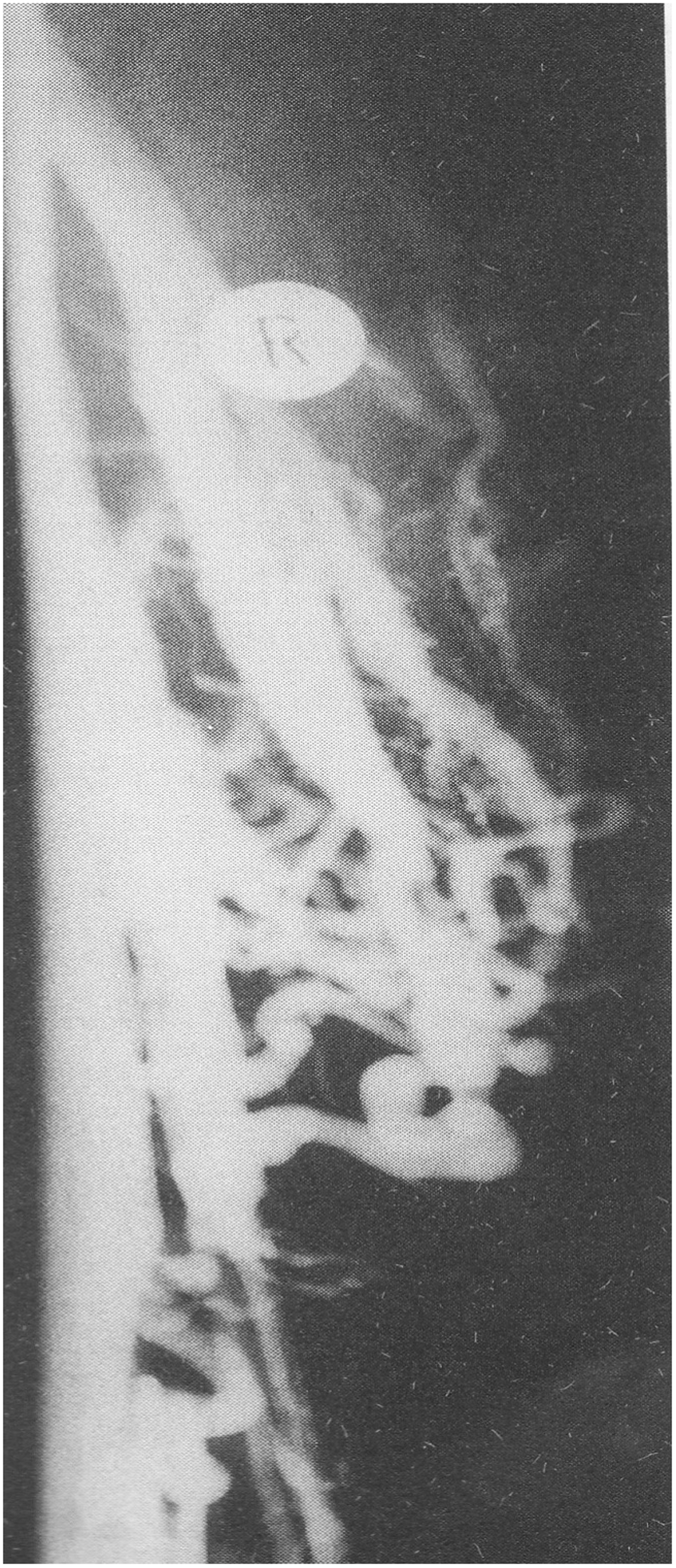
A close relation obviously exists between the amount of saphenous reflux and the size of calf perforators. Although no study has as yet statistically assessed the correlation between the intensity of saphenous reflux and the diameter of calf perforators, it can be reasonably presumed that the intensity of saphenous reflux determines the size of calf perforators. In any case, the diameter of calf perforators significantly diminished after elimination of saphenous reflux.
Conclusion
There is a bidirectional flow within calf perforators both in varicose vein patients and in healthy people. The outward flow in calf perforators is no reflux; it is the exact opposite of reflux. Whereas reflux is a diastolic, centrifugal, pathological, within the calf perforators inward into deep lower leg veins oriented flow inducing ambulatory venous hypertension, the outward component of the bidirectional flow is a systolic, centripetal streaming propelled by the calf pump via the GSV in the physiological direction toward the heart. It neither interferes with the physiological decrease in venous pressure nor causes any hemodynamic disorder. The calf pump has a double-barreled outflow: the main one through the popliteal-femoral axis, and the additional one through the GSV.
Simultaneous pressure recordings in the PTV and GSV proved that calf perforators enabled free pressure transmission between deep and superficial veins of the leg, which resulted in quick pressure equalisation; this evidences that deep and superficial veins of the lower leg form conjoined vessels, both in primary varicose veins and in healthy persons. Venous pressure measurements as well as plethysmographic findings evidenced that large incompetent calf perforators did not cause ambulatory venous hypertension. Selective abolition of large incompetent calf perforators did not improve the hemodynamic disorder.
Thigh and lower leg perforators feature different hemodynamic behaviour. Thigh perforators are connected to the higher pole of the ambulatory pressure gradient and become the source of reflux when incompetent. In contrast to that, calf perforators are connected to the lower pole of ambulatory pressure gradient and cannot become the source of reflux. The size of calf perforators is highly influenced by the intensity of saphenous reflux.
Footnotes
Declaration of Conflicting Interests
The author(s) declared no potential conflicts of interest with respect to the research, authorship, and/or publication of this article.
Funding
The author(s) received no financial support for the research, authorship, and/or publication of this article.