
Abstract
Recurrent lower limb venous insufficiency is often a challenge in clinical practice and is most commonly due to incompetent perforators. Many of these patients do not have adequate symptom relief with compression and require some form of treatment for incompetent perforator interruption. Various treatment methods have been tried with different efficiencies.
Objective
To evaluate the feasibility, efficiency and safety of an outpatient combined cyanoacrylate adhesion–sodium tetradecyl sulphate sclerotherapy for treatment of patients with symptoms of persistent or recurrent lower limb venous insufficiency secondary to incompetent perforators.
Methods
Eighty-three limbs of 69 patients with symptoms of persistent or recurrent lower limb venous insufficiency secondary to incompetent perforators were treated with cyanoacrylate embolization of incompetent perforators and sclerotherapy of dilated collateral veins (surface branch varicose veins). Technical success, procedural pain, perforator occlusion, venous occlusion, clinical improvement and ulcer healing were assessed. Follow-up was done three- and six-month post-procedure.
Results
Procedure could be successfully performed in all patients. One hundred and ninety-one perforators were treated in total. Perforator and varicose veins occlusion rate was 100%. Deep venous extension of cyanoacrylate occurred in four (4.8%) patients, with no adverse clinical outcome. Venous clinical severity score improved from a baseline of 8.18 ± 3.60 to 4.30 ± 2.48 on three-month follow-up and 2.42 ± 1.52 on six-month follow-up (p < 0.0001). All ulcers showed complete healing within three months. Significant prolonged thrombophlebitis occurred in 38.5% of limbs.
Conclusion
Combined cyanoacrylate adhesion and setrol sclerotherapy is technically easy, has a lot of advantages including being an outpatient procedure and highly efficacious but with a guarded safety profile.
Background
Recurrent lower limb venous insufficiency is a commonly faced clinical problem. Higher recurrence rates are found in patients who underwent surgical ligation or stripping as their primary treatment compared to endovenous thermal ablation, with five-year recurrence rates ranging between 20 and 60% after surgery and between 27 and 33% after radiofrequency ablation.1,2 No clear data are available on post-laser recurrence. A study by Bush et al. 3 has documented a 3-year recurrence rate of 7% following thermal ablation, with higher rate after radiofrequency compared to laser ablation. Perforator incompetence has been found to be the most common cause of recurrence and persistent or recurrent leg ulcers.3–5 Various treatment modalities have been employed for perforator interruption including open ligation, subfascial endoscopic perforator surgery, sclerotherapy, thermal ablation, coil embolization and Sapheon glue embolization.5–10 Open ligation and subfascial endoscopic perforator surgery are not routinely performed due to invasive nature and high complication rate, thermal ablation techniques are successful but require anaesthesia and may cause inadvertent nerve damage, and coil embolization has been found ineffective.6,9,10 Sapheon glue embolization has been shown to cause satisfactory perforator occlusion but requires insertion of large vascular sheaths into the perforator and special application devices to deliver Sapheon glue. 10 We explore in this study, the feasibility of an outpatient procedure combining cyanoacrylate glue embolization of perforators and sclerotherapy of collateral veins for the treatment of recurrent varicose veins.
Methods
Institutional ethics review board approval was obtained for this study. All patients referred to our interventional radiology clinics with recurrent venous pathology after previous surgery or thermal ablation of varicose veins were evaluated with Duplex ultrasound, and patients with incompetent perforators as the cause of recurrent or persistent venous insufficiency were selected for this treatment. Duplex ultrasound criteria for labelling a perforator as incompetent were defined as perforator reflux lasting for more than 0.35 s and a diameter of more than 3 mm at fascia level.11,12 Patients with smaller perforator but with significant reflux, if adjacent to an ulcer or directly communicating with enlarged symptomatic superficial veins were also selected for treatment. Patients with other causes of recurrent or persistent venous insufficiency including incomplete ablation or recanalization of ablated saphenous system, accessory saphenous reflux or small saphenous reflux were subjected to laser ablation and excluded from this study. Patients with incompetent perforator more than 7 mm size were excluded and subjected to laser ablation. Patients with deep venous obstruction, immobility, significant arterial insufficiency, active cellulitis, known hypercoagulable state, gravid status and patients unwilling for follow-up were also excluded from the study.
Written informed consent was taken from all patients in this study explaining the procedure, its benefits and risks involved. All patients in this study were treated in the period between October 2015 and May 2016.
Procedure
The procedures were performed in a clean ultrasound suite. A thorough Duplex ultrasound (using L12-3 Linear Array transducer, CX50 Compact Xtreme Ultrasound System, Philips Healthcare) was performed to identify and map the incompetent perforator veins and dilated collateral channels. Patients were positioned either supine or prone based on the position of the incompetent perforators and distribution of dilated superficial varices. The skin site and the linear transducer were cleaned with 2% chlorhexidine solution. Cyanoacrylate embolization of perforators was carried out followed by sclerotherapy of collateral veins in all patients.
Cyanoacrylate glue (Endocryl, Samarth Pharma Pvt Ltd), composed of n-butyl cyanoacrylate is a low viscosity liquid which firmly adheres to cellular surfaces and polymerizes immediately on contact with any ionic solution. The aim of treatment of perforator with cyanoacrylate glue was to obliterate perforator reflux by introducing cyanoacrylate glue either into the perforator or till the perforator–superficial vein junction and immediately providing external compression to cause occlusive adhesion of the vessel walls. Perforator veins are classified anatomically into direct and indirect depending on whether they drain into an axial deep vein or into calf venous sinuses, respectively.
13
Technique of embolization was different for direct and indirect perforators. Cyanoacrylate glue was injected through a 22 G winged infusion set with its tip percutaneously placed using ultrasound guidance either within an indirect perforator or inside a superficial collateral vein 1–2 cm from a direct perforator (Figure 1). Intravenous placement was confirmed by aspiration of blood through the infusion set, after which the infusion set was irrigated thoroughly with 5% dextrose solution until all traces of blood within the infusion set and in its hub were cleared. A total of 0.3 ml (for direct perforator embolization) or 0.5 ml (for indirect perforator embolization) of cyanoacrylate glue and 0.4 ml of air were taken in a 1 ml syringe. Cyanoacrylate glue followed by air was injected quickly into the winged infusion set followed by immediate firm external compression at the injection site for 30 s while the infusion set was withdrawn (0.4 ml of air is taken to fill the intraluminal dead space of infusion set and push the full dose of injected cyanoacrylate glue into the cannulated vein). In patients with oedematous or obese limbs where the intended veins to be treated were deep seated, a 22 G lumbar puncture needle was used to inject cyanoacrylate glue, and volume of air used to fill the needle dead space was 0.1 ml instead of 0.4 ml.
(a) Incompetent perforator (P) from posterior tibial vein (PTV) showing reflux. (b) Percutaneous needle (arrows) placement within the perforator away from the deep venous junction. (c) Post-cyanoacrylate glue injection – compression ultrasound showing hyperechoic glue (*) occluding the perforator.
Cyanoacrylate glue was not directly injected into direct perforators to avoid deep venous extension of injected cyanoacrylate glue which might occlude the deep vein, unless the needle tip could be placed at least 3–5 cm away from the perforator–deep vein junction; and additionally, a lower dose of 0.3 ml was always used. If, after injecting cyanoacrylate glue within the superficial vein near a direct perforator, the perforator showed reflux, another venous puncture was made further proximal to the perforator or within the perforator (more than 2 cm from the perforator–deep vein junction) and another similar injection with a lower dose of cyanoacrylate glue (usually 0.2 ml) and external compression was performed till the perforator showed no reflux. Cyanoacrylate glue in the embolized veins appeared hyperechoic and cast a post-echoic shadow (Figure 1). After every direct perforator embolization, the connecting deep vein was evaluated for patency by checking augmentation response to foot compression in the deep vein cranial to the level of the perforator, since direct visualization of the deep venous segment near the perforator could be difficult due to post-acoustic shadowing of overlying cyanoacrylate glue (Figure 2).
Evaluating patency of the deep veins after glue embolization of perforator, which casts a post-acoustic shadow (*) by longitudinal view imaging of the deep veins cranial and caudal to the embolized glue (a) and looking for augmentation response to distal compression (b).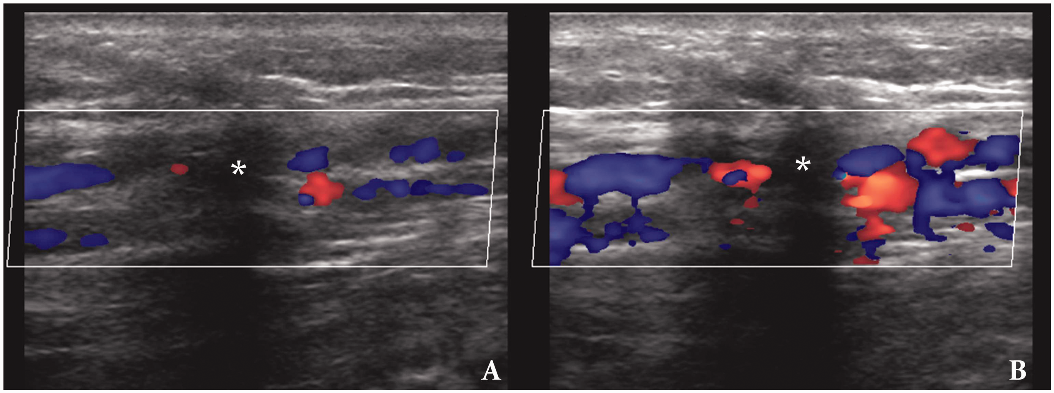
After all incompetent perforators were embolized, foam sclerotherapy was performed for dilated surface branch varicose veins. Ultrasound guidance was used to cannulate collateral veins and sodium tetradecyl sulphate (Setrol, Samarth Pharma Pvt Ltd) foam made by agitating a mixture of 2 cc setrol and 6 cc of air across a partially open three-way stopcock was injected. The injected limb was elevated to prevent proximal deep venous flow of sclerosant and the patient was asked to alternately plantar and dorsiflex the foot to increase deep venous flow to wash away any sclerosant which might have flowed into the deep veins. Ultrasound was used to confirm presence of hyperechoic foam in all abnormally dilated superficial dilated collateral veins. Compression stocking (class II, 30–40 mmHg) was applied in the treated limb after sclerotherapy.
Intra-procedural pain was documented using visual pain scale. Patients were mobilized immediately after the procedure. The patients were instructed to not remove the stockings for 24 h after the procedure and wear compression stocking during daytime for six weeks. Post-procedure pain was managed with oral analgesics for one to two weeks, and cold fomentation as and when required.
Follow-up
Patients were asked to visit for follow-up after three and six months after treatment. Venous clinical severity score was used for objective assessing of clinical improvement. Ultrasound was used to evaluate occlusion of perforators and varicose veins.
Statistics
Basic statistical methods were used to express patient demography, perforator occlusion rate, ulcer healing rate and visual pain scale data. Venous clinical severity score was plotted on Box and Whisker plot and the significance of its change from baseline values to follow-up values was calculated using Friedman test using VassarStats statistics software (Vassar College, Poughkeepsie, NY, USA).
Results
Patients’ demographic and clinical details.
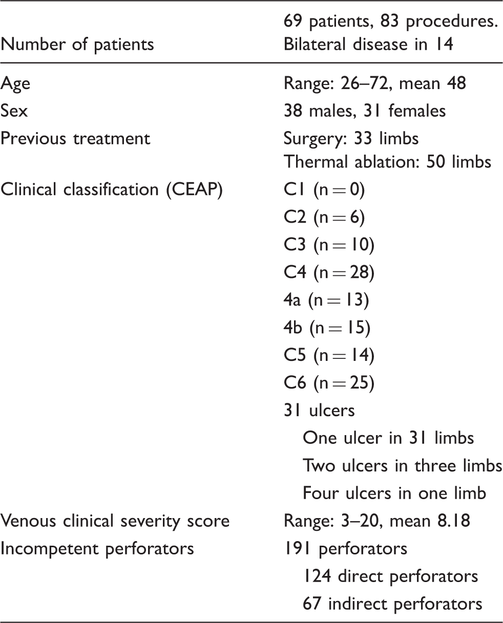
CEAP: clinical-etiology-anatomy-pathophysiology.
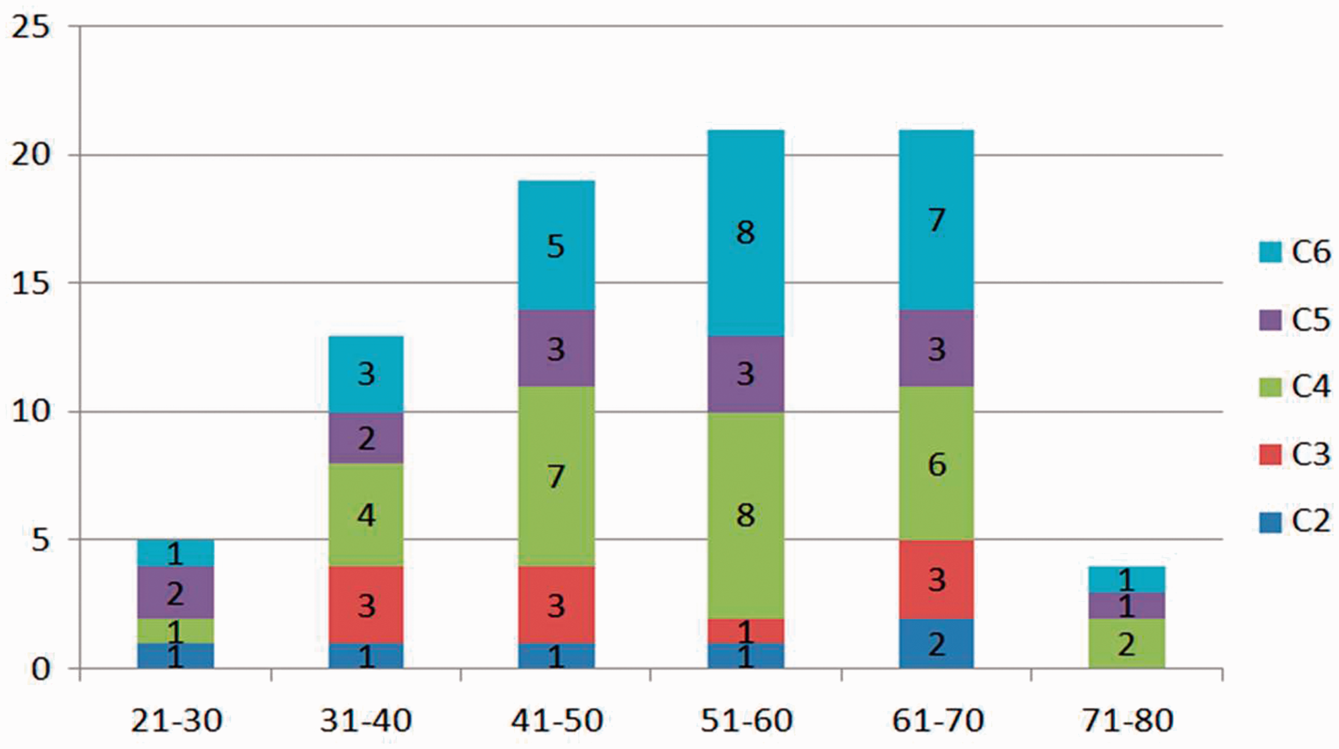
Stacked column bar chart showing age-wise distribution of treated limbs with respect to CEAP clinical classification system. CEAP: clinical-etiology-anatomy-pathophysiology.
Follow-up could be obtained in all 83 instances, three and six months after the procedure.
Ultrasonographic response
All incompetent perforators treated with cyanoacrylate glue were occluded and did not show reflux on follow-up Doppler ultrasound performed three and six months after procedure. It was also noted that thrombotic occlusion and organization of entire perforator till the perforator–deep vein junction occurred even if the cyanoacrylate glue reached only till the perforator–superficial vein junction or midway into the perforator (Figure 4). The dilated subcutaneous collaterals treated with sodium tetradecyl sulphate foam showed complete occlusion and no recanalization in three- and six-month follow-up in all cases. Twelve-month follow-up was available in 14 of the treated cases, which showed persistent occlusion of all treated perforators. De novo incompetent perforator and focal varices were found in one patient who presented with recurrent oedema and was treated the same way using cyanoacrylate glue and sodium tetradecyl sulphate foam uneventfully.
Follow-up Doppler image of a glue (G) embolized perforator showing occlusion of entire course of perforator (arrows) till its junction with the deep vein even when glue is seen only in its superficial segment.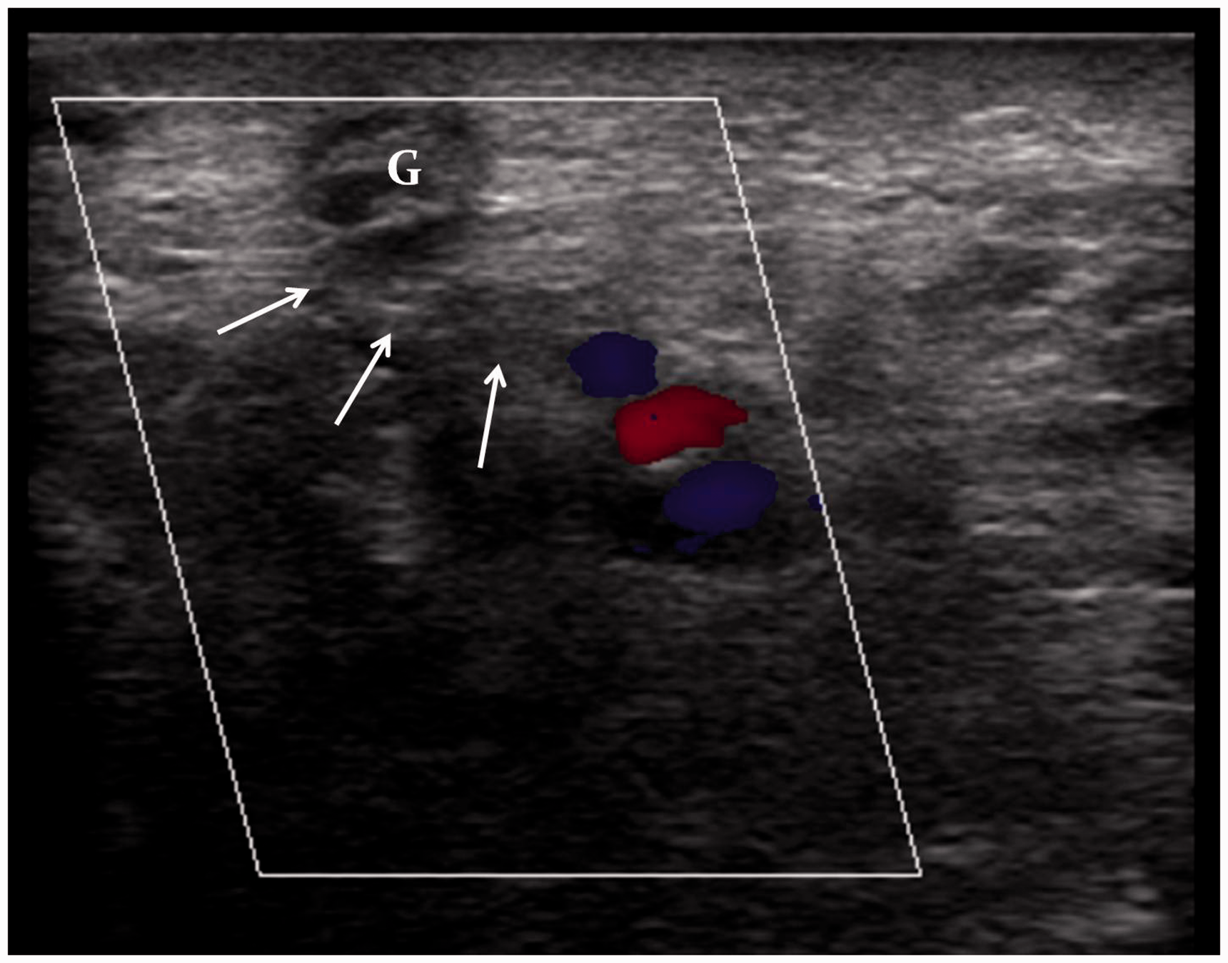
Clinical response
All patients showed clinical improvement. Venous clinical severity score improved from a baseline of 8.18 ± 3.60 to 4.30 ± 2.48 on three-month follow-up and 2.42 ± 1.52 on six-month follow-up (p < 0.0001) (Figure 5). All twenty-five patients with ulcer showed complete healing of all ulcers (100% ulcer healing rate).
Box and whisker plot showing distribution of Venous Clinical Severity Scores at baseline and three and six months after the procedure. Horizontal line within the box indicates the median, top and bottom margins of the box show first and third quartile and the whiskers represent the minimum and maximum score without outliers.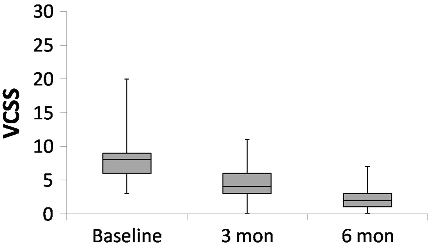
Complications
Deep venous extension of cyanoacrylate glue occurred in four patients in our initial stages. Although this was clinically silent, it is a major complication tantamount to deep venous thrombosis. Significant pain and thrombophlebitis lasting more than three weeks requiring oral analgesics occurred in 32 (38.5%) instances but had subsided by the third month follow-up visit. Skin darkening over the treated veins was noted in many patients but subsided within six months and was not quantitated. No infection or neurological damage occurred in any of the patients. No deep venous thrombosis was seen other than the four cases with deep venous extension of cyanoacrylate.
Discussion
The significance of perforators, their contribution to venous symptoms including ulcer and role of treating them in case of primary varicose veins is disputable. Many studies favour the fact that incompetent perforators contribute to chronic venous disease and need treatment,5,14–18 while some disagree.19,20 The role of incompetent perforators in recurrent or persistent disease and requirement of their treatment are less controversial and there are many supporting studies, some of which have shown incompetent perforators to be the most common cause of recurrence.3–5,21,22 Treatment of saphenous veins has been shown to cause changes in perforator haemodynamics resulting in incompetence and recurrence.10,23,24 Incompetent perforators are best evaluated using Duplex ultrasound and are diagnosed if perforator reflux lasts more than 0.35 s and has diameter of at least 3 mm at fascia level.3,11,12 Though a study successfully explores the use of non-contrast computed tomography venography for varicose vein evaluation, it is not usually or widely used. 25 Multiple treatment options are available for incompetent perforator vein occlusion, many of which have a good clinical success rate and some, like coil embolization and cryoablation, have proved to be ineffective.9,26 This study explores the feasibility, safety and efficiency of an outpatient treatment procedure for recurrent varicose veins due to incompetent perforators using cyanoacrylate glue embolization of perforators and sodium tetradecyl sulphate sclerotherapy of dilated leg collaterals.
Cyanoacrylate glue is a low viscosity liquid which rapidly polymerizes on contact with ionic solutions like blood. 27 They produce an immediate cast of the vessel and cause occlusion. Polymerization of cyanoacrylate glue is an exothermic reaction but heat produced is insufficient to cause endothelial damage. 28 If injected into a vessel and compressed, they cause adhesion of the vessel layers. There are various successful applications of cyanoacrylate glue, including varicose veins, where modified cyanoacrylate with slow polymerization properties has been injected through special delivery devices to occlude varicose veins.29–31 A study by Toonder et al. 10 describes a successful usage of Sapheon cyanoacrylate glue for incompetent perforators; however, the procedure requires a hospital operating room and introduction of large vascular sheaths into perforators which is technically difficult. 10
In the present study, routinely available cyanoacrylate (Endocryl) has been used for embolizing incompetent perforators, exploiting its adhesive and occlusive properties. Injection into veins followed by immediate external compression resulted in partial collapse and occlusion of the venous segment at and around the injection site. Because of low viscosity of cyanoacrylate glue, the actual length to which the cyanoacrylate glue might extend and cause occlusion is difficult to predict, and hence, while embolizing direct perforators, to prevent cyanoacrylate glue extension into the tibial veins, puncture was made distal from the perforator–deep vein junction, lower volume of cyanoacrylate glue used and cyanoacrylate glue extension into even a short segment of perforator was considered satisfactory. Such care was not followed while embolizing an indirect perforator. In spite of posterior tibial vein extension in four cases, no clinical untoward effect occurred. This is probably because of involvement of only one of the posterior tibial veins with preserved patency of the other. Involvement of larger veins from popliteal vein upwards by cyanoacrylate extension could be potentially catastrophic, and hence this procedure should be performed by practitioners experienced with handling cyanoacrylate.
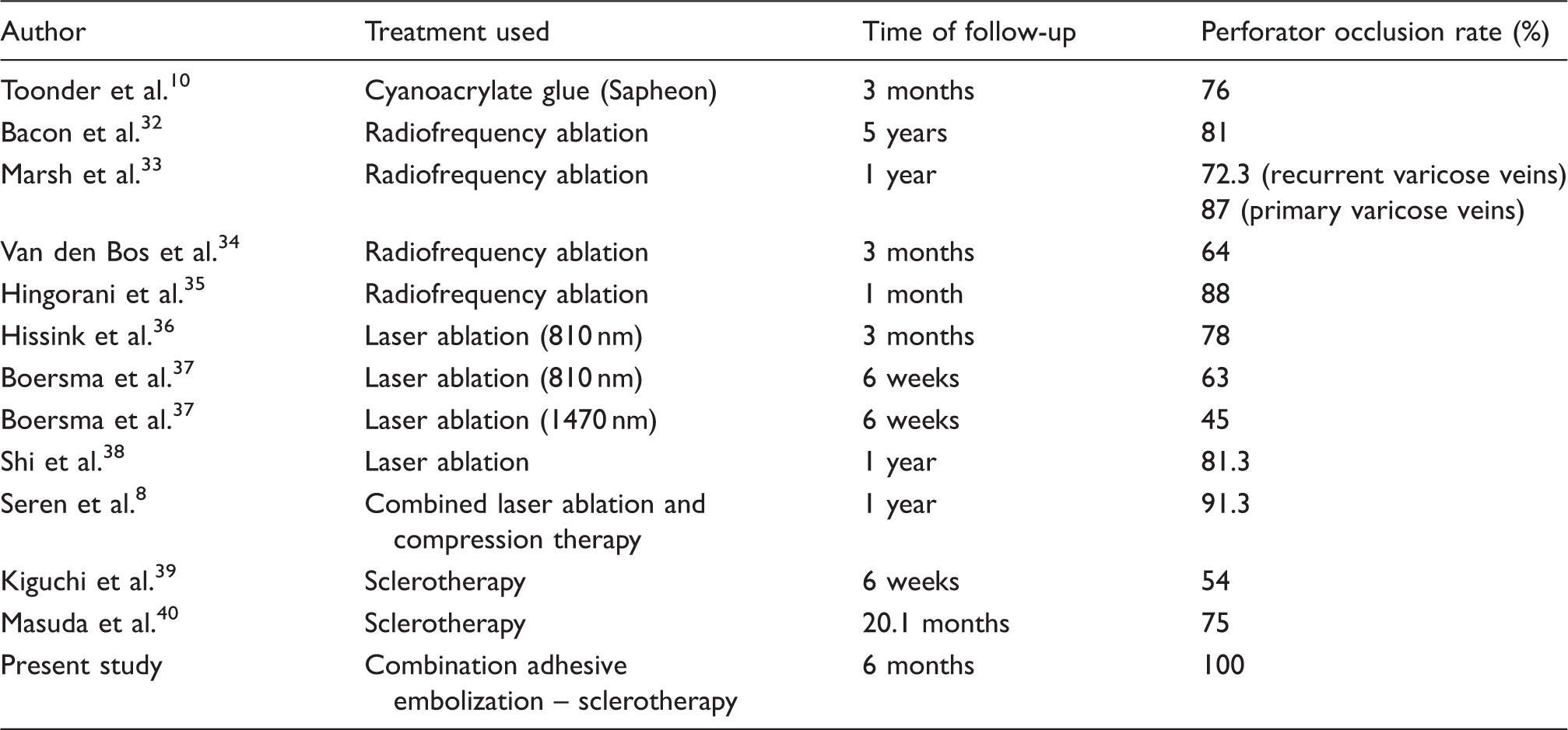
Experience of multiple authors using different treatment methods on incompetent perforators.
The advantages of the described combination treatment for recurrent varicose veins can be summarized as easy affordability; universal availability of drugs and materials required; outpatient nature of procedure not requiring patient admission, immobilization or anaesthesia; high efficiency in occluding incompetent perforators and dilated subcutaneous collateral veins; significant clinical improvement in all treated patients; high ulcer healing rate; less and overall tolerable procedural pain; absence of neurological complications and deep venous thrombosis; no requirement of perivenous tumescence and shorter procedural time. The disadvantages are decreased controllability of extent of spread of injected cyanoacrylate glue causing occasional inadvertent deep vein occlusion (although clinically silent in our study, and seen in the early days of our experience), higher rate of persistent pain and thrombophlebitis after the procedure and requirement of compression stockings after the procedure. Due to the potential of this procedure to cause permanent deep venous occlusion, it should be performed only by practitioners with adequate experience with handling cyanoacrylate glue.
Conclusion
Combined cyanoacrylate glue embolization of perforators and sclerotherapy of dilated subcutaneous collateral veins as treatment of recurrent lower limb venous insufficiency secondary to incompetent perforators is highly efficacious and can be performed as an outpatient procedure with technical ease, but has a guarded safety profile due to its potential to cause permanent deep venous occlusion and hence should be performed by physicians experienced with handling cyanoacrylate.
All procedures performed in studies involving human participants were in accordance with the ethical standards of the institutional and/or national research committee and with the 1964 Helsinki declaration and its later amendments or comparable ethical standards.
Footnotes
Contributorship
KPBP conceived the idea of this procedure and applied in many patients. BJ made the study protocol, helped obtain ethical approval and assisted KPBP in many of the procedures. AT and TS assisted KPBP in many of the cases and helped in making the manuscript especially in collecting references. All authors reviewed and edited the manuscript and approved the final version.
Declaration of Conflicting Interests
The author(s) declared no potential conflicts of interest with respect to the research, authorship, and/or publication of this article.
Funding
The author(s) received no financial support for the research, authorship, and/or publication of this article.
Ethical approval
Hospital ethics committee approval was obtained for this study.
Guarantor
KPBP.