






Abstract
Objective
To investigate the effect of diadynamic currents administered prior to exercises on pain and disability in patients with osteoarthritis of the knee.
Design
A randomized-controlled trial.
Setting
Special Rehabilitation Services in Taboão da Serra.
Participants
Patients with bilateral knee osteoarthritis.
Intervention
Participants were randomly allocated to Group I (diadynamic currents and exercises; n = 30, 60 knees) or Group II (exercises alone; n = 30, 60 knees) and were treated three times a week for 8 weeks.
Main outcome measures
The primary outcome measures were change in knee pain evaluated by visual analog scale and disability Index Score (Lequesne). Secondary outcomes included change in mobility (Timed Up and Go test), range of motion (goniometer), muscle strength (dynamometer), a composite score for pain and disability (Western Ontario and McMaster Universities Osteoarthritis questionnaire), and a drug diary to measure consumption of rescue pain medication (paracetamol). All measurements were collected at baseline, 8 weeks, and 6 months from baseline (follow-up).
Results
There were 60 participants with a mean (SD) age of 63.40 (8.20) years. Between-group differences in the follow-up (8 weeks and 6 months) were observed for pain at rest, pain during activities of daily living and disability. There was improvement in Group I that was maintained for the three variables 6 months after treatment. Mean difference for pain at rest was −3.08 points (95% confidence interval −4.13; −2.02), p < 0.01 with an effect size of 1.4; mean difference for pain during activities of daily living was −2.40 points (95% confidence interval −3.34; −1.45), p < 0.01 with an effect size of 1.24; and mean difference for disability was −4.08 points (95% confidence interval −5.89; −2.26), p < 0.01 with an effect size of 1.04.
Conclusion
Patients with symptomatic knee osteoarthritis receiving 8 weeks of treatment with diadynamic currents as an adjunct to a program of exercises had significantly greater improvements in pain and disability than those receiving exercises alone. Beneficial effects were sustained for 6 months.
Keywords
Introduction
Osteoarthritis represents failed repair of joint damage resulting from stresses initiated by any joint or periarticular tissue abnormality. The rate of progression varies among persons and within a knee over time. The symptoms and signs of knee osteoarthritis include pain, stiffness, reduced joint motion, and muscle weakness. Long-term consequences can include reduced physical activity, deconditioning, impaired sleep, fatigue, depression, and disability. 1 As disease advances, there are alterations in nociceptive processing, such as increased peripheral and central sensitization, that worsen pain, especially during movement, and this contributes to a decline in activities of daily living. 2
Reductions in activity to avoid pain (kinesiophobia) interferes with the ability to undertake rehabilitation strategies such as regular exercises. There is high-quality evidence that education and exercise improve function in individuals with knee osteoarthritis.3,4
A systematic review of randomized controlled clinical trials of therapeutic exercise in patients with knee osteoarthritis found that exercise can significantly reduce pain and improve physical function and quality of life. 5 Furthermore, exercise may improve cardiorespiratory function, increase muscle strength, stabilize posture, and improve psychological health. 6 Thus, exercise has an important role in the treatment of knee osteoarthritis, although adherence to exercise protocols remains a challenge. 7
It has been suggested that an analgesic intervention given before undertaking exercises may improve adherence and treatment outcome.8–12
Transcutaneous electrical nerve stimulation (TENS), interferential currents, and diadynamic currents are electrophysical agents used to alleviate pain associated with musculoskeletal conditions. 13 Transcutaneous electrical nerve stimulation is commonly used under the supervision of health care professionals in clinical settings or self-administered by patients at home. Diadynamic currents are low-frequency monophasic sinusoidal pulsed currents, up to 100 Hz. It has been suggested that diadynamic currents may alleviate pain by stimulating peripheral sensory and motor nerves which reduces excitability and activity of second order nociceptive neurons in the central nervous system (i.e. 'gating'). Other beneficial physiological actions include vasodilatation and hyperemia. Short periods, long periods, fixed diphase, fixed monophase, and syncopal rhythms are different characteristics of diadynamic currents utilized during treatment.14,15
Both dynamogenic and inhibitory actions of diadynamic currents are used in treatment of various ailments. There is a paucity of studies investigating the efficacy of diadynamic currents to alleviate pain. A single-treatment session of diadynamic currents usually does not take longer than 12 mins with a rapid onset of analgesic effect.13,16 Although the analgesic effects of TENS is well known, there is a lack of evidence on the analgesic effect of diadynamic currents on knee osteoarthritis. Therefore, the aim of the present study was to investigate the medium-term effects of diadynamic currents combined with exercise on pain and disability in patients with osteoarthritis of the knee.
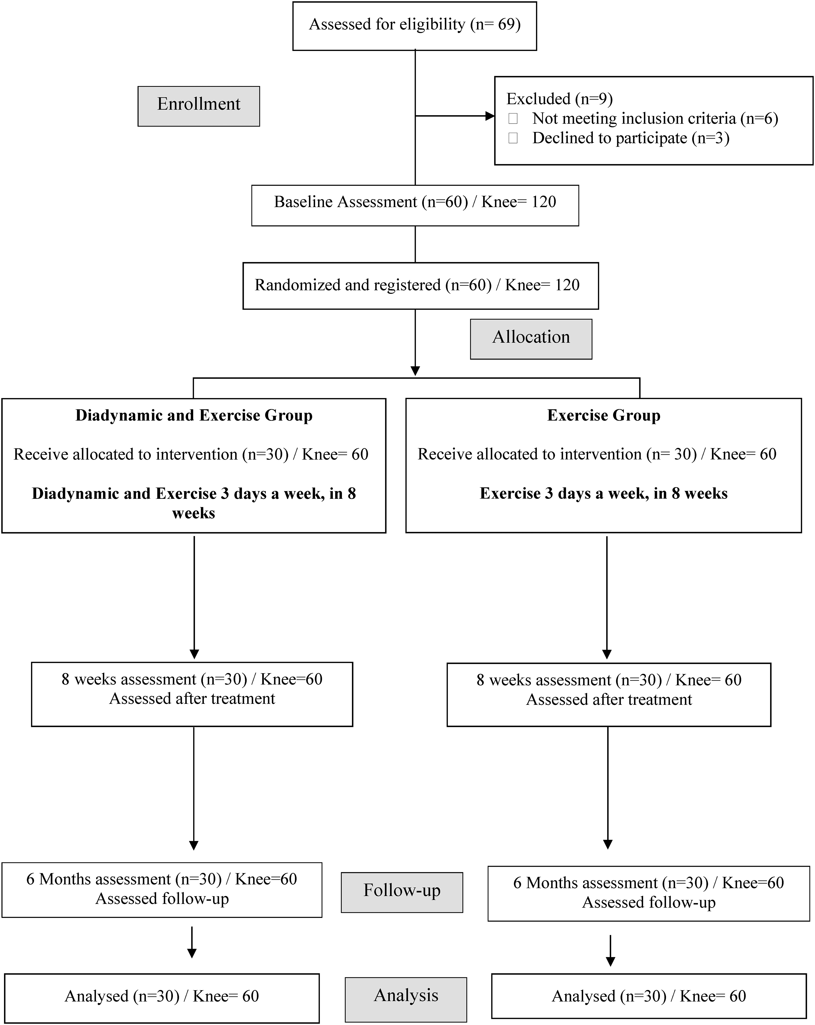
Participant flow diagram.
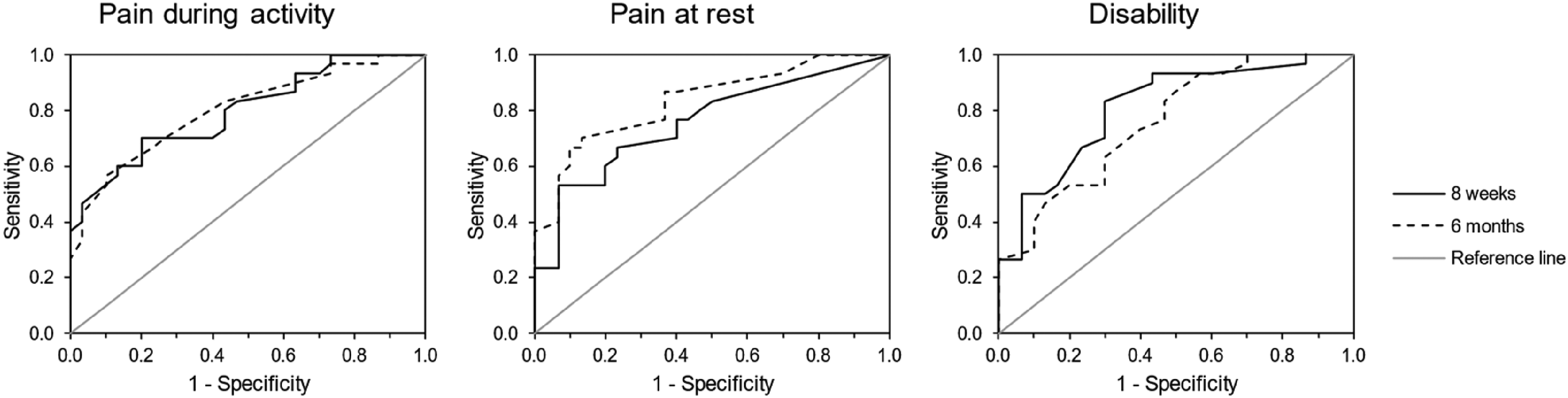
Comparison of ROC curves for pain during activity, pain at rest and disability scales at 8 weeks and 6 months.
Exercise program conducted over the 8 weeks of treatment.

Methods
This randomized-controlled trial was registered in the Brazilian Clinical Trials Register (CT01306435) and approved by the local Ethics Research Committee (protocol n° 23475614.3.0000.5512). The study was conducted according to the CONSORT recommendations for non-pharmacological trials, 17 and data collected from January 2017 to December 2019.
Participants were patient volunteers who attended the Special Rehabilitation Services in Taboão da Serra-SP and had been diagnosed with knee osteoarthritis by an independent rehabilitation specialist. Participants fulfilled the following inclusion criteria: (a) aged between 50 and 75; (b) symptomatic knee osteoarthritis for at least 3 months; (c) visual analog scale score above 3 out of 10 (0 = no pain, 10 = worst pain imaginable); 18 and (d) grades two-ten according to Kellgren–Lawrence. 19 The exclusion criteria were as follows: cancer, diabetes, symptomatic hip osteoarthritis, or used antidepressants, anti-inflammatory medications or anxiolytics during 6 months prior to enrollment.
Patients who had already been evaluated by an orthopedic doctor, had x-ray examinations, received a medical diagnosis of knee osteoarthritis and were awaiting physiotherapeutic care at the Taboão da Serra Rehabilitation Service were referred for the study. All the participants provided written informed consent prior to taking part in the study. Basic demographic information, including medical history and physical examination results, was provided by an investigator not involved in the study. Patients who met our inclusion criteria were randomly divided into Group I (diadynamic currents and exercises; n = 30, 60 knees) and Group II (exercises alone; n = 30, 60 knees). We used a computer-generated random sequence block of four without stratification for the randomization process. The concealment was performed by placing the treatment assignment into sealed envelopes until the initiation of intervention.
Anthropometric and demographic data, the duration of knee pain, the use of pain relief medications, the knee range of motion, and a variety of patient-reported outcomes were collected at baseline (before randomization), 8 weeks from baseline and 6 months from baseline (follow-up) by the same blinded assessor.
The primary outcomes were pain intensity measured by the visual analog scale with a minimal clinically important change set at two points 18 and disability measured using the Lequesne questionnaire, 20 which consists of 11 questions about pain, discomfort, and function. Scores range from 0 to 24 (0 = “no” to 24 = “extremely severe” dysfunction).
Secondary outcomes included medication intake (paracetamol) for relief of knee pain, mobility and balance, range of motion, muscular strength, and activity. Mobility and balance were evaluated by the Timed Up and Go test. 21 The Timed Up and Go test, a measure of functional mobility, quantifies in seconds the time that the individual needs to stand up from a chair, walk 3 m, turn back toward the chair, and sit down again. Range of motion of the knees was measured with a universal goniometer according to the methods described by Marques. 22 Muscular strength was estimated at maximal isometric force for the quadriceps, using a portable dynamometer (Lafayette, USA). Under stabilized conditions, patients, sitting with knees flexed at 60 (measured by a goniometer), 23 were asked to extend the legs as far as they could. Three attempts were conducted, and the mean value was obtained. Muscular strength was estimated at maximal isometric force for the quadriceps, using a portable dynamometer. Under stabilized conditions, patients, sitting with knees flexed at 10, 60, and 90 degrees (measured by a goniometer), 23 were asked to extend the legs as far as they could. The mean value of three attempts was calculated. Physical activity was measured using the Western Ontario and McMaster Universities Osteoarthritis questionnaire, 24 which is self-administered and measures pain, stiff joints, and physical activity. Increased scores suggest decreased activity.
All participants had osteoarthritis of both knees, and therefore both knees were treated with the allocated treatment.
Following initial assessment, participants received diadynamic currents prior to exercises (Group I) or only exercises (Group II) three times a week for 8 weeks.
The treatment with diadynamics was performed with the use of a Stymat S-210 apparatus. Stationary plate electrodes measuring 4 × 9 cm were put in medial and lateral side of the knee. The duration of the treatment amounted to 8 minutes on each side and was a sequence of different diadynamic currents: diphase currents 4 minutes and long period 4 minutes. The intensity depended on the participant's individual reactions and on average amounted to 15 mA. The duration of the treatment was established conforming to the methodology of Bernard's currents. Due to secondary inhibition, the maximum duration was 10 minutes.
The exercise intervention was administered as three, 45 min sessions per week for 8 weeks (Table 1).8,9 Each session consisted of:
Ten minutes warming-up (treadmill, ergometer bike or rowing machine). Thirty minutes two–three sets with Phase-1, Phase-2, and Phase-3. Five minutes stretching (hamstrings, quadriceps, adductors, and gastrocmenius).
Participants were instructed not to use analgesic medications except paracetamol (maximum of 500 mg/day) or non-steroid anti-inflammatory drugs during the study, and not to change their regular physical exercise and activities during the study.
Sample size was calculated to detect a difference between two independent means (groups) assuming that participants would receive diadynamic currents prior to exercises (Group I) or only exercises (Group II) three times a week for 8 weeks, and the primary outcomes would be pain intensity measured by the visual analog scale and disability measured using the Lequesne questionnaire (quantitative variables). We hypothesized an effect size of Cohen's D = 0.8 with α = 0.05 and the power = 0.8 resulting in a sample size of 52 participants (G*Power software, version 3.1.9.7). Considering potential attrition, we set the target sample size at 60 participants.8,9
The Shapiro–Wilk test was used to test normality of continuous variables. The age, weight, height, and body mass index of volunteers between groups were compared using Student's t-test for independent samples. To investigate the effect of the treatment on the pre- and post-evaluation, as well as the interaction of this effect between the groups, the General Linear Models with mixed design (evaluations time×groups) were applied. Tukey's post hoc tests were used to analyze the effects of the interactions. Receiver operating characteristic curve analysis was used to compare and evaluate the accuracy of discrimination thresholds of pain during activity, pain at rest and disability scales at 8 weeks and 6 months. Sensitivity and specificity of each scale was made according to the best Youden's index. Two essential statistics obtained from the ROC curve were the area under curve and minimal clinically important difference (MCID). MCID is the point on the ROC curve that is nearest to the diagram's upper left corner. All analyzes were performed using the statistical program R version 3.1.3 using the R Commander graphical interface or Statistical Package for the Social Sciences® (SPSS IBM Corp., Armonk, US) version 21. All analyses were performed considering an alpha error of 5%.
Results
Patients
The flow of participants through the study is presented in Figure 1. In total, 69 participants were considered for inclusion in the study. Of these 69, nine were excluded because they did not meet the inclusion criteria or declined to participate. The remaining 60 participants were randomly allocated to Group I or Group II. Sixty participants (120 knees) completed the trial per protocol. Thus, the per protocol analysis involved 60 participants (30 participants in each group).
The participant's characteristics are described in Table 2.
Clinical and demographic characteristics of the participants in both groups.

N: number; Kg: kilograms; M: meters; SD: standard deviation; BMI: body mass index; OA: osteoarthritis.
Primary outcomes
Between-group differences in the follow-up (8 weeks and 6 months) were observed for the variable pain at rest, pain during activities of daily living and disability. Improvement in outcomes were observed for Group I compared with Group II for the three variables, with the largest size of effect occuring at the 6 months time point after treatment. Estimated mean differences at the 6 months time point were; pain at rest −3.08 points (95% confidence interval −4.13; −2.02), p < 0.01 with an effect size of 1.40, pain during activities of daily living −2.40 points (95% confidence interval −3.34; −1.45), p < 0.01 with an effect size of 1.24, and disability −4.08 points (95% confidence interval −5.89; −2.26), p < 0.01 with an effect size of 1.04 (Table 3).
Mean (standard deviation), between-group differences (95% confidence interval), p-value to test differences between groups in each evaluation calculated through Tukey–Kramer adjustment test and Cohen's D (effect size).

Pain rest: pain during rest; pain ADL: pain during activities of daily living; values shown are mean and standard deviation (SD), estimated mean differences between groups (95% CI) and effect size. *p < 0.05.
Secondary outcomes
There was a greater reduction in the use of medication (paracetamol) in Group I compared with Group II after 8 weeks, where the number of days of analgesic medication use for relief of knee pain was significantly reduced (p < 0.01).
Table 4 shows that Group I also presented higher values in all subscales of the activity compared with Group II (p < 0.01). Significant statistical differences were also observed in mobility and range of motion at the 8-week follow-up and this was maintained at 6 months (p < 0.01).
Mean (standard deviation), between-group differences (95% confidence interval), p-value to test differences between groups in each evaluation calculated through Tukey–Kramer adjustment test, and Cohen's D (effect size).

RoM: range of motion; Values shown are mean and standard deviation (SD), estimated mean differences between groups (95% CI) and effect size. *p < 0.05.
Receiver operating characteristic curve analyses were performed with the objective of evaluating the impact of treatment with diadynamic currents associated with exercise for the sensitivity and specificity of three different measures (pain during activity, pain at rest and disability); see Figure 2. Sixty individuals participated, 30 in the group treated with diadynamic currents and exercise (Group I) and 30 in the group treated with exercise alone (Group II). After 8 weeks, receiver operating characteristic curve analysis demonstrated significant accuracy for “pain during activity” (area under the curve: 0.79, 95% confidence interval: [0.68;0.90], p < 0.01), “pain at rest” (area under the curve: 0.75, 95% confidence interval: [0.63;0.88], p < 0.01) and “disability” (area under the curve: 0.81, 95% confidence interval: [0.70;0.92], p < 0.01). In other words, if patients were randomly selected, at least 75% of individuals in Group II would have higher scores than individuals in Group I, which is an acceptable level of discrimination according to Hosmer et al. 25 The best cutoff point (minimal clinically important difference, MCID) is the one nearest to the diagram's upper left corner, and as such: the sensitivity and specificity of each scale was 0.70 and 0.80, respectively, for the cutoff point of five on “pain during activity” scale; 0.67 and 0.77, respectively, for the cutoff point of two on “pain at rest” scale; 0.80 and 0.70, respectively, for the cutoff point of 6 on “disability” scale. After 6 months, receiver operating characteristic curve analysis also showed significant accuracy for “pain during activity” (area under the curve: 0.80, 95% confidence interval: [0.69;0.91], p < 0.01), “pain at rest” (area under the curve: 0.83, 95% confidence interval: [0.73;0.93], p < 0.01) and “disability” (area under the curve: 0.75, 95% confidence interval: [0.64;0.87], p < 0.01). The sensitivity and specificity of each scale was 0.70 and 0.73, respectively, for the cutoff point of six on “pain during activity” scale; 0.70 and 0.87, respectively, for the cutoff point of four on “pain at rest” scale; 0.73 and 0.60, respectively, for the cutoff point of seven on “disability” scale.
Discussion
The administration of electric currents across the intact surface of the skin to alleviate pain and improve function and quality of life associated with musculoskeletal conditions is becoming increasingly popular. 26 The efficacy of diadynamic currents combined with an exercise program on pain and disability in patients with knee osteoarthritis was assessed in this study. The addition of diadynamic treatment to an exercise program three times a week for 8 weeks was found to be more effective in improving pain and disability when compared to the group that performed exercises without diadynamic currents during the same period.
Some people report the intensity of polarized diadynamic currents to be painful. Therefore it is possible that diadynamic currents are acting via counter-irritation, with concurrent release of endorphins, to reduce knee pain.16,27 Can et al. 28 found that there were no statistically significant differences between TENS and diadynamic currents in relief of pain, knee function or activity level in patients with patellofemoral syndrome. Can et al. argued that data was suggestive that diadynamic currents may be marginally better than TENS in alleviating pain because of counter-irritation effects; TENS is usually administered at comfortable intensities below pain threshold. The benefit of diadynamic currents found in the present study includes reduction of symptoms and improvement of the execution of the movements.
It has been suggested that strength gains in patients with knee osteoarthritis are not beneficial unless they are accompanied by an increase in function.29,30 In the present study, a greater gain in muscle strength and improvement in physical activity was found in the group that received diadynamic currents prior to exercises, and this gain was maintained to the 6 months follow-up.
Rafjur et al. 31 found that some electrical therapies (e.g. TENS, interferential currents, and high voltage currents) appear to be effective in treating chronic low back pain. Diadynamic currents appear to be less beneficial for degenerative proliferative disease of the spine. However, Ratajczak et al. reported that both diadynamic current and TENS can relieve pain and improve functional abilities in patients with lumbar discopathy on completion of the therapy. 13 Sayilir et al. 14 found that both diadynamic current and TENS relieved pain at the end of 1 month of therapy and argued that this demonstrates that diadynamic current has direct benefits for the relief of chronic pain in the medium-term and indirect benefits such as improving functional, personal, occupational, and social outcomes associated with pain. Our findings are consistent with Sayilir et al.
Strengths of this study include adherence to CONSORT principles in design and reporting, meeting the recruitment target estimated a priori by a sample size calculation, per protocol analysis with no dropouts or withdrawals at the 6-month follow-up time point, and blinded assessment of outcomes.
Administering diadynamic currents prior to exercises also reduced the consumption of analgesic medication, with the potential to lower drug-related adverse effects. 31
A limitation of the study was the absence of a placebo control for the diadynamic current intervention, i.e. the absence of a sham diadynamic current combined with exercise. This was due to the logistical challenges of creating a sham diadynamic current device and constraints resourcing additional personnel to administer the placebo diadynamic current intervention. We acknowledge that this reduces confidence in our ability to attribute outcomes to diadynamic currents per se, and that other aspects of the diadynamic treatment intervention, such as patient–therapist interaction, and expectations associated with receiving any type of treatment, may have contributed to beneficial effects. Nevertheless, our findings support claims that diadynamic current has potential for clinical utility and therefore we recommend follow-up studies to isolate effects associated with the electrical currents of diadynamic treatment with greater precision. A significant limitation of the present study lies in the choice of an effect size of 0.8 as the basis for sample size calculation. We acknowledge that this value is considered high, based on specific considerations within the context of our study and on previous guidelines in the literature, notably those proposed by Cohen 32 for behavioral studies. Cohen's interpretations should not be deemed universal. Effect size, or how large the difference is, can impact statistical power. This means that when there is a larger effect size, there is a greater difference between groups. It is crucial to recognize that the use of a higher effect size can directly influence the required sample size to achieve adequate statistical power. However, we understand that this choice may lead to discussions regarding the generalization of results, considering the magnitude of the effect compared to similar interventions. Importantly, after the analyses, the values obtained for the effect size were greater than 1.0. This observation is critical and may indicate a more substantial effect magnitude than initially anticipated.
In conclusion, patients with symptomatic knee osteoarthritis receiving 8 weeks of treatment with diadynamic currents as an adjunct to an exercise program reported clinically significant improvements compared to those receiving exercise alone. Beneficial effects were seen in pain, disability, and medication intake and were sustained for 6 months.
For people with knee osteoarthritis, adding diadynamic current to an 8-week program of strength exercises improves pain and disability, which is maintained at 6-month follow-up. For people with knee osteoarthritis, adding diadynamic current to an 8-week program of strength exercises reduced the number of days of analgesic medication use (paracetamol). Benefits were observed when diadynamic current was administered for 4 min on each knee before commencement of exercises and using a sequence of different diadynamic currents: diphase current 2 minutes and long period current 2 minutes. The intensity depended on the patient's individual reactions with the average current administered being 15 mA.
Footnotes
Author contributions
All authors have made substantial contributions to all three of the sections below:
Conception and design of the study or acquisition of data, or analysis and interpretation of data. Draughting the article or revising it critically for important intellectual content. Final approval of the version to be submitted.
Specifics:
PPA: conception and design, collection of data, analysis and interpretation of the data, and drafting of the article; MIJ: conception and design, interpretation of the data, and critical revision of the article for intellectual content; JMB: conception and design, interpretation of the data, and critical revision of the article for important intellectual content; GBP and WSJ: analysis and interpretation of the data, and statistical expertise; RAC: conception and design, interpretation of the data, and critical revision of the article for intellectual content. All authors read and approved the final manuscript. The primary author: Patricia Pereira Alfredo takes responsibility for the integrity of the work as a whole, from inception to finished article.
Competing interest statement
The authors certify that the grant sponsor is not involved in study design, collection, analysis, and interpretation of data; in the writing of the manuscript; and in the decision to submit the manuscript for publication.
Declaration of conflicting interests
The authors declare no potential conflicts of interest with respect to the research, authorship, and/or publication of this study. Mark I. Johnson's declares that in the previous 5 years his employer has received income for expert consultancy services that he has undertaken for GSK plc, TENSCare, and LifeCare Ltd. that lie outside of the submitted work. Mark I. Johnson also declares book royalties from Oxford University Press.
Funding
The authors received no financial support for the research, authorship, and/or publication of this article.
