
Abstract
Chimeric antigen receptor T (CAR-T) cells have made brilliant achievements in the treatment of many kinds of malignant tumors, and six kinds of CAR-T products have been approved by the Food and Drug Administration (FDA), bringing new hope for the treatment of diseases. However, the complications associated with CAR-T cell therapy should not be ignored. Neurological complications often jeopardize patients’ lives, including immune effector cell-associated neurotoxicity syndrome, cerebrovascular accidents, movement and neurocognitive treatment-emergent adverse events. The current knowledge of the mechanism and treatment of these complications is still insufficient, which is a direction that needs to be solved in the future.
Keywords
Introduction
Chimeric antigen receptor T (CAR-T) cell therapy is a rapidly developing cellular immunity technology that plays an important role in the treatment of various malignant tumors. It can specifically recognize target antigens in vivo, rapidly proliferate, and kill tumor cells. However, despite its unique advantages in disease treatment, it may cause serious complications such as cytokine release syndrome (CRS) and immune effector cell-associated neurotoxicity syndrome (ICANS), which can be life threatening.1,2 About 40.8% of patients treated with CAR-T cells reported new neurological symptoms, and while these neurological toxicities are generally manageable, severe acute neurotoxicity and delayed hypokinesia can also occur. 3 With the gradual expansion of the applications of CAR-T cells, these neurological complications brought about by them have attracted increasing attention (Figure 1). This article unfolds the complications of CAR-T cell therapy, summarizing the possible neurological complications during CAR-T cell therapy and progress in countermeasures.
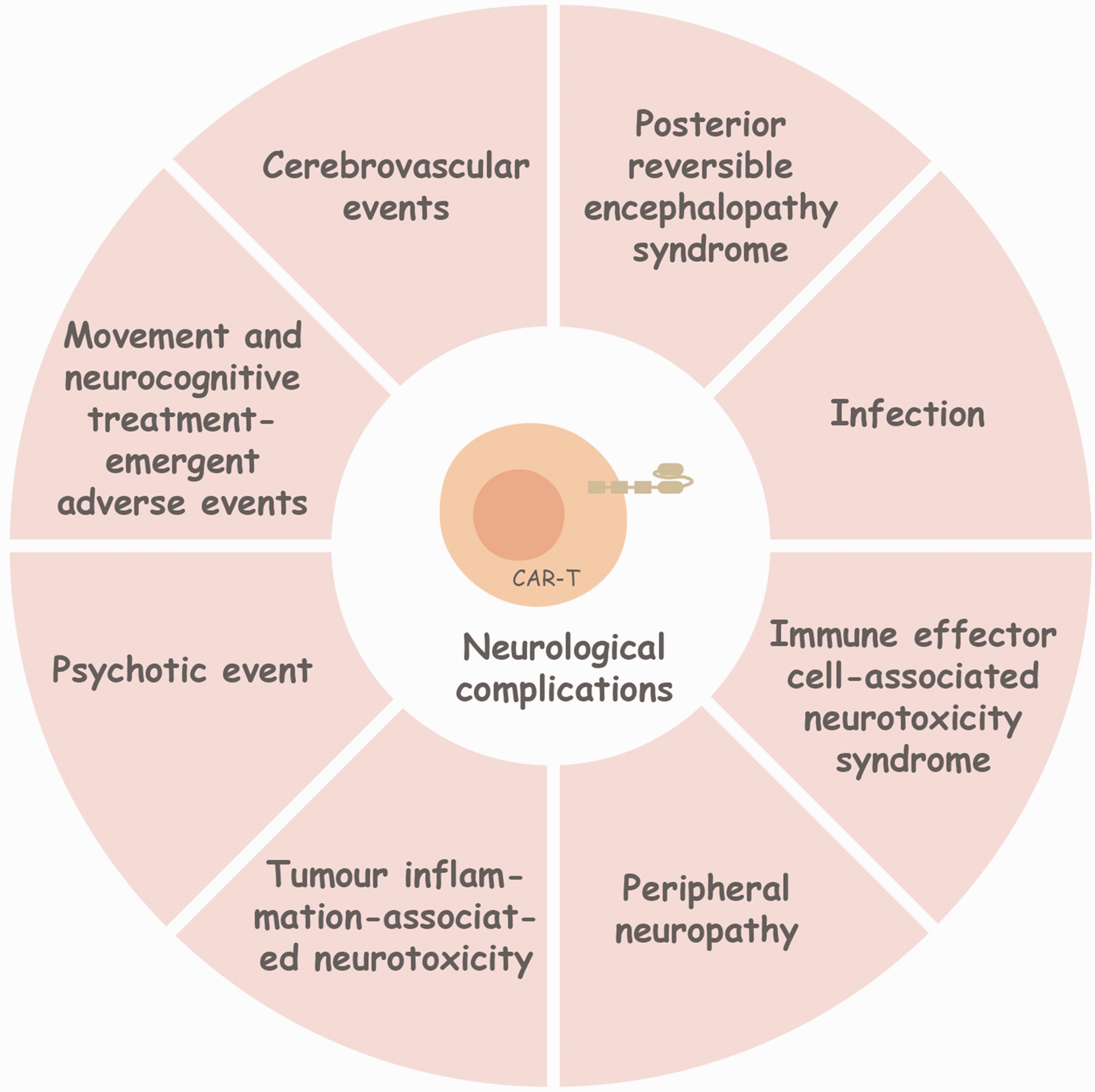
Neurological complications after the infusion of CAR-T cells. CAR – T cell therapy has shown great potential in the field of cancer treatment, but it is also accompanied by a series of neurological complications. These complications pose challenges to the neurological health of patients, suggesting that close monitoring and management are required in clinical applications.
Immune effector cell-associated neurotoxicity syndrome
ICANS is among the most common complications of CAR-T cell therapy, and its incidence is second only to that of CRS. Several CAR-T products currently available in the market cause ICANS to varying degrees (Table 1), and those who develop high-grade ICANS have a poor prognosis; 4 therefore, it is particularly important for preventing and treating ICANS.
Incidence and management of ICANS in marketed CAR-T products.
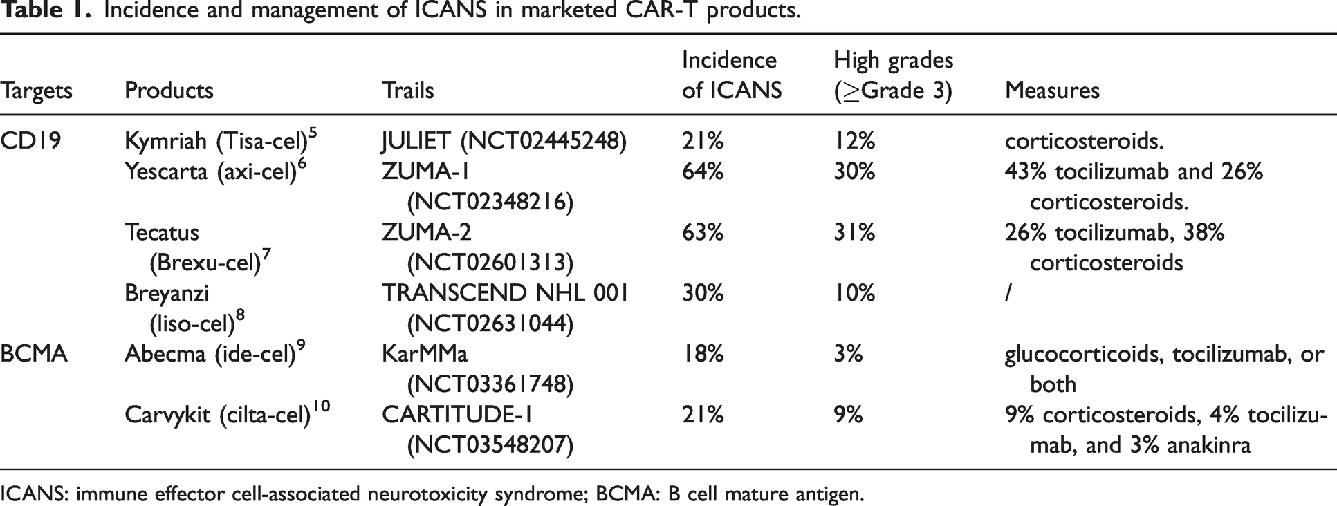
ICANS: immune effector cell-associated neurotoxicity syndrome; BCMA: B cell mature antigen.
Definition and clinical presentation
The American Society for Transplantation and Cellular Therapy defines ICANS as “a disease characterized by a pathological process involving the central nervous system following immunotherapy that results in the activation or engagement of endogenous or infused T cells and/or other immune effector cells”. 11 ICANS often occurs approximately 5 days after CAR-T cell infusion and lasts for approximately 10 days. 12 Signs or symptoms may be progressive, with early manifestations of altered handwriting, tremor, and a mild state of blurred consciousness. 13 Other features include fever, aphasia, disorientation, and myoclonus; in severe cases, seizures, sustained epileptic states, and cerebral edema may occurr.14 –16 Early recognition of these neurological signs helps recognize and manage ICANS. 17
Pathogenesis
However, the pathogenesis of ICANS remains unclear. Studies have shown that ICANS occurs because of multiple causal donor effects (Figure 2). 18
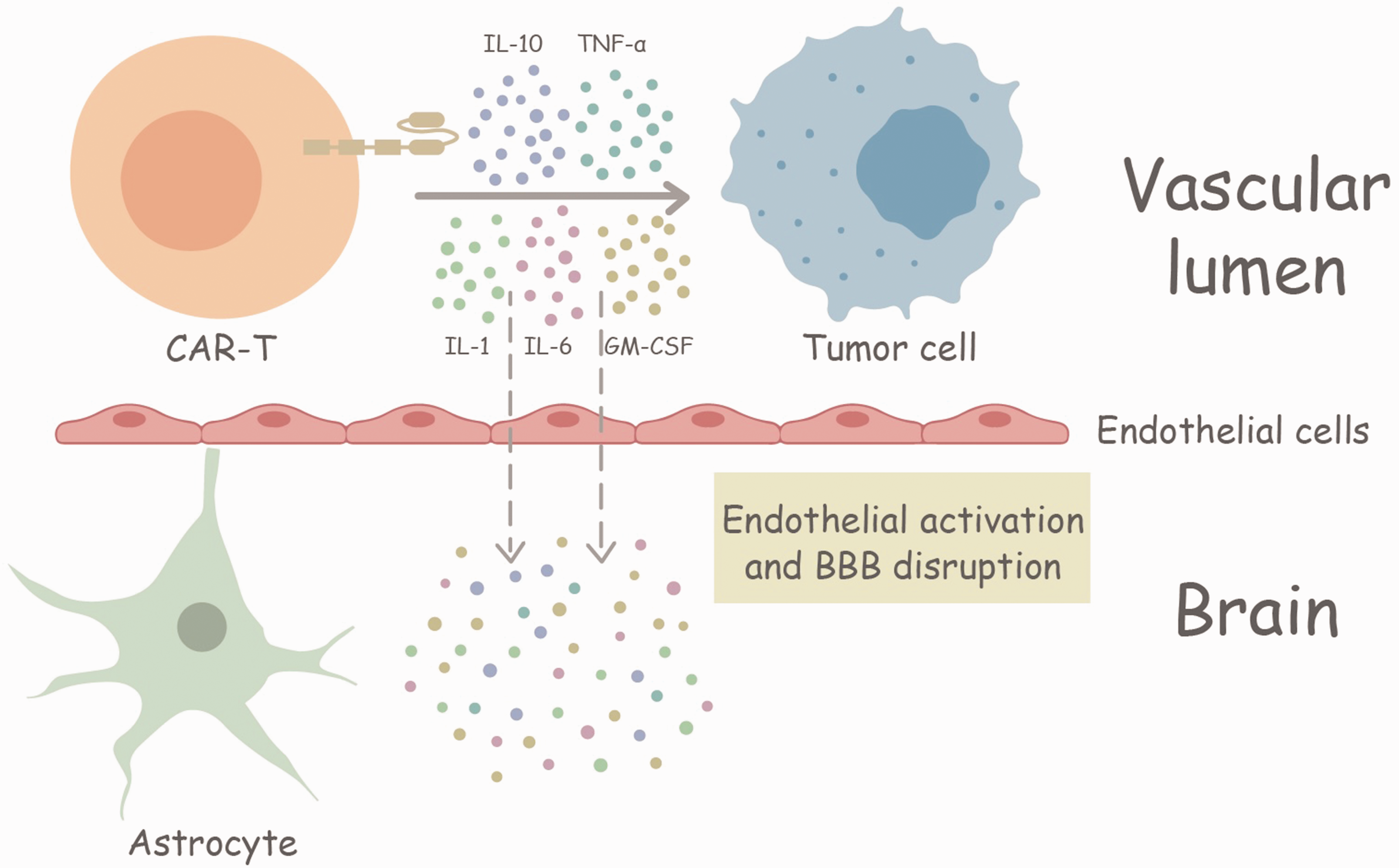
Pathogenesis of ICANS. CAR-T cells: chimeric antigen receptor T cells; ICANS: immune effector cell-associated neurotoxicity syndrome; BBB: blood brain barrier. CAR-T cells secrete cytokines targeting tumor cells. These cytokines also act on vascular endothelial cells, activating them and disrupting the blood – brain barrier, which often causes neurological toxicity.
Cytokine-mediated toxicity
Guo et al. found that serum cytokine concentrations were significantly elevated during acute neurotoxicity. High concentrations of cytokines activate cerebral perivascular cells as they cross the blood–brain barrier (BBB), leading to the secretion of IL-6 and vascular endothelial growth factor, which activates endothelial cells, causing an increase in BBB permeability. 19 The close correlation between ICANS and the severity of CRS suggests that systemic inflammatory response is an important causative factor for ICANS. Further, interleukin 1 (IL-1), IL-2, IL-6, IL-10, interferon γ (IFN-γ), and granulocyte-macrophage colony-stimulating factor (GM-CSF) are all closely associated with neurotoxicity. 20 In a study by Cohen et al., increased peak serum levels of IL-6, IFN-γ, and macrophage inflammatory protein-1α were the most closely associated with ICANS. 21 In a study by Shalabi et al., patients suffering from ICANS had significantly elevated serum IL-6, tumor necrosis factor α (TNF-α), and IL-15 levels. 22 In addition, IL-18 is also associated with the development of ICANS. 23 Increased BBB permeability renders the CNS unimmune to high systemic concentrations of cytokines; thus, ICANS is associated with an early onset of CRS as well as with a rapid elevation of inflammatory cytokines in the serum and CNS.
Endothelial cell activation with BBB damage
Under normal conditions, angiopoietins 1 (ANG1) and 2 maintain normal ratios to prevent endothelial cell activation. In ICANS, ANG2 levels and the ANG2/ANG1 ratio were significantly elevated, indicating endothelial activation and damage to the BBB. 24 Mural cells are integral components of the neurovascular unit. 25 CD19 may also be expressed on the surface of mural cells. 26 Thus, CAR-T targeting CD19 may lead to impairment of the BBB, which in turn may allow for the influx of immune cells (including CAR-T cells) to flow into the CNS; cytokines in the serum (such as IL-6, IL-10, and IFN-γ) activate pericytes and thus endothelial cells, and BBB permeability is increase.19,27 Gust et al. analyzed a patient with grade 4 ICANS who had died of neurotoxicity and found that the biomarkers of endothelial activation were significantly higher in the patient after treatment compared to those before treatment. 19 Thus, endothelial activation and the resulting impairment of the BBB, that is increased permeability, is another possible cause of ICANS.
Others
Vinnakota et al. have suggested that microglia play an important role in the development of ICANS. CAR-T cells targeting CD19 (CAR19T cells) lead to the activation of microglia, which causes cognitive deficits. The transforming growth factor β-activated kinase-1 (TAK1)-NF-κB-p38mitogen activated protein kinases (MAPK) pathway was activated in microglia after CAR19T cell infusion. Although microglial depletion ameliorates neurocognitive deficits, depletion of TAK1 improves neurocognitive function, and TAK1 inhibitors enhance the anti-lymphoma activity of CD19-targeted CAR-T cells. 28 Thus, microglia also play an integral role in the development of ICANS through the TAK1-NF-κB-p38MAPK axis, providing an opportunity for ICANS treatment by inhibiting the TAK1.
Nie et al. reported the case of a patient with MCL treated with bre-cel who developed grade 4 ICANS and ultimately died of cardiac arrest after receiving a CAR-T cell infusion. A subsequent autopsy revealed multifocal demyelinating encephalopathy throughout the brain tissue and confirmed region-specific oligodendrocyte spectrum cell loss as a potential cellular and pathophysiological correlate of severe ICANS. 29
In addition, preexisting frontal lobe dysfunction may also be a causative factor of ICANS. Fontanelli et al. analyzed 16 patients treated with CAR-T cells and found that ICANS occurred in only two patients who exhibited subtle clinical signs of frontal lobe damage at baseline; the neurological manifestations of these two patients, that is, speech and language deficits and corticofrontal clonus, were all due to dominant frontal lobe dysfunction. 30 Thus, baseline frontal lobe dysfunction is associated with the development of ICANS and may predict ICANS development; however, studies should be conducted with a larger sample size.
Influencing factors
Biological characteristics of tumor
Biological characteristics of tumors are important factors affecting ICANS. Less aggressive diseases, such as follicular lymphoma (FL), are less neurotoxic than highly aggressive diseases, such as B-cell acute lymphoblastic leukemia (B-ALL), diffuse large B-cell lymphoma (DLBCL), and mantle cell lymphoma (MCL). 31 Similarly, Jacobson et al. demonstrated that the incidence of grade ≥3 ICANS in patients with FL (15%) was lower than that in patients with marginal zone lymphoma and DLBCL (38% and 32%, respectively). 32 Thus, tumor biology is an important factor that influences ICANS after CAR-T cell therapy.
Structure of CAR-T cells
The structure of CAR T cells is closely associated with the occurrence of ICANS. CAR-T cells targeting CD19 may cause more severe ICANS, because CD19 is expressed in brain mural cells. 26 Different CAR-T cells targeting CD19 may induce different neurotoxicity owing to their different co-stimulatory structural domains. Currently, the CD28 domain and 4-1BB are most frequently chosen as co-stimulatory domains. CAR-T cells with CD28 structural domains (tisa-cel and liso-cel) were more likely to cause ICANS, which occurred earlier and with higher severity than CAR-T cells expressing 4-1BB structural domains (axi-cel and liso-cel), whereas ide-cel, a CAR-T cell with 4-1BB structural domains targeting BCMA caused ICANS with lower incidence and severity.33,34 CAR-T cells incorporating the CD28 co-stimulatory domain generally demonstrate rapid cytotoxicity and potent anti-tumor efficacy. However, they are more susceptible to T-cell exhaustion and tend to have limited persistence. Conversely, CAR-T cells featuring the 4-1BB co-stimulatory domain exhibit enhanced proliferative potential, reduced risk of exhaustion, and superior long-term durability. 33 In preclinical studies, T cells expressing CARs with the CD28 co-stimulatory domain were shown to release higher levels of cytokines compared to those expressing CARs with the 4-1BB co-stimulatory domain. This difference in cytokine release may contribute to the variability in the incidence of ICANS. 35 Therefore, the target of CAR-T cells and the structure of CARs are important factors influencing the occurrence and severity of ICANS. Further exploration is needed to develop CAR-T cell constructs with lower toxicity. 1
Cytokine release syndrome
ICANS is also significantly associated with the presence and severity of CRS. In the study by Santomasso et al., all 33 patients presenting with ICANS had CRS before the onset of neurologic symptoms and there was a strong correlation between severe ICANS and severe CRS. 12 This suggests that ICANS usually occurs after CRS and that alleviation or prevention of severe CRS may reduce the incidence of severe neurological toxicity.
Others
The systemic inflammatory response prior to CAR-T cell infusion may also influence the occurrence of ICANS. DeMatteis et al. analyzed the association between systemic validation prior to lymphatic depletion (LD) and ICANS. The results showed that a lower percentage of CD3+CD8+ lymphocytes and higher levels of C-reactive protein before LD were associated with ICANS, and the percentage of CD8+CD45RA+CD57+ senescent cells was significantly increased in patients with ICANS. 36 This suggests that systemic inflammation prior to CAR-T cell infusion is associated with ICANS, and that ICANS may be a possible cause of CAR-T cell infusion prior to the initiation of the final event. In addition, patients with CNS involvement immediately prior to CAR-T cell infusion were more likely to develop any grade of neurotoxicity, prior drug therapy such as isocyclophosphamide, disease load, and meningeal involvement were all risk factors for the development of ICANS.37 –40
Moreover, age is not a contraindication for CAR-T cell therapy. Johnson et al. divided patients into two groups: the 75+ age group and the 65–74 age group. The incidence of ICANS in the two groups was 38.5% and 48.7% (p = 0.24), whereas that of grade ≥3 ICANS was 16.9% and 21.1% (p = 0.49), respectively, which was not statistically different. 41 Thus, age does not have a significant effect on the occurrence of ICANS, and advanced age is not a contraindication for CAR-T cell therapy.
Diagnostic methods and assessment
Diagnosis
Magnetic resonance imaging (MRI), lumbar puncture, and electroencephalography (EEG) are common adjunct tests to diagnose ICANS. 11 Mauget et al. found that of the 91 patients who developed ICANS, 71 underwent MRI, with normal results in 80% of the patients; the most common abnormal result was a nonspecific high signal; 43 patients underwent lumbar puncture, with abnormal results in 86%; 51 patients underwent EEG, 82% had abnormal results, and 16% had a change in treatment. However, in their study, abnormal lumbar puncture results led to the initiation of antimicrobial drugs in three patients with undiagnosed infections and unnecessary hepatotoxicity in one patient. 42 Therefore, Mauget et al. concluded that EEG is the most therapeutically oriented diagnostic method among the three ancillary tests. Huby et al. found that frontal intermittent rhythmic δ activity is a CAR-T cell infusion and a useful diagnostic tool after ICANS. Their study included 38 patients who received CAR-T cell infusions, 17 of whom developed ICANS. EEG of all patients on days 6–8 revealed that frontal intermittent rhythmic δ activity was only observed in patients with ICANS, with a sensitivity of 88% (15/17) and a negative predictive value of 100%. Further, frontal intermittent rhythmic δ activity disappeared after ICANS subsided, usually after steroid treatment. 43 Thus, frontal intermittent rhythmic δ activity is a reliable tool for diagnosing ICANS; however, further expansion of the sample size is needed to confirm this finding. In addition, continuous EEG monitoring is crucial in the pathogenesis of ICANS in the presence of nonconvulsive seizures; in patients presenting with ICANS treated with Axi-Cel, nonconvulsive seizures were present in 75% of patients with seizures, which supports the use of continuous EEG in patients with ICANS. 44
[18F]fluorodeoxyglucose (FDG) positron emission tomography (PET) may also aid in the diagnosis of ICANS. Morbelli et al. found that ICANS was characterized by frontal hypometabolic features after whole-body FDG-PET in 21 patients with refractory DLBCL. 45 Therefore, FDG-PET may also aid in the diagnosis of ICANS, and further exploration is needed to validate its role in clinical and pathophysiologic evaluation.
Neurofilament light chain (NfL) is a marker of neural axonal injury. Schoeberl et al. found that serum NfL levels were significantly elevated after CAR-T cell infusion and correlated with ICANS severity. 46 Therefore, serum NfL levels can be used to assess ICANS severity and improve CAR-T cells as a predictive biomarker for patient monitoring after cell infusion. However, this preliminary result needs to be validated in a larger prospective patient cohort and a related clinical trial (NCT04614987) is currently ongoing.
Assessment of ICANS
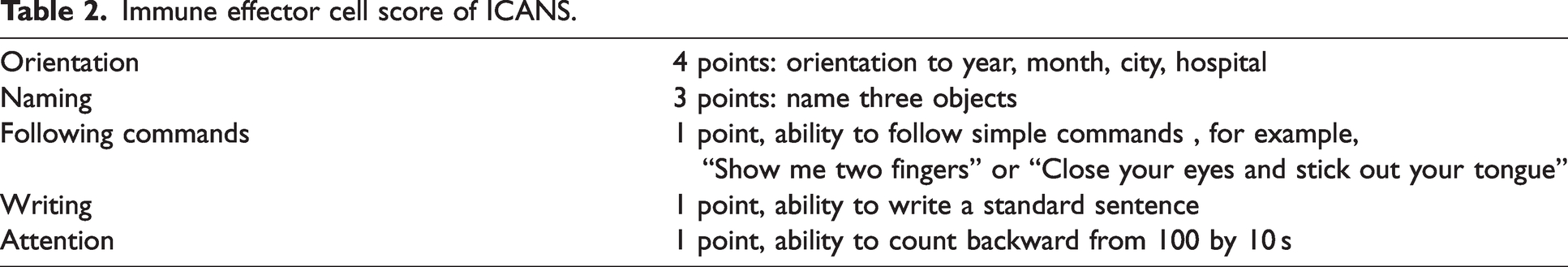
The immune effector cell score is one of the most commonly used scales for assessing ICANS (Table 2), and its inclusion of the five domains of naming, orientation, writing, attention, and following commands in receptive aphasia is an important tool to aid in the grading of ICANS. 34
Immune effector cell score of ICANS.
The endothelial activation and stress index (EASIX) is widely used to assess the degree of endothelial inflammatory injury. A correlation between endothelial injury and ICANS has been demonstrated. 47 Therefore, EASIX may be a predictive method for the occurrence of severe ICANS after CAR-T. Zhao et al. obtained modified EASIX scores in patients with AML treated with CAR-T cells on the day before LD, the day before CAR-T cell infusion, and the day after CAR-T cell infusion: C-reactive protein (mg/L) × lactate dehydrogenase (U/L)/platelets (109/L). 48 The results showed that there was a significant difference in the EASIX scores of patients who developed mild (grades 1-2) and severe ICANS (grades ≥3) at the three time points, indicating that the m-EAISX score is a reliable prognostic indicator for CAR T-cell therapy after severe ICANS. In addition, several methods are available for predicting ICANS. The HMM+PLR model developed by Amidi et al. predicts the risk of post-infusion ICANS and the time course after the onset of ICANS; 49 the visual EEG-ICANS (VE-ICANS) score, a physiologically grounded rating scale developed by Jones et al., is strongly correlated with the severity of ICANS and is a promising biomarker for ICANS in practical applications, enabling a more accurate assessment of ICANS severity. 50 There are also several ongoing clinical trials assessing ICANS, including those using MRI to assess ICANS (NCT05510596) and those assessing neurotoxicity through pupil visualization (NCT06144151), which provide fresh ideas for ICANS diagnosis.
Treatment
For grade 1 ICANS, mainly supportive symptomatic treatment is used; for grade ≥2 ICANS, cortisol hormone is used; and for grade ≥3, patients need to be treated in the ICU with close testing.51,52 The early initiation of hormone and tolizumab therapies prevents excessive CAR-T cell therapy toxicity. 53 Therefore, early recognition and initiation of therapy are extremely important for ICANS (Table 3).
Classification and treatment measures of ICANS.
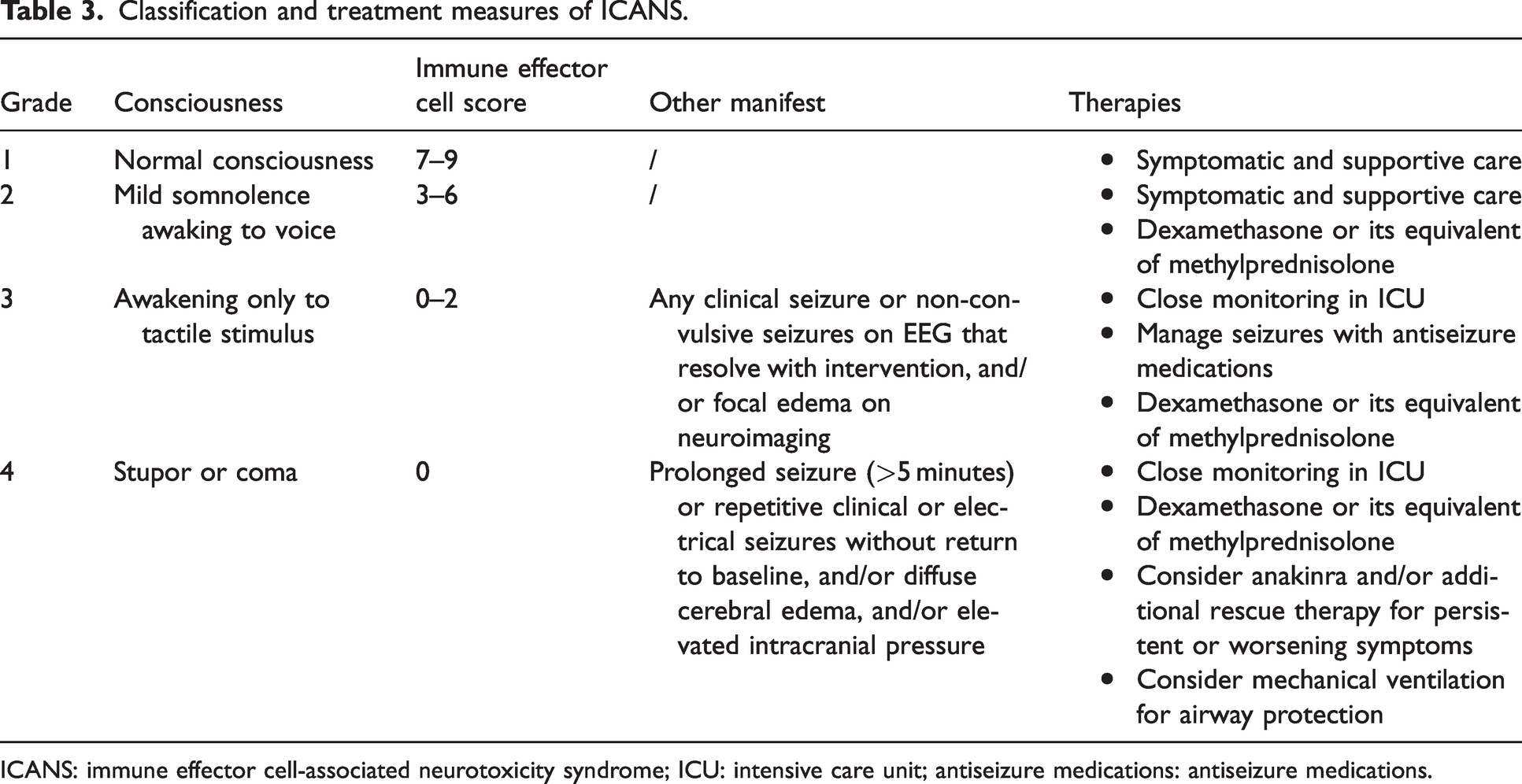
ICANS: immune effector cell-associated neurotoxicity syndrome; ICU: intensive care unit; antiseizure medications: antiseizure medications.
Symptomatic treatment
The aim of a clinical trial by Pensato et al. was to evaluate the timing of prophylactic use of antiseizure medications. They divided the patients treated with CAR-T cells into two groups: one with antiseizure medications immediately before CAR-T cell infusion (PP group) and another with antiseizure medications preemptively at the onset of neurological symptoms (PET group). The PP group included two patients who experienced transient seizures with severe neurotoxicity associated with progressive severe encephalopathy. The PET group included three patients with epileptic events. All patients tolerated antiseizure medications well, and there was no need for long-term antiseizure medications use. 54 Antiseizure medications are safe and effective as a prophylactic measure for epilepsy in CAR-T cell therapy. However, the timing of its use is currently not preferable, and further randomized controlled trials are needed for validation.
Patients who develop fatal cerebral edema are critically ill and require urgent management. In a report by Pensato et al., a 35-year-old female patient who developed diffuse cerebral edema after receiving CAR-T cell infusion died after treatment with hormones, mannitol, and hypertonic saline. 55 Currently, common measures for patients who develop cerebral edema after CAR-T cell infusion include hyperventilation, hormones, and treatment with hypertonic saline and require multidisciplinary management, including neurosurgery, to manage it. 56
Hormones
Corticosteroids are the most commonly used drugs to control grade ≥2 ICANS. They suppress the neuroinflammatory response, and a short course of use reduces the duration of symptoms without affecting the efficacy of CAR-T cells.57,58 Therefore, it is the drug of choice for ICANS treatment.
Intrathecal injections may also be an effective approach for patients for whom systemic hormone use is ineffective. In a study by Shalabi et al., six patients were treated with systemic steroids after developing severe ICANS. Five of these patients received intrathecal hydrocortisone because of rapid progression of neurotoxic symptoms. Severe ICANS symptoms in all five patients subsided rapidly and significantly within 24 h of administration. 59 Overall, intrathecal hormone injection can be effective in controlling severe ICANS symptoms when the systemic use of hormones is ineffective. However, the study included a small sample size, and further prospective studies are needed to determine the effect of intrathecal hormone injections on the efficacy of CAR-T cells and their therapeutic effect on severe ICANS.
Wang et al. categorized patients treated with CAR-T cells into a GC (glucocorticoids)-used group (GC group) and a non-GC-used group (NGC group) and compared the duration of remission, progression-free survival (PFS), and overall survival (OS) to determine the effect of cortisol on CAR-T cell efficacy. The results showed that the overall remission rate (ORR) and complete remission rate (CRR) were not significantly different between the two groups, and there were no differences in PFS, OS, and duration of remission. 60 Therefore, Wang et al. concluded that long-term and high-dose use of cortisol hormones had no effect on PFS, OS, and CAR-T cell proliferation.
However, high cumulative doses of corticosteroids may inhibit the function and persistence of CAR-T cells, resulting in poor overall and progression-free survival. 58 In view of this, Katsin et al. used intrathecal chemotherapy (methotrexate in combination with cytarabine and dexamethasone) for the rapid control of ICANS, which resulted in the discontinuation of intravenous glucocorticosteroids and ensured the CAR-T cells. 61 Asawa et al. also reported two cases of hormone-refractory ICANS treated with intrathecal chemotherapy. Both patients developed cortisol hormone-refractory ICANS after CAR-T cell infusion, and the combination of intrathecal chemotherapy with methotrexate and hydrocortisone led to a rapid improvement in their neurological functio. 62 Thus, intrathecal chemotherapy may be an effective method for rapidly controlling ICANS; however, a larger sample size is needed to confirm its safety and efficacy. A clinical trial of intrathecal dexamethasone with simvastatin is also underway (NCT04514029), and no trial data have been published; therefore, we look forward to the latest results.
Anakinra
Anakinra is an IL-1 receptor antagonist that is often used in patients with hormone-refractory ICANS. In preclinical models, anakinra attenuated immunotoxicity induced by CAR-T cell therapy targeting CD19 without affecting the efficacy of CAR-T cell treatment efficacy. 63
Wehrli et al. retrospectively analyzed 14 patients with ICANS treated with anakinra in a single-center study. Anakinra was associated with improvements in clinical and laboratory markers of inflammation and the modulation of cytokine levels, but the effects on factors related to CRS were more pronounced than those on neurotoxicity, and the use of anakinra did not result in a rapid hormonal taper. 64 Gazeau et al. evaluated the safety of anakinra for refractory CRS and ICANS. The indications for anakinra in their study were grade ≥2 ICANS with worsening or no improvement in symptoms after high-dose corticosteroid therapy (n = 40) and grade ≥2 CRS with worsening of symptoms after treatment with tolizumab (n = 3). Thirty patients (70%) were initiated on anakinra at the time of peak ICANS severity and were receiving concomitant cortisol hormone therapy, seven patients (16%) started anakinra therapy after peak severity, and six patients (14%) experienced peak toxicity following therapy. Higher anakinra doses and shorter times from CAR-T cell infusion and CRS/ICANS onset to anakinra initiation were associated with lower treatment-related mortality, whereas higher anakinra doses were not associated with lower treatment-related mortality. 65 Anakinra may be an effective adjunct to cortisol hormones in the treatment of CAR-T cell-induced CRS and/or steroid-refractory ICANS. In short, anakinra in combination with cortisol is a reliable regimen for the effective control of ICANS, with a limited impact on CAR-T cell efficacy.
In addition, Park et al. conducted a phase II trial (NCT04148430) to evaluate the efficacy of the prophylactic use of anakinra in the prevention of severe ICANS caused by CD19-targeted CAR-T cells. The results of this study showed that anakinra is rapidly distributed in the serum and cerebrospinal fluid, thereby inhibiting IL-1-related cytokines (including those in the cerebrospinal fluid), and that prophylactic subcutaneous administration of anakinra reduces the incidence of high-grade ICANS in patients who receive CD19-targeted CAR-T cell therapy and does not affect the efficacy of CAR-T cells. 66 The results of the study showed that anakinra is effective in preventing severe ICANS in patients who receive CD19-targeted CAR-T cell therapy but not in preventing severe ICANS in patients who have received CD19-targeted CAR-T cells. Therefore, the prophylactic use of anakinra is an important tool for preventing high-grade ICANS during CAR-T cell therapy.
Regarding the use of anakinra in the prophylaxis and treatment of ICANS, there are several other clinical trials underway, including NCT04205838, NCT04359784, NCT04148430, NCT04359784, and NCT04148430 exploring the preventive role and NCT04359784 exploring the therapeutic role, and we look forward to the publication of additional trial data.
Lenzilumab
GM-CSF is another key cytokine involved in ICANS development. lenzilumab, a monoclonal antibody to GM-CSF, is among the possible drugs for ICANS treatment. In vitro, it promoted the proliferation of CAR-T cells without loss of function; it also enhanced the antitumor activity of CAR-T cells in a xenograft model and ameliorated CRS and ICANS induced by CAR-T cell therapy. 67 Based on the unique advantages of lenzilumab demonstrated in vivo and ex vivo, a clinical trial of lenzilumab and CAR-T cell combination therapy (NCT04314843) is underway, bringing new hope for the prevention and treatment of ICANS.
Others
Certain treatments during CAR-T cell manufacturing are also feasible in preventing ICANS, including the selection of suitable targets and the construction of a reasonable CAR structure as described above. Gong et al. proposed another option: they generated a polymer by in situ concatenation of polyethylene glycol (PEG) with the surface of CAR-T cells (termed PEGylation) to produce a polymeric spacer, which blocks the cell-to-cell interactions between CAR-T cells and tumor cells and monocytes, thereby reducing cytokine secretion and attenuating CRS and ICANS without affecting the efficacy of the CAR-T cells. 68 Therefore, in situ PEGylation of CAR-T cells is a possible method for attenuating ICANS and clinical translational results are expected. method, and the results of its clinical translation are expected. In addition, clinical trials have been conducted on editing CAR-T cells to reduce ICANS (NCT03696784).
The activation of the TGFβ-activated kinase-1 (TAK1)–NF-κB–p38 MAPK pathway in microglia has been implicated in the pathogenesis of ICANS. Studies have shown that TAK1 inhibitors, such as takinib, and CSF1R inhibitors can reduce the severity of ICANS in animal models. These findings suggest that TAK1 inhibitors and CSF1R inhibitors could be potential therapeutic options for ICANS and warrant further exploration. 28
Siltuximab, an IL-6 antagonist, is currently being used for the treatment of CRS in patients refractory to tocilizumab. Retrospective studies have indicated that patients with ICANS treated with siltuximab achieved a response rate as high as 60%. 69 However, the use of corticosteroids was not standardized in this cohort, which may have confounded the assessment of siltuximab’s impact on ICANS symptoms. Further dose-optimization studies and prospective clinical trials are needed to evaluate the feasibility of siltuximab for treating ICANS.
Tolizumab is not effective in treating and preventing ICANS because of its large size, which does not cross the blood–brain barrier and may promote the transfer of IL-6 from the bloodstream to the CNS and is mostly used in the treatment of CRS. 57 Dasatinib, a tyrosine kinase inhibitor, is used in the treatment of chronic lymphocytic leukemia and is currently being conducted for its combination with CAR-T cell therapy for the prevention of CRS with ICANS (NCT04603872). Siltuximab, which is also an IL-6 antagonist, is currently undergoing a clinical trial for its treatment of CRS and ICANS after CAR-T cell infusion (NCT04975555). Interferon-Beta-1a (FP-1201) prevents the targeting of CD19 CAR-T cell-induced toxicity in a clinical trial (NCT05936229) is also underway. These clinical trials open endless possibilities for the treatment of ICANS, and we look forward to the publication of their results.
The aforementioned treatments for ICANS are primarily applicable to adult patients. In pediatric patients, the incidence of ICANS is relatively lower and the symptoms are generally milder. Treatment mainly focuses on supportive care and the use of corticosteroids (such as methylprednisolone) to control moderate to severe symptoms (such as aphasia, cognitive impairment, or seizures). During the treatment process, it is essential to closely monitor changes in the patient’s symptoms and adjust the treatment plan according to individual circumstances to ensure safety and efficacy. 70
Cerebrovascular events
Cerebrovascular events are rare but life-threatening complications of CAR-T cell therapy; however, the pathogenesis remains unclear, and most existing studies are case reports.
Wang et al. reported a patient with relapsed refractory MM who was treated with CD19 - and BCMA-targeted CAR-T cells. The patient responded to CAR-T cell therapy but developed neurological symptoms following CRS. Head computed tomography (CT) and MRI suggested multiple cerebral infarctions and bilateral anterior cerebral artery infarction. 71 In a retrospective analysis by Lemonie et al., of 957 patients treated with axi-cel or tias-cel, 3 died from stroke and 3 died from intracerebral haemorrhage. 72 Kersten et al. reported a patient who developed neurological symptoms after CAR-T cell infusion. The patient was suspected to have ICANS; however, imaging suggested a venous sinus embolism, and the symptoms disappeared after treatment with low-molecular-weight heparin. However, a few days later, the patient developed ICANS symptoms that disappeared after treatment with ASMs and corticosteroids. 73
When studying the role of MRI in the diagnosis of ICANS, Mauget et al. found that ischemic stroke was accidentally found on MRI in 4 of 33 patients with severe ICANS, but all of them were limited and did not cause corresponding clinical symptoms. 74 In a study by Rubin et al., out of 100 patients who received CAR-T cell infusion, 2 had ischemic stroke and 2 had subarachnoid hemorrhage. 75 Cordeiro et al. reported three cases of cerebrovascular accidents and one case of transient ischemic attack when studying the late events caused by CD19-targeting CAR T cells. 76 Lefebvre et al. also reported two patients who developed cerebrovascular disease after CAR T cell infusion. 77 Gust et al. also found one rare case of an intracranial hemorrhage in their study. 19 In the simultaneous treatment of MM with CAR-T cells targeting CD9 and BCMA, one patient died of cerebral hemorrhage; however, the researchers speculated that the disease was due to thrombocytopenia. 78
Cerebrovascular events, as a serious condition, occur occasionally in patients receiving CAR-T cell therapy, albeit with a low incidence rate. This may be associated with thrombocytopenia and coagulation dysfunction, but further comparisons with larger sample sizes are still needed to explore the underlying mechanisms of occurrence. Since ICANS is the second most common complication of CAR-T cell therapy, the differential diagnosis of cerebrovascular events (including hemorrhage and infarction) after CAR-T cell therapy is particularly important, as it is easily confused with ICANS. When neurological symptoms occur after CAR-T cell infusion, relevant auxiliary examinations should be performed to rule out cerebrovascular events. 79
Movement and neurocognitive treatment-emergent adverse events
Movement and neurocognitive treatment-emergent adverse events (MNT) are another neurological complication of CAR-T cell therapy. The symptoms of MNT overlap with those of ICANS but occur after ICANS and are characterized by progressive aggravation, long duration, and ineffective cortisol-hormone therapy. Cortisol hormones, chemotherapy (cyclophosphamide), and intrathecal injections (methotrexate combined with cytarabine) are typically used; however, their efficacy is limited. Management of MNT includes enhanced bridging therapy to reduce the baseline tumor load, early treatment with CRS and ICANS, and extended neurotoxicity monitoring/reporting beyond 100 days after infusion. 16 Preventing the occurrence of MNT is the most important method. At present, our understanding of the pathogenesis and treatment of MNT is not perfect, and further studies with larger sample sizes are require.
Long-term toxicities following CAR-T cell therapy may also manifest as neurocognitive and motor dysfunction. VanOekelen et al. reported a case of progressive dyskinesia with features of Parkinson’s syndrome approximately 3 months after cilta-cel infusion, and benzodiazepine and levodopa did not respond to treatment. They found that BCMA was expressed on the surface of both the basal ganglia and astrocytes in this patient and that the patient’s symptoms may be related to the persistence of CAR-T cells in the blood and cerebrospinal fluid, as well as the infiltration of basal ganglia lymphocytes. 80 During the CARTITUDE’s 2-year follow-up, six patients with signs and symptoms of Parkinson’s syndrome were also identified; one patient had late onset (914 days after cilta-cel infusion), and one patient eventually died of Parkinson’s syndrome. 81 In the study by Tost et al., CAR-T cells were used to treat central nervous system lymphoma. Nine patients experienced neurotoxicity, mainly manifested as cognitive impairment, balance disorders, and motor deficits. Among them, three patients had neurological damage lasting more than three months, and one patient eventually died from neurotoxicity. 82 The mechanisms underlying these late-onset neurotoxicities are still unclear. BCMA is the most commonly used target of CAR-T cells for MM treatment; however, the expression of BCMA in the neural tissues of some patients may affect its applicability in MM. Therefore, it is necessary to identify other therapeutic targets for treating MM. The target genes under study are GPRC5D, FcRH5, among others. 83 We are looking forward to the publication of additional data.
Infection
Infection is another common complication of CAR-T cell therapy; however, few studies have investigated the association between CNS infections and CAR-T cell therapy.
Progressive multifocal leukoencephalopathy (PML) is a demyelinating infection of the central nervous system caused by the reactivation of the JC virus (JCV) and is a life-threatening CNS infection. Sdrimas et al. reported the first case of a patient with relapsed DLBCL who developed PML 7 months after receiving targeted CAR-T cell therapy. The patient’s condition stabilized after combination therapy with mefloquine, mirtazapine, and intravenous immunoglobulin. 84 Cytomegalovirus (CMV) encephalopathy is also a serious infection that endangers a patient’s life. Cousin et al reported a patient who developed cytomegalovirus encephalopathy after CAR-T infusion. The patient had positive CMV serology before the infusion but had no antiviral therapy. On day 39 after infusion, the patient had developed aphasia but rapidly improved spontaneously. A few days later, he developed equilibrium disorders and confusion, and the cerebrospinal fluid test showed lymphocytic aseptic meningitis. Seven months after CAR-T infusion, the patient developed progressive memory disorders, MRI suggestive of viral encephalitis, CMV found in CSF, eventually confirmed CMV encephalopathy and died at 8.5 months. 85
A prospective study of human herpes virus 6 B (HHV6B) testing for up to 12 weeks after a CAR-T infusion was conducted by Kampouri et al. Of the 88 patients investigated, 6 experienced HHV 6 B reactivation. In the retrospective analysis of 626 patients, only one patient may have HHV6 B encephalitis, the incidence was 0.17%.86,87 Li et al. reported three patients with encephalitis/myelitis caused by the acute reactivation of HHV6B after CAR-T cell infusion. Neuropsychiatric symptoms of varying degrees were observed in three patients after infusion. Metagenomic next-generation sequencing showed high HHV6B loads in the cerebrospinal fluid of all three patients. Only one patient recovered after intravenous shock therapy with highly active antiviral drugs and immunoglobulins, whereas the other two died of HHV6B encephalitis. 88
Kersten et al. reported a case of cerebral toxoplasmosis following CAR-T cell therapy. The patient developed an abnormal mental status after CAR-T cell infusion, and brain MRI indicated toxoplasmosis; however, toxoplasmosis serological tests were negative. Following treatment with pyrimethamine, folate, and sulfadiazine, the patient’s neurological symptoms improved and resolved within a few days. 73
Various factors may lead to nervous system infections after CAR-T cell infusion, such as immune dysfunction caused by the primary disease itself, lymphocyte depletion chemotherapy before CAR-T cell infusion, and the use of immunosuppressants in the course of treatment complications after CAR-T cell infusion.89,90 In addition, intracranial infection may be closely related to ICANS, because the occurrence of ICANS is often related to the abnormal activation of endothelial cells and the resulting damage to the blood-brain barrier and increased permeability. Therefore, the relationship between intracranial infection and ICANS-related pathophysiological changes still needs further clinical studies to verify. Therefore, CAR-T cell infusion should prevent infection caused by various pathogens, including bacteria, fungi, viruses, and parasites, and the prophylactic use of antibiotics and intravenous immunoglobulins are reliable preventive measures. Clinical trials of intravenous immunoglobulins to prevent infection after CAR-T cell infusion are currently underway (NCT05952804) and we look forward to their success.
Others
Tumor inflammation-associated toxicity (TIAN) is a neurological symptom associated with CAR-T cell-mediated inflammation that occurs at the site of disease when CAR-T cells are used to treat CNS tumors and is an important toxicity in neuroimmuno-oncology. TIAN is generally divided into two categories: intracranial hypertension secondary to inflammation-induced tissue edema and cerebrospinal fluid circulation obstruction and primary dysfunction of the brain or spinal cord structure caused by inflammation.91,92 The former typically requires emergency treatment, whereas the latter requires conservative management. Therefore, when treating CNS tumors with CAR-T cells, it is necessary to pay attention to this neurological complication and carefully identify its type to implement appropriate treatment measures.
Posterior reversible encephalopathy syndrome (PRES) is a reversible toxicity that is closely associated with CAR-T cell therapy. 91 It is characterized by headaches, altered mental status, visual disturbances, and seizures. 93 Garfall et al. reported a patient with multiple myeloma (MM) who developed neuropsychiatric symptoms after receiving CAR-T cell infusion (CART-BCMA) targeting the BCMA. After the administration of tolizumab and corticosteroids, the patient’s signs and symptoms quickly disappeared, and owing to its rapid reversibility and imaging findings, the patient was diagnosed with PRES. 94 In this case, PRES occurred when systemic CRS improved, and there were high levels of CRS-related cytokines in the cerebrospinal fluid; therefore, PRES was considered to be CNS CRS. Currently, our understanding of the pathogenesis of PRES is insufficient, and further research is needed.
Cordeiro et al. also reported eight patients with psychiatric events requiring intervention after CAR-T cell infusion: four with newly diagnosed mood disorders and four with worsening preexisting mood disorders (depression and anxiety). 76 Therefore, psychiatric events after CAR-T cell therapy are neurological complications that require attention.
Acute myelopathy is a possible emergency after CAR-T therapy, and early treatment can prevent its long-term neurological sequelae. Diorio et al. reported 2 cases of acute myelitis in children after targeted CD22 CAR-T therapy, both of which showed high T2 sequence signal in the posterior spinal cord on MRI. 95 Deschenes-Simard et al. found that acute myelopathy may be caused by a variety of reasons, including secondary to severe ICANS, spinal infarction, infection, etc. 96 For acute myelopathy following CAR-T infusion, glucocorticoids are the preferred treatment, and avoidance of neurotoxic drugs is essential. 96
Attention should also be paid to the non-central nervous system neurotoxicity associated with CAR-T cell therapy. Kuboki et al. reported a case of peripheral nerve disturbance following tisa-cel treatment. The DLBCL patient developed CRS on day 3 after tisa-cel infusion, which rapidly resolved after treatment with tolizumab and methylprednisolone, and lower extremity motor weakness appeared on day 7. Lower extremity nerve conduction studies showed that decreased action potential amplitude of complex muscles was accompanied by increased motor weakness. Dexamethasone and tolizumab were ineffective, and motor weakness gradually improved after autologous peripheral blood stem cell transplantation. 97 Cordeiro et al. also reported a patient who developed peripheral neuropathy 17 months after CAR-T cell infusion. However, the possible etiologies included diabetes, inflammatory response, and drug action, and the relationship with CAR-T cells remains unclear. 76 There are few studies on the peripheral neurotoxicity of CAR-T cell therapy, and its pathogenesis remains unclear and requires further exploration.
Conclusion
With the brilliant achievements of CAR-T cell therapy in hematologic tumors, the complications caused by CAR-T cell therapy have also received increasing attention. Neurological complications are among the most common, including ICANS, cerebrovascular events, and neurocognitive and motor dysfunctions. However, our understanding of the pathogenesis of these complications remains limited. The prevention and treatment of these complications remain a significant challenge. Most of the relevant studies were retrospective analyses; prospective studies are lacking. Therefore, the prevention and treatment of neurological complications caused by CAR-T cell therapy is currently a problem that needs to be solved, and it is necessary to constantly develop new drugs and improve the structure of CAR-T cells.
Footnotes
Funding
The author(s) disclosed receipt of the following financial support for the research, authorship, and/or publication of this article: This review was supported by the National Natural Science Foundation of China (No.81873444, No. 82070213 and 82370196 to Yi Xiao).
Declaration of conflicting interests
The author(s) declared no potential conflicts of interest with respect to the research, authorship, and/or publication of this article.
Authors’ contributions
S.Y. and M.Z. were the major contributors to the writing of the manuscript. S.Y. and M.Z. contributed equally to the study. Y.X. and X.Z. supervised the writing of the article and provided the critical review. All the authors have read and approved the final manuscript.