
Abstract
Background
Interstitial brachytherapy (IBT) has been introduced as treatment for unresectable pancreatic cancers to maximize local dose and minimize irradiation of surrounding normal tissue. MicroPET-CT systems provide excellent anatomic and molecular information.
Purpose
To use 18F-FDG micro-positron emission tomography (PET)/computed tomography (CT) to evaluate the therapeutic effect of 125I interstitial brachytherapy on transplantation tumor of human pancreatic carcinoma in Balb/c-nu mice.
Material and Methods
Xenograft models were created by subcutaneous injection of Swl990 human pancreatic cancer cell suspensions into the immunodeficient Balb/c-nu mice. The study was randomly divided into three groups: control group (n = 6), empty seed implant group (n = 6), and 125I seed implant group (n = 6), respectively. Before and 1 week after treatment, 18F-FDG microPET-CT scan was performed. In-vivo cell proliferation and apoptosis were monitored by thymidine kinase 1 (TK1) immunostaining and Dutp-biotin nick end labeling (TUNEL) assay.
Results
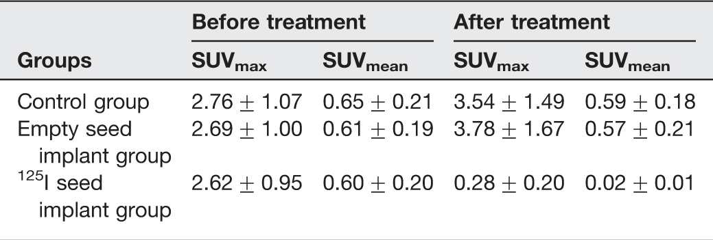
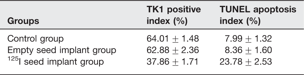
Our results showed that before treatment the SUVmax and SUVmean values among three groups had no significant statistical difference. One week after treatment the SUVmax and SUVmean in 125I seed implant group were significantly lower than before, while for the empty seed group and control group there were no significant difference compared with before treatment. Immunohistochemical analysis of tumor tissue revealed significantly less TK1 positive cells in 125I seed implant group than in empty seed group and control group. The index of apoptosis was slightly higher in 125I seed implant group than in empty seed group and control group as evaluated by TUNEL assay.
Conclusion
These results suggest that 18F-FDG microPET-CT may be useful as a non-invasive imaging modality to assess early response to 125I seed brachytherapy in a pancreatic carcinoma Xenograft.
Pancreatic cancer is a gastrointestinal malignancy that is a serious threat to human health. The 5-year survival rate of pancreatic cancer is <5%, giving pancreatic cancer patients the poorest prognosis among the malignant cancers (1, 2). Pancreatic cancer is often difficult to diagnose in an early stage; only 10–15% of patients are eligible for surgical treatment at diagnosis. Current clinical treatments for pancreatic cancer have limited efficacy, highlighting the need for improved treatment strategies to prolong patient survival (3, 4). Implantation of radioactive isotopes for the treatment of pancreatic carcinoma has been used for the past several decades as it can maximize local dose and minimize irradiation of surrounding normal tissue (5–7).
PET/CT systems provide excellent anatomic and molecular information. Applying imaging modalities in small animals can accelerate the development of new imaging markers and treatment methods as well as increase our understanding of pathophysiologic processes. The purpose of this study was to use 18F-FDG microPET-CT to assess the early effect of 125I interstitial brachytherapy on pancreatic carcinoma xenografts.
Material and Methods
Tumor model
Animal care and all experimental procedures were performed under the approval of the Animal Welfare Council. Male Balb/c-nu mice (20 g, 4–6 weeks old) were purchased from Chinese Academy of Sciences Shanghai Experimental Animal Center and allowed to acclimatize for 1 week in the animal facility before any intervention was initiated. The human pancreatic carcinoma cell line SW1990 was acquired from American Type Culture Collection (ATCC, Manassas, VA, USA). 1 × 10¹⁰/L cells were injected subcutaneously into the right hind limbs of the BALB/c-nu mice. The tumor size was between 8–10 mm after 2 weeks.
125I seed
125I seeds were provided by Shanghai GMS pharmaceutical limited company (XinKe Pharmaceutical Ltd., Shanghai, China). A single seed is 0.8 mm in diameter, 4.5 mm long, a half-life of 59.6 d, and main transmission of 27.4–31.4 Kev X-ray and 35.5 Kev γ-ray. The activities of each seed were 0.8 mCi. The radiation distance of 125I seed is only 1.7 cm, which eliminates the potential injury to the physician, staff, and family. The internal irradiation was relatively long-acting, which can last up to 180 days.
Experimental design
Three groups of mice were grouped as follows (n = 6 in each group): control group; empty seed implant group; and 125I seed implant group. The treatment period was 1 week. Before treatment the mice were scanned with 18F-FDG (Siemens Healthcare, Erlangen, Germany) to determine the baseline level of tracer uptake, and then the scan was repeated 1 week after treatment.
MicroPET-CT imaging
Mice were injected with 7.4 MBq 18F-FDG intravenously. One hour after tracer injection mice were anesthetized and fixed on a bed in presence of three fiducial markers allowing fusion of PET and CT pictures. A 20-min PET scan was acquired using a MicroPET (Siemens Healthcare, Erlangen, Germany). After data acquisition, PET data were reconstructed with the maximum a posteriori (MAP) reconstruction algorithm. The MAP reconstruction algorithm is an interative algorithm commonly used in nuclear medicine image reconstruction (8, 9). The pixel size was 0.866 × 0.866 × 0.796 mm and in the center field of view the resolution was 1.4 mm full-width-at-half-maximum.
Following the microPET scan, a micro-CT scan was acquired. A 10-min CT scan was performed with following parameter settings: 360 rotation steps, tube voltage 80 kV, tube current 500 µA, binning 4, and exposure time 310 ms. The pixel size was 0.091 × 0.091 × 0.091 mm.
MicroPET-CT images were fused in the software (Siemens Medical Solutions, Erlangen, Germany). Before fusion region of interests (ROIs) were drawn on the CT pictures manually by qualitative assessment covering the whole tumors and subsequently tumor volume and tracer uptake, assessed by standard uptake values (SUV) mean and maximum, were generated by summation of voxels within the tomographic planes.
TK1 immunohistochemistry
Three micrometer thick tissue sections were prepared from formline-fixed, paraffin-embedded specimens. The sections were deparaffinized and rehydrated. In order to unmask antigen activity in the tissues, sections were incubated with citric acid buffer (pH 6.0) for 30 min at +90℃ in a water bath. Parts of the sections were also treated in a microwave oven adjusted to +100℃ water. Sections were incubated for 30 min using 3% H202 to block the endogenous peroxidase. Non-specific binding sites were blocked by incubation with normal sera (normal donkey serum). Slides were incubated with an anti-TK1 chicken IgY antibody, raised against a synthetic peptide, residue 161–183 of the human TK1 sequence, diluted in PBS to 100 ug/mL (SSTK Inc., Shenzhen, China) (10). Biotinylated secondary antibodies (Donkey anti-chicken IgG antibody for TK1 staining, diluted in 1:400, Jackson Immuno-Research Lab, West Grove, PA, USA) were applied for 40 min at room temperature. The ABC reagent (Vector Lab., Burlingame, CA, USA) in PBS, 0.1% Tween 20 was added and incubated for 40 min at room temperature. Diaminobenzidine was used as a chromogen and the slides were counterstained with haematoxyline. Staining without the primary TK1 antibody was performed in parallel and served as negative control.
The specificity of the anti-TK1 reaction was tested by means of a competition experiment with additions of a 300-fold molar excess of the peptide corresponding to the residue 161–183. PBS was used instead of peptide in the experiments as negative controls.
TK1 positive cells in the tissue sections were defined as at least 100 cells per microscopic field of 10 fields at × 200–400 magnification by two pathologists independently. Fields were accepted for evaluation if they did not contain areas of extensive necrosis, non-specific background staining, or sectioning artifacts. The number of stained cells within the tumors was scored as follows: no staining (−), 1–19% (+), 20–49% (++), and 50% (+++).
TUNEL assay
TUNEL-positive cells were analyzed using the in situ Cell Death Detection Kit (Roche, Mannheim, Germany) according to the manufacturer's instructions. Briefly, formalin-fixed tissues were permeabilized with proteinase K (30 min, 37℃) and peroxidase blocked in methanol containing 0.3% H2O2. Fluorescent nucleotides mixed with terminal deoxynucleotide transferase were added for 60 min at 37℃, followed by incubation with converter-peroxidase (POD) conjugated anti-fluorescein antibody (provided in the kit) for 30 min at 37℃. Slides were developed using diaminobenzidine (DAB) substrate for 10 min and counterstained using methylene green (7.5%, 7 min at room temperature).
Statistical analysis
All data were plotted as mean ± standard deviation. Statistical analysis was performed with SPSS 17.0 software (SPSS Inc., Chicago, IL, USA). One-way ANOVA, Student's t test, and Pearson correlation were used for comparisons. Differences were considered significant when P was <0.05.
Results
PET metrics
PET parameters assessment before and after treatment among the three groups (n = 18)
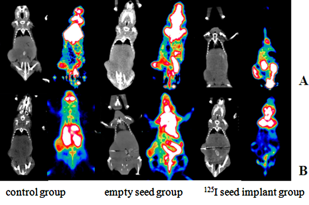
Representative PET and CT images of pancreatic cancer xenograft mice before (a) and after (b) treatment
Characterization and expression of TK1 immunostaining and TUNEL assay
TK1 and TUNEL assay among three groups (n = 18)
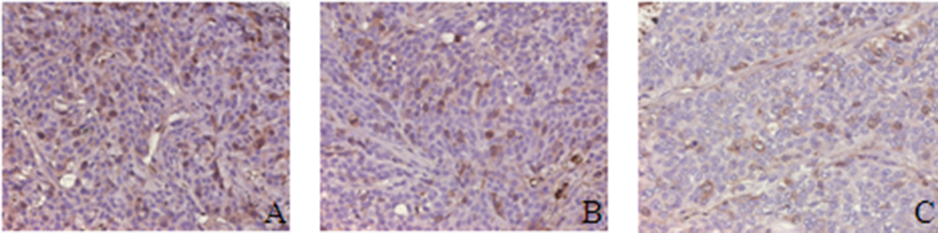
Demonstration of the cytosolic immunoreaction of TK1: (a) control group; (b) empty seed implant group; (c) 125I seed implant group

TUNEL assay for apoptotic: (a) control group; (b) empty seed implant group; (c) 125 I seed implant group
Discussion
In the current experimental setting, the 125I seed showed significant antitumoral effects. Possible advantages of 125I seeds over other forms of radiotherapy were as follows: (i) radiation from seeds was characterized by attenuation over a short distance, which can keep a higher accumulative dose (up to 160 Gy) within the tumor; (ii) 125I seeds could kill tumor cells continuously by keeping cells in the resting period and causing tumor stem cell apoptosis; (iii) the radiation distance was only about 1.7 cm, which eliminated any potential injury to the physician, staff, and family; (iv) the internal irradiation were relatively long-acting, which could last up to 180 days; and (v) the distribution of 125I seeds could be arranged selectively according to the asymmetrical growth of tumor.
Cytosolic TK1 levels in tumor extracts are most likely to be directly related to the proliferation rate of the tumor, as observed in large studies of breast tumor samples (11). Necrotic areas on TUNEL assay were defined as a sign of tumor destruction. SUV values could be useful for quantificative changes in tumor FDG tracer uptake in the setting of therapy response assessment. In our study, SUV values continued to rise in control group and empty seed implant group accompanied by high TK1 positive index and low TUNEL apoptosis index. Conversely, SUV values in 125I implant group showed significant decreased accompanied by low TK1 positvie index and high TUNEL apoptosis index. The degree of TK1 immunostaining and TUNEL assay correlated with SUVmax values which reflect the consequence of proliferation and necrosis of the tumor.
Image detection played an important role in pancreatic cancer studies. During the past years, several imaging modalities have been investigated for their potential ability in providing non-invasive biomarkers for tumor growth. With the increasing use of small animals for research, such imaging must have sufficiently high spatial resolution to allow anatomic localization as well as sufficient specificity and sensitivity to provide an accurate description of the molecular distribution and concentration. Hybrid PET/CT can be used to define the anatomic distribution (spatial priors) corresponding to the target organs, thereby further improving spatial resolution and contrast recovery in submillimeter structures (12). These enhancements will continue to improve the sensitivity, precision, and quantitative accuracy of measured organ perfusion rates, cell survival, and proliferation rates obtained by small-animal PET.
Several articles have hinted the potential application of 18F-FDG microPET-CT in clinical research. Christoforidis et al. (13) performed intravitreal injection with I-124 bevacizumab and ranibizumab in rabbit model to determine whether bevacizumab and ranibizumab remain confined within the vitreous cavity after intravitreal injection and to determine the pharmacokinetic properties of these agents within the vitreous cavity, evaluating by a microPET-CT scanning. Park et al. (14) used microPET-CT to assess a novel low-dose radiotherapy regimen for treating glioblastoma multiforme. They found that 0.2-Gy pulses with 3-min interpulse intervals radiation treatment was more efficacious than the standard 2-Gy fractionation treatment and was associated with less normal tissue damage.
Spatial resolution is determined by detector size, ring diameter, positron range, detector interactions, and reconstruction smoothing (15). Advanced image reconstrucion improved spatial resolution (16). In a study by Cheng et al. (9), MAP reconstruction algorithm produced more accurate activity concentration estimates than filtered-backprojection (FBP) or ordered subsets expectation maximization (OSEM) due to improved spatial resolution leading to lesser partial volume effect. MAP reconstruction algorithm used in our study showed significant statistical difference in SUVmax and SUVmean values among the three groups 1 week after treatment.
In conclusion, the current study suggests that non-invasive molecular imaging 18F-FDG microPET-CT might be a useful tool to assess early response to 125I seed brachytherapy in a pancreatic carcinoma Xenograft. The different radiation dose of 125I seed implantation was yet to be clarified in future study.
Footnotes
Acknowledgements
This work was supported by the Youth Foundation of Shanghai Public Health Bureau (2010Y010); the Postdoctoral Science Foundation of China (2012M511110); the National Natural Science Foundation of China (81271682, 30830038, 30970842, 81071180, and 81071281); the “973” Project (2012CB932604); the fund of Science and Technology Commission of Shanghai Municipality (10ZR1419800, 11JC1407400, 10JC1410000, 10441902002, and 10JC1410900); and the Shanghai Leading Academic Discipline Project (No. S30203).