






Abstract
Background
Although the use of polytetrafluoroethylene (PTFE)-covered biliary stents has proven to be feasible for the treatment of benign and malignant biliary disease, less is known regarding the outcomes of percutaneous placement of a covered stent in patients with malignant duodenobiliary obstruction.
Purpose
To investigate the technical and clinical efficacy of the percutaneous placement of a PTFE-covered biliary stent in patients with malignant duodenobiliary obstruction.
Material and Methods
From April 2007 to September 2012, the medical records of 45 consecutive patients with malignant duodenobiliary obstruction were retrospectively reviewed. All percutaneous biliary stent deployment was performed using PTFE-covered stents, whereas duodenal stent insertion was performed either fluoroscopically or endoscopically using covered or uncovered stents.
Results
Biliary stent deployment was technically successful in all patients. None of the stents migrated after deployment. Procedure-related minor complications, including self-limiting hemobilia, occurred in three (7%) patients. Successful internal drainage was achieved in 39 (87%) of the 45 patients. The median survival time after biliary stent placement was 62 days (95% confidence interval, 8–116 days), and the cumulative stent patency rates at 1, 3, 6, and 12 months were 96%, 92%, 75%, and 38%, respectively. The causes of biliary stent dysfunction included stent occlusion caused by a subsequently inserted duodenal stent (n = 7), food impaction (n = 3), and sludge incrustation (n = 1). One patient developed acute cholecystitis 131 days after biliary stent placement and underwent percutaneous transhepatic gallbladder drainage.
Conclusion
Percutaneous insertion of a PTFE-covered stent is a safe and effective method for palliative treatment of patients with malignant duodenobiliary obstruction. If possible, subsequent biliary stent insertion is preferable in order to prevent possible biliary stent dysfunction caused by subsequent insertion of a duodenal stent.
Introduction
Malignant biliary and duodenal obstruction is a common complication of gastroduodenal or pancreaticobiliary malignancy (1–4). In patients with periampullary cancers, duodenal invasion is not uncommon, and gastric outlet obstruction develops in 5–25% of patients with pancreatic cancer (2). As most of these malignancies are unresectable at the time of diagnosis, the best option is palliative management (1–4). Simultaneous insertion of biliary and duodenal stents or consecutive insertion of these stents is known to be a safe and effective palliative method (2–4).
Although the use of polytetrafluoroethylene (PTFE)-covered biliary stents has proven to be feasible for the treatment of benign and malignant biliary disease (5–11), less is known regarding the outcomes of percutaneous placement of a covered stent in patients with malignant duodenobiliary obstruction. Therefore, the purpose of this study was to investigate the technical and clinical efficacy of the percutaneous placement of a PTFE-covered biliary stent in patients with malignant duodenobiliary obstruction and who underwent combined duodenal stent insertion.
Material and Methods
Patient population
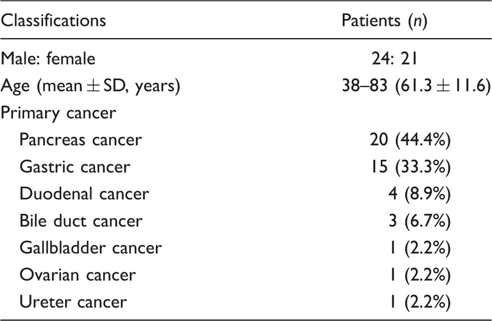
Based on the medical records from April 2007 to September 2012, 45 consecutive patients with malignant duodenobiliary obstruction were included in our study. Demographic, clinical, and laboratory data were collected from the medical records of all 45 patients or from the electronic patient information database. Accurate and definite follow-up data were obtained for all 45 patients. This retrospective study was approved by the institutional review board of our medical institution, and written informed consent was waived.
Baseline demographics and clinical data of the 45 study patients.
Technique
Percutaneous, biliary, covered-stent insertion was performed after percutaneous transhepatic biliary drainage (PTBD). PTBD and stent insertion were performed under conscious sedation using intravenous pethidine hydrochloride (Demerol, Keukdong Pharmaceutics, Seoul, Korea) and local anesthesia using intramuscular lidocaine (Jeil Pharmaceutics, Taegu, Korea). Antibiotics were administered intravenously 24 h before each procedure and for at least 48 h afterward. An intrahepatic bile duct was punctured using a 21-gauge Chiba needle (Cook, Bloomington, IN, USA), under fluoroscopy guidance or ultrasound guidance. Based on standard biliary drainage procedures, an 8.5-F biliary drainage catheter (Cook) was initially placed.
All biliary stent deployment was performed using PTFE-covered stents (GD stent [TaeWoong Medical, Kimpo, Korea] or a ComVi stent [TaeWoong Medical]). Stents were in diameters of 10 mm and in lengths of 7, 8, 9, and 10 cm. The GD was used for the management of extrahepatic bile duct obstruction, whereas the ComVi stent was used for duodenal extension in patients who had previously undergone duodenal stent insertion. Duodenal stent insertion was performed either fluoroscopically or endoscopically. All stents used under fluoroscopic guidance were expandable, nitinol dual stents (Hercules SP Pyloric, S&G Biotech, Sungnam, Korea) consisting of an outer, partially covered stent and an inner, bare nitinol stent (12,13). All endoscopic duodenal stents were bare stents (Boston Scientific, Galway, Ireland) because outer diameter of delivery system of dual stents is too large to insert via endoscopy (14,15).
If there was simultaneous duodenobiliary obstruction, a biliary stent was inserted within several days following insertion of the duodenal stents. In cases of biliary obstruction that developed prior to duodenal stent insertion, as biliary obstruction developed near the papilla or involved the papilla in all patients, all biliary stents were placed with the distal end in the duodenum. In cases of biliary obstruction that developed after duodenal stent insertion in patients with type II duodenal obstruction, the distal end of the extrahepatic biliary stent (GD stent) was inserted into the duodenal stent lumen through the mesh of the duodenal uncovered stent (Fig. 1) or into the space between the duodenal covered stent and the duodenal wall (Fig. 2). If bile flow through the biliary stent was insufficient due to duodenal uncovered stent luminal narrowing or extrinsic compression of the biliary stent caused by the duodenal covered stent, an additional covered stent (ComVi stent) was subsequently inserted into the non-stented, distal duodenal lumen (Figs. 1c, 2c, and 2d). The percutaneous biliary drainage catheter was left in place for 3–5 days following biliary stent insertion and was subsequently removed if free contrast-agent flow through the stent into the duodenum was documented and if there was no symptom or sign of cholangitis (16).
Percutaneous transhepatic cholangiograms obtained in a 64-year-old man. Type II duodenal obstruction caused by pancreatic cancer and had been treated with a duodenal uncovered stent 1 month previously. (a) Cholangiogram via the right PTBD shows a malignant distal CBD obstruction (black arrow). The duodenum is cannulated with a metal-tip diagnostic catheter through the mesh of the duodenal uncovered stent (white arrow). (b) A 7-mm balloon catheter (white arrows) is advanced via the right PTBD, and the mesh of the duodenal uncovered stent is dilated. (c) Cholangiogram obtained after placement of two, biliary covered stents (10 mm × 8 cm, white arrow; 8 mm × 9 cm, black arrow) shows good communication via the stent grafts. To provide sufficient internal drainage through the duodenal stricture, a second stenting (8 mm × 9 cm, black arrows) overlapping the first stent (10 mm × 8 cm, white arrows) was done. One week after the biliary stent placement, tubography (not shown) showed good contrast passage and no stent migration. The PTBD catheter was then removed. The patient died due to disease progression 61 days after the biliary stent placement, although there was no evidence of stent dysfunction until then. Percutaneous transhepatic cholangiograms obtained in a 58-year-old woman with type II duodenal obstruction caused by pancreatic cancer and treated using aduodenal covered stent 2 months previously. (a) Cholangiogram via the right PTBD shows a malignant distal CBD obstruction (black arrow). (b) Placement of the first biliary covered stent (10 mm × 10 cm, white arrows) into the space between the duodenal covered stent and the duodenal wall, was done. (c) To improve the internal drainage, a second biliary covered stent placement was planned overlapping the first stent (black arrow). Radiopaque linear gold markers (white arrows) were incorporated into both ends of the second stent. (d) After placement of the second stent (8 mm × 10 cm, white arrow), the cholangiogram shows fluent contrast passage into the duodenum. Six days after biliary stent placement, tubography (not shown) showed good contrast passage and no stent migration. The PTBD catheter was then removed. The patient died due to disease progression 173 days after the biliary stent placement, although there was no evidence of stent dysfunction until then.

Study endpoint and statistical analysis
The study endpoints included the assessment of technical success, complications, successful internal drainage, patient survival, and stent patency after biliary stent insertion. Technical success was defined as placement of the biliary stent in an adequate position without migration and good contrast passage through the stent into the duodenum. Complications were classified as major and minor according to the guidelines of the Society of Interventional Radiology Standards of Practice Committee (17). Successful internal drainage was defined as successful removal of the temporary drainage catheter and a decrease in the serum bilirubin level to <75% of the pretreatment value within the first month after biliary stent insertion. Patient survival was defined as the interval between the initial biliary stent insertion and a patient’s death or last follow-up. If the patient was alive at the time of the last follow-up, survival was considered equal to the follow-up duration. Stent patency was defined as the interval between the initial biliary stent insertion and the occurrence of stent dysfunction. If dysfunction was not evident during a patient’s life, stent patency was considered as equal to the time of the patient’s survival. A stent was assumed to be patent at the time of a patient’s death if the serum bilirubin levels were normal or only mildly increased (<3 mg/dL). If a patient was obviously jaundiced or had higher bilirubin levels, although terminal stages of disease and replacement of liver parenchyma by tumor could lead to global hepatic dysfunction without stent dysfunction, the stent was assumed to be obstructed.
The paired-sample Student t test was used to compare the serum bilirubin levels before and after biliary stent insertion. Patient survival and stent patency rates were calculated using the Kaplan-Meier method. All statistical analyses were performed using PASW software (version 18, SPSS In., Chicago, IL, USA), with P value <0.05 indicating statistical significance.
Results
Duodenobiliary obstruction
A simultaneous biliary and duodenal obstruction occurred in 16 study patients. In the remaining 29 patients, duodenal obstruction occurred before biliary obstruction in 15 patients and after in 14 patients. The median interval between the onset of biliary and duodenal obstruction was 5.3 months (range, 1–18.5 months). Duodenal obstruction occurred proximal to the duodenal papilla and without involvement of the papilla (type I) in 21 patients, at the second part of the duodenum with involvement of the papilla (type II) in 19, and distal to the papilla without involvement of the papilla (type III) in five patients. The level of extrahepatic biliary obstruction was the upper CBD in seven patients, the lower CBD in 17 patients, and the entire CBD in 21 patients.
Technical and clinical outcomes of biliary stenting
Biliary stent deployment was technically successful in all patients. A single stent was sufficient to relieve malignant biliary obstruction in 26 patients with type I or III duodenal obstruction. Among eight of the 19 patients with type II duodenal obstruction, a single biliary stent was inserted before duodenal stent placement. In the remaining 11 of these 19 patients, a single biliary stent was inserted through the mesh of the duodenal uncovered stent or into the space between the duodenal covered stent and the duodenal wall. However, an additional covered stent was necessary for sufficient internal drainage in five patients due to duodenal uncovered stent luminal narrowing (n = 2) or severe biliary stent compression caused by the duodenal covered stent. Therefore, the 45 patients included in our study received a total of 50 biliary covered stents. None of the stents migrated after deployment.
Procedure-related, minor complications occurred in three (7%) patients. Three patients experienced self-limiting hemobilia that completely resolved 1–3 days following stent placement and without transfusion. One patient developed acute cholecystitis 131 days following the biliary stent placement and underwent percutaneous cholecystostomy tube placement.
Follow-up cholangiography 3–6 days after biliary stent placement showed adequate decompression of the biliary ducts in 39 patients. The temporary drainage catheters were removed from these 39 patients if contrast agent flow through the stent into duodenum was documented. The remaining six patients with type II (n = 3) or type III (n = 3) duodenal obstruction showed immediate stent dysfunction due to extrinsic compression by the duodenal covered stent in the three patients with type II duodenal obstruction or caused by food impaction in the three patients with type III, and their temporary drainage catheters could not be removed due to either their refusal of additional stenting or recurrent food reflux. The mean serum bilirubin level which was 8.2 mg/dL ± 6.7 before drainage, decreased significantly to 3.4 mg/dL ± 4.3 1 month after biliary stent placement (P = 0.027). Therefore, successful internal drainage was achieved in 39 (87%) of 45 patients.
Patient survival
Clinical follow-up until death or the end of the study was available for all patients, and the cut-off date for data analysis was 31 December 2012. During the median follow-up period of 132 days (range, 8–920 days), 41 patients died and four patients remained alive. Twelve (27%) patients died within 30 days following biliary stent placement, although their deaths were not directly related to the procedure. According to the Kaplan-Meier analysis, the median survival time of these 41 patients after biliary stent placement was 62 days (95% confidence interval, 8–116 days) (Fig. 3).
The Kaplan-Meier curve shows the survival rate of the study patients. Cross hatches indicate censored events.
Stent patency
According to the Kaplan-Meier analysis, the cumulative stent patency rates at 1, 3, 6, and 12 months were 96%, 92%, 75%, and 38%, respectively. During the follow-up period, stent dysfunction occurred in five of 39 patients (12.8%) after a mean of 147 days (Fig. 4). Among these five patients, stent dysfunction occurred in one patient with type I duodenal obstruction due to sludge incrustation and in four patients with type II duodenal obstruction due to extrinsic compression caused by the subsequent insertion of the duodenal stents (uncovered stent [n = 1] and covered stent [n = 3]). Four of these five patients were treated with PTBD, and one patient was treated using endoscopic ultrasound-guided hepatico-esophagostomy. In the four patients who underwent PTBD, additional biliary stent placement could not be performed due to failure of the guidewire passage through the occluded biliary stent (n = 1) or their poor general condition caused by the disease progression (n = 3).
The Kaplan-Meier curve shows the stent patency rate of the study patients. Cross hatches indicate censored events.
Therefore, overall biliary stent dysfunction occurred in 11 (24.4%) of 45 patients. The two, common causes of biliary stent dysfunction were stent occlusion due to a subsequently inserted duodenal stent (n = 7) in patients with type II duodenal obstruction and food impaction (n = 3) in patients with type III.
Discussion
In this study, the technical success rate (100%), complication rate (7%), successful internal drainage rate (87%), and median patient survival time (132 days) were in good agreement with the results of previous studies of combined biliary and duodenal stent insertion (3,4,18). In those studies, the technical success rate was in the range of 88–100%, the overall complication rate was in the range of 13–22%, and the median patient survival time was in the range of 81–195.5 days.
Several studies have suggested that biliary stents would be more successfully inserted prior to duodenal stent insertion because of the difficulty either in passing the duodenal stricture with the duodenal stent or in accessing the papilla through the mesh of the duodenal stent (1,19,20). Moreover, an endoscopic approach for biliary stenting might be impossible because the major papilla could not be identifiable when using a longstanding duodenal stent. Although endoscopic biliary stenting might be possible when using a duodenal stent, percutaneous biliary stenting is a more effective method (1). In addition, the use of PTFE-covered biliary stent can prevent tumor ingrowth and tend to yield higher patency rates (5,7,21,22). In our cases, subsequent insertion of a biliary covered stent was technically successful through the mesh of the duodenal uncovered stent or in the space between the duodenal covered stent and the duodenal wall in all 19 patients with type II duodenal obstruction, and it did not disturb internal contrast flow. If contrast passage through the initial biliary stent is not fluent, an additional covered stent insertion to bridge the distal unstented duodenal lumen was effective in patients with duodenal stent in situ.
In this study, we found that the stent dysfunction rate was 13% and the most important mechanism causing stent dysfunction was external compression of the biliary stent by the subsequently inserted duodenal stents in patients with type II duodenal obstruction and food impaction due to duodenobiliary reflux in patients with type III duodenal obstruction. One problem possibly associated with combined duodenal and biliary stent insertion is obstruction of the biliary stent caused by the subsequently inserted duodenal stent, itself. Hamada et al. (2) showed that duodenal stent placement is a risk factor for biliary stent dysfunction. In our cases, biliary stent dysfunction occurred immediately after duodenal covered stent insertion in seven of 19 patients with type II duodenal obstruction, due to extrinsic compression by the subsequently inserted duodenal covered or uncovered stent. However, we did not observe dysfunction of the subsequently inserted biliary stent in any of the 19 type II patients who underwent prior duodenal stent insertion. Therefore, we suggest that biliary stents would be better inserted after duodenal stent insertion as it is not technically difficult via the percutaneous route and could thus prevent immediate biliary stent dysfunction after subsequent duodenal stent insertion in patients with duodenobiliary obstruction.
As a biliary obstruction develops near the papilla in most patients with malignant duodenobiliary obstruction, a biliary stent should be placed with one end in the duodenum. Therefore, duodenobiliary reflux is the major cause of biliary stent dysfunction. In our study, immediate dysfunction of the biliary stents due to food impaction occurred in three of five patients with type III duodenal obstruction. Two possible processes can be considered as causes of food reflux, i.e. increased duodenobiliary reflux due to narrowing of the stented duodenal lumen and reduced duodenal peristalsis which might not be sufficiently improved by the use of a duodenal stent.
In this study, we did not observe any biliary stent dysfunction resulting from tumor ingrowth. Covered stents using PTFE have been found to be effective for preventing tumor ingrowth and have been associated with relatively low complication rates (5–11). In one comparative study of PTFE-covered and uncovered stents, PTFE-covered stents were significantly superior to uncovered stents in terms of stent patency, and there was no significant difference in the complication rate between the PTFE-covered and uncovered stents (7). In the studies using PTFE-covered stents, the 6-month patency rates were in the range of 55.5–92.6%, whereas the stent occlusion rates were in the range of 9–19% (5–11). It is difficult to compare our results with those of these previous studies as each treated patient had a different type, location, and stage of disease; however, the patency and stent occlusion rates we observed were similar to those in studies using PTFE-covered stents.
This study has several limitations including its retrospective design and inclusion of a relatively small number of patients. Moreover, because differences among the two types of duodenal stents used may have influenced the biliary stent patency, further prospective, randomized comparisons are necessary.
In conclusion, percutaneous insertion of a PTFE-covered stent was seen to be a safe and effective method for the palliative treatment of patients with malignant duodenobiliary obstruction. Percutaneous insertion of a subsequent biliary covered stent was technically feasible after duodenal stent insertion. If possible, subsequent biliary stent insertion is preferable in order to prevent possible biliary stent dysfunction caused by subsequent insertion of a duodenal stent, especially in patients with type II duodenal obstruction.
Footnotes
Funding
This research received no specific grant from any funding agency in the public, commercial, or not-for-profit sectors.