






Abstract
Background
For the evaluation of severely injured trauma patients a variety of total body computed tomography (CT) scanning protocols exist. Frequently multiple pass protocols are used. A split bolus contrast protocol can reduce the number of passes through the body, and thereby radiation exposure, in this relatively young and vitally threatened population.
Purpose
To evaluate three protocols for single pass total body scanning in 64-slice multidetector CT (MDCT) on optimal image quality.
Material and Methods
Three total body CT protocols were prospectively evaluated in three series of 10 consecutive trauma patients. In Group A unenhanced brain and cervical spine CT was followed by chest–abdomen–pelvis CT in portovenous phase after repositioning of the arms. Group B underwent brain CT followed without arm repositioning by a one-volume contrast CT from skull base to the pubic symphysis. Group C was identical to Group A, but the torso was scanned with a split bolus technique. Three radiologists independently evaluated protocol quality scores (5-point Likert scale), parenchymal and vascular enhancement and artifacts.
Results
Overall image quality was good (4.10) in Group A, more than satisfactory (3.38) in Group B, and nearly excellent (4.75) in Group C (P < 0.001). Interfering artifacts were mostly reported in Group B in the liver and spleen.
Conclusion
In single pass total body CT scanning a split bolus technique reached the highest overall image quality compared to conventional total body CT and one-volume contrast CT.
Introduction
Computed tomography (CT) imaging in trauma has become increasingly important and prevalent (1–8). Recently the beneficial effect of total body CT scanning on mortality in severe trauma patients was reported when compared to selective CT scanning after conventional work-up (9). Although rapid diagnosis led to an increase in probability in survival, also some criticism followed for this approach, focusing on the increased radiation exposure in this overall relatively younger population (10–12).
In most centers performing total body CT, the technique comprises a multidetector CT (MDCT) of the head, cervical spine, chest, and abdomen. Despite this consensus in the scanned volume, a considerable variation in the scan protocols is seen in different trauma centers around the world (2,4–9,13,14). This variation includes contrast timing and number of phases. Of note, the term pass reflects merely the acquisition of a single scan series, whereas contrast phase points at a certain time after administration of contrast medium targeted at the specific enhancement of organs of interest. When up to four passes through the body (non-contrast, arterial, portovenous, and excretion phase) are used this could lead to higher and unnecessary radiation exposure. However, only a paucity on data on the quality of protocols exist (13,14), and no prospective study so far for 64-slice CT.
In this study we evaluated three different scan protocols (portovenous contrast phase, with and without arm repositioning and split bolus contrast technique) with respect to quality and scan times. The aim of this study was to evaluate three protocols for single pass total body scanning in 64-slice MDCT on optimal image quality.
Material and Methods
The present study was a single center prospective study performed at a level 1 university trauma center in The Netherlands before the start of a multicenter, randomized trial on total body CT in trauma patients (April 2011). Our trauma resuscitation room has a sliding gantry 64-slice CT-scanner (Sensation 64, Siemens Medical Solutions, Forchheim, Germany) with a multifunctional, radiolucent trauma resuscitation table (15,16). All consecutive polytrauma patients who were admitted during daytime were eligible. Inclusion criteria for total body CT scanning were: life-threatening problems (respiratory rate >29 or <10/min, or pulse >120/min, systolic blood pressure <100 mmHg, refill >4 s or exterior blood loss >500 mL, or Glasgow Coma Score ≤13 or abnormal pupils), or clinical signs of flail chest, open chest, multiple rib fractures, pelvic fracture, unstable vertebral fractures, spinal cord compression, or fractures from at least two long bones. Exclusion criteria were: age <18 years, known pregnancy, patients referred from other hospitals or any patient judged too unstable to undergo scanning and requiring resuscitation or immediate operation. All patients were followed up for the complete hospital stay. The study was approved by the local ethics committee, with a waiver for informed consent.
Imaging protocol
Three different trauma scan protocols were evaluated. Inclusion took only place during office hours when at least one of the investigators was present to control the work flow and perform time registration. Three series of 10 patients were included. No randomization was performed; after every 10 consecutive inclusions the protocol was changed for the next 10 patients.
All patients received a CT of the brain, cervical spine, chest, and abdomen / pelvis; scan parameters were equal in the three groups: collimation 64* 0.6 mm with 120 kV and 380 mAs for brain, reference mAs of 250 for cervical spine and 200 for body, rotation time 1.0 s for brain and cervical spine and 0.5 s for chest / abdomen, with standard pitch of 0.85, 0.9, and 1.4, for the respective body parts (median DLP of the torso was 1125, 1125, and 1128 mGycm for the three respective groups). The protocols for cervical spine and torso used a 4D automatic tube current modulation (CARE dose 4D Automatic Exposure Control, Siemens, Forchheim, Germany). During scanning no gantry tilt was used. Intravenous contrast (Optiray® 350 125 ml Pre fill, Covidien Mallinckrodt, Cincinnati, OH, USA) was administered via an 18 G peripheral cannula in the right antecubital vein. Preset contrast medium protocols were programmed in the injection device (Optivantage DH injector, Covidien Mallinckrodt). In all three groups administration of intravenous contrast medium was followed by a saline chase of 40 mL at 4 mL/s. Brain reconstruction was in axial planes with 5 mm head kernel and 1 mm bone kernel, cervical spine in 1 mm bone kernel in axial, sagittal, and coronal planes. Torso was reconstructed at 3 mm axial and coronal slices in soft and bone kernel.
The following scanning protocols were compared:
Group A: Conventional total body trauma CT. Non-contrast enhanced CT brain and cervical spine with arms alongside the patient, after which arms were elevated and positioned alongside the head followed by CT of chest / abdomen / pelvis after administration of 100 mL intravenous contrast medium at a rate of 4 mL/s in the venous phase, started after 60 s. Group B: One-volume contrast CT. Non-contrast enhanced CT of the brain, followed by a contrast-enhanced volume CT from skull base until the pubic symphysis, 4 mL/s with fixed delay of 30 s and arms alongside the body. Cervical spine was included into this torso scan, with the upper abdomen generally scanned in a late arterial phase, Group C: Split bolus. Equal to Group A, but with split bolus technique: non-contrast enhanced CT of the brain and cervical spine, followed by repositioning of the arms alongside the head and scanning the torso with a fixed delay split bolus: at 60 s before start of the CT 80 mL intravenous contrast medium at a rate of 4 mL/s and saline chase, followed at 20 s before start of the CT by 40 mL contrast medium at a rate of 5 mL/s and saline chase.
Evaluation
Subjective image quality was assessed on a standardized form independently by three radiologists with 6, 8, and 12 years of experience in trauma imaging. These observers were blinded for patient data and scanning protocols. All studies were evaluated using a picture archiving and communication system PACS (Impax 4.5, AGFA Gevaert, Mortsel, Belgium). Qualitative image assessment focused primarily on organ / vessel delineation in combination with its homogeneity of enhancement. A lower score was attributed when artifacts significantly hindered this evaluation. Subjective scores for image quality were recorded for the overall quality of the total body CT scan and on specific body regions: brain, cervical spine, thoracolumbar spine, lung parenchyma, mediastinum, liver, spleen, kidney, pelvis, and aortic arch, abdominal aorta at the level of the superior mesenteric artery, and portal vein. For this assessment a 5-point Likert scale was used: 1, non-diagnostic image quality; 2, poor image quality; 3, satisfactory quality; 4, good image quality; and 5, excellent image quality. Hounsfield Units (HU) attenuation determined by setting a region of interest (ROI) half of the vessel caliber for the aortic arch, abdominal aorta, and the portal vein were registered, as well as in the parenchyma of the liver, spleen, and renal cortex using a 1 cm ROI. In case artifacts were present the type of artifact, location, and interference with evaluation were noted.
Several time points were registered: time of admission; start and end of CT acquisition (scout view and last axial image, respectively); time of diagnosis for treatment planning; and time of departure from the trauma resuscitation room.
Statistical analysis
Differences between patient series were assessed by Fisher’s exact test (gender) and one-way analysis of variance (age, injury severity score [ISS]). Differences between the three investigated protocols in image quality scores were assessed by balanced univariate analyses of covariance with adjustment for differences among radiologists. Differences between protocols by contrast enhancement values and acquisition times were assessed by balanced univariate analysis of variance. A value of P < 0.05 was considered statistically significant. Bonferroni correction was applied during post hoc comparisons.
The intra-class correlation coefficient (ICC) was used to measure inter-observer absolute agreement among the three reviewers on image quality. The ICC values with a 95% confidence interval (CI) were calculated using a two-way mixed-effects model with single measures. The ICC is an index of concordance that indicates the degree of agreement beyond that expected by chance alone, and is appropriate when assessing agreement between two or more observers. ICC values higher than 0.8 were considered to represent almost perfect concordance, values between 0.61 and 0.8 as substantial, between 0.41 and 0.6 as moderate, between 0.21 and 0.4 as fair, between 0.0 and 0.2 as slight, and below 0.0 as poor according to the Landis and Koch classification (17). Statistical analysis was performed using the Statistical Package for the Social Sciences (SPSS version 18.0, SPSS Inc. Chicago, IL, USA).
Results
The preset 30 consecutive patients were included, of which the demographics are displayed in Table 1. The three groups differed by age, with elder patients in Group C and younger patients in Group B. There were no significant differences in sex, mechanism of injury, or ISS between the patients in each scanning protocol group. Of every group an axial and coronal reformatted image is displayed (Fig. 1).
Axial and coronal reformatted images (3 mm soft kernel) of three trauma patients of Group A (conventional total body trauma CT with CT scanning of the body in venous phase after repositioning of the arms), Group B (one-volume contrast-enhanced CT from skull base until the pubic symphysis) and Group C (split bolus technique). Demographics for conventional (Group A), one-volume contrast (Group B) and split bolus protocol (Group C) total body trauma CT. Data are expressed as median (interquartile range) unless otherwise indicated. F, female; ISS, injury severity score; M, male; SCI, spinal cord injury; TBI, traumatic brain injury.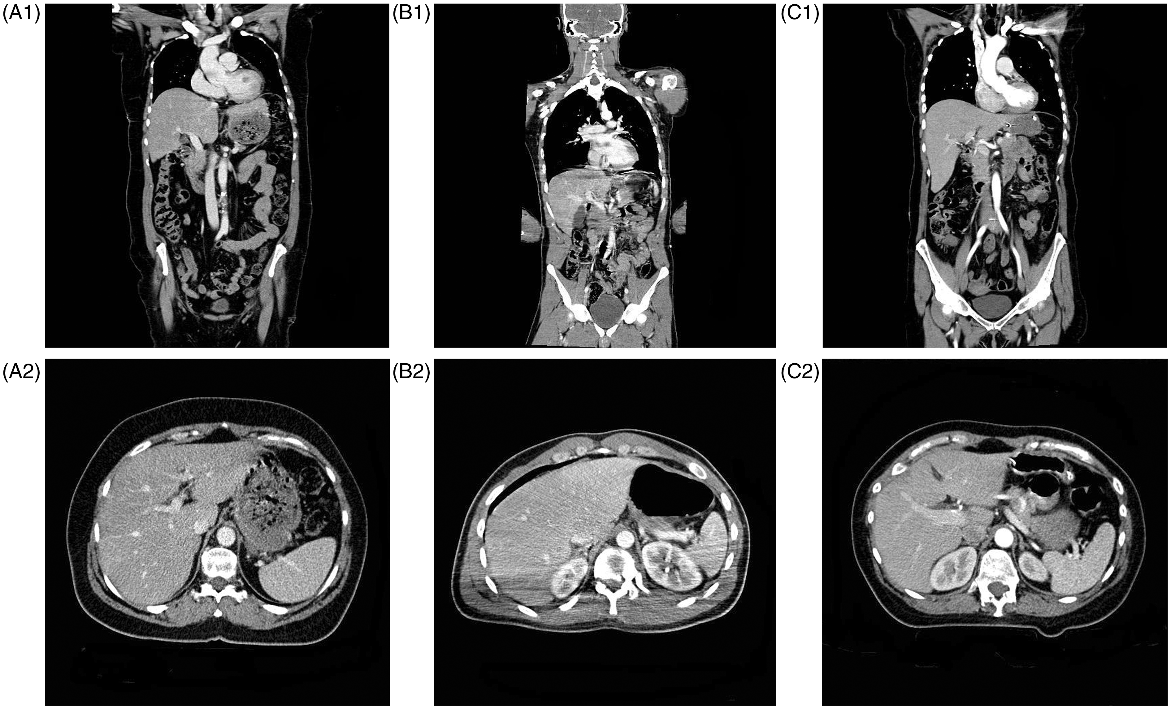

Quality evaluation and main vessel enhancement in total body CT using conventional (Group A), one-volume contrast (Group B), and split bolus protocol (Group C).
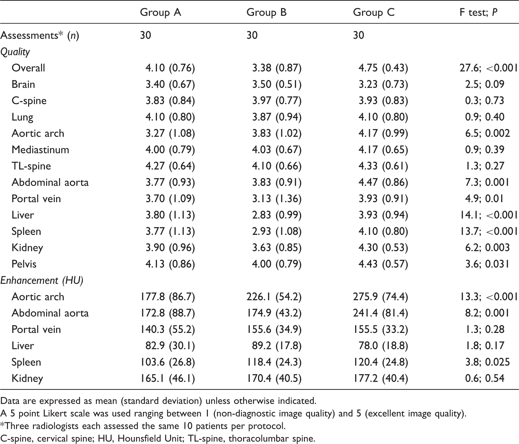
Data are expressed as mean (standard deviation) unless otherwise indicated.
A 5 point Likert scale was used ranging between 1 (non-diagnostic image quality) and 5 (excellent image quality).
Three radiologists each assessed the same 10 patients per protocol.
C-spine, cervical spine; HU, Hounsfield Unit; TL-spine, thoracolumbar spine.
Contrast enhancement values for the aortic arch were highest in Group C, followed by Group B and Group A. HU values in the spleen (P = 0.039) and abdominal aorta (P = 0.002) were lower in Group A compared to Group C. Lower HU values compared to Group C were observed for Group B in the abdominal aorta (P = 0.003). Otherwise, no significant differences in contrast enhancement values were observed.
In one patient in group A and one patient in group B breathing artifacts were reported. In one patient in group A evaluation of the neck was disturbed by a necklace. Interfering beam hardening artifacts were noted in three patients in the upper abdomen in Group B, more precisely the liver and spleen, due to the proximity of the arms.
Time intervals for total body CT scanning using conventional (Group A), one volume-contrast (Group B) and split bolus scan protocol (Group C).
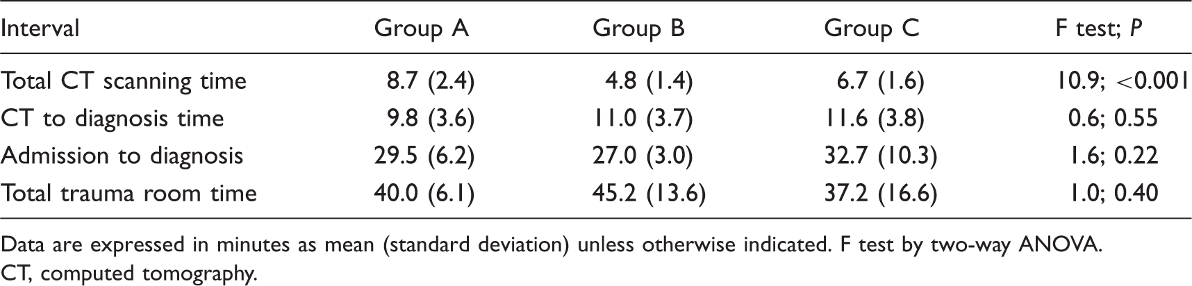
Data are expressed in minutes as mean (standard deviation) unless otherwise indicated. F test by two-way ANOVA.
CT, computed tomography.
Inter-observer absolute agreement among the three observers on image quality in total body CT using conventional (Group A), one-volume contrast (Group B), and split bolus protocol (Group C).

Data are expressed as intraclass correlations coefficients (95% confidence interval).
C-spine, cervical spine; TL-spine, thoracolumbar spine.
Discussion
This study shows that the split bolus technique for single pass total body CT scanning had the highest overall image quality, compared to the conventional total body trauma CT and the one-volume contrast CT protocol. A split bolus technique combines different contrast phases into one acquisition, thereby diminishing radiation exposure with only limited increase of the amount of contrast medium. It most frequently is used in renal imaging (18,19). In trauma only three reports on a multi-phasic scan protocol in MDCT have been published (20–22). Loupatatzis et al. compared in 16-slice MDCT a tri-phasic injection scheme (70 mL at 3 mL/s, followed by 0.1 mL/s for 8 s, and 70 mL at 4 mL/s) to their standard CT angiography protocol (20). The tri-phasic protocol achieved similarly high image quality for arteries compared to standard CT angiography protocol, parenchymatous organs had better image quality compared to specialized protocols. As in our study arm artifacts reduced the enhancement of spleen and liver parenchyma. In a retrospective study Yaniv compared the same contrast protocol set-up for 64-slice MDCT with an arterial-phase contrast-enhanced CT of the thorax and a portovenous scan of the abdomen and pelvis, but added a preceding unenhanced CT of the abdomen (21). The tri-phasic injection protocol enabled better vascular and abdominal parenchymal imaging, although mean enhancement values in the aorta were significantly greater with the conventional protocol. Nguyen et al. (22) compared a standard injection protocol with an one-volume acquisition from the circle of Willis to the pubic symphysis in 16-slice MDCT using a biphasic (150 mL at 6 and 4 mL/s) or mono-phasic (110 mL at 4 mL/s, 400 mg I/mL) injection. No significant differences were found in mean enhancement values in the aorta, liver, spleen, and kidney for the three protocols. Quality scores were significantly higher for liver, spleen, and kidney with the arms above the head compared to arms alongside the body. Single-pass protocols had significantly shorter median acquisition times than the conventional protocol. Hence, our findings for 64-slice MDCT are in concordance with these previous CT studies.
In blunt abdominal trauma an arterial phase is more sensitive for the detection of intrasplenic pseudoaneurysms, but a portal venous phase is more sensitive for the detection of parenchymal injuries and active bleeding of the spleen (23). Dual phase CT can be considered as a complete work-up as it has better overall diagnostic performance than single phase CT. However, this is at the cost of doubling radiation exposure to the abdomen. As group B is scanned in a late arterial phase, it could be inferior in case of active bleeding and parenchymal injuries. Whether the theoretical superposition of an arterial on a portal venous phase in split bolus provides a comparable complete performance as dual phase imaging has to be proven in a large prospective study.
Total body CT can also be useful to diagnose blunt cerebrovascular injuries (24–26). Only protocol B offered the possibility to screen for these injuries. When started at clinoidal level the split bolus protocol can also fulfill these requirements, though was not investigated in our study. It was, however, satisfactorily used in later patients if during CT acquisition a skull base or upper cervical spine fracture was seen. Although injuries of the supraaortic vessels are rare after blunt trauma, these cerebrovascular injuries are associated with fractures of the cervical spine or skull base (27).
Time is essential in trauma, as underlined by the ATLS philosophy “time is life”. We observed a time difference between the protocol without repositioning of the arms and the other two protocols with arm repositioning. Remarkably, only few reports have focused on this important work flow aspect in correlation with scan protocols (14,28–30). Whether the slight time gain can outweigh the observed lower diagnostic quality remains to be seen.
We did not focus on radiation exposure in our study. However, Brink et al. found that scanning with the arms alongside the body resulted in a 45% increase in radiation exposure and a decrease in image quality compared to scanning patients with a cranial position of the arms (31). Recently this has been confirmed by two other studies (32,33).
Our study has several limitations. First, there are different combinations and settings possible when considering contrast medium phases (non-enhanced, arterial, venous, delayed phases) and body regions (head, neck, thorax, abdomen / pelvis, and extremities). We only studied three different contrast protocols, and therefore we cannot make a statement on other variations. Second, patients’ BMI or weight was not known. Since contrast enhancement of the liver parenchyma is influenced by body weight, differences in liver enhancement and subsequently in quality of liver imaging may be caused by differences in body weight between patients. For the split bolus group ROI triggering in the thoracic aorta probably could have resulted in higher arterial enhancement values. On the other hand, targeted planning by the technician would have resulted in a more complicated procedure and longer examination times. Fixed delay appears to be more practical in these stressful circumstances and therefore less error-prone. Further, diagnosis time, admission to diagnosis time, and total room time are not only influenced by the type of CT scan protocol but also by the specific types of injuries and by emergency room management factors. Total CT scanning time is therefore the most relevant time factor comparing groups A, B, and C. A final limitation concerns the restricted sample size for the assessments of differences among the imaging protocols. After checking whether the data fulfilled the test conditions, it was noted that homogeneity of variances could not be assumed: (i) for the contrast enhancement values for portal vein and liver, and (ii) the acquisition time between admission and diagnosis. The corresponding P values of the ANOVA tests are well above 0.05, but should nevertheless be interpreted with some caution. Although unlikely, these results may be false negative. Of note, in our series no important missed diagnosis during follow-up was recorded. Since the end of the study we used the split bolus technique in all following cases as part of an international multicenter randomized trial on total body CT in severe trauma patients (34). Further experience requiring more prospective studies are needed to evaluate its clinical accuracy.
In conclusion, evaluation of three scanning protocols for single pass total body CT in severe trauma patients showed that the split bolus technique reached the highest scores in image quality and vascular and parenchymal enhancement. The one-volume contrast CT protocol was quickest and can probably be used in selected cases were time gain of some minutes could potentially outweigh the reduced image quality.
Footnotes
Conflict of interest
None declared.
Funding
This research received no specific grant from any funding agency in the public, commercial, or not-for-profit sectors.