
Abstract
Background
Generalized anxiety disorder (GAD) has been related to functional brain activities and structural brain abnormalities.
Purpose
To investigate the neural mechanism on working memory dysfunction in patients with GAD in terms of the combined functional and morphological brain abnormalities.
Material and Methods
Patients with GAD and healthy controls matched for age, sex, and education level underwent high-resolution T1-weighted (T1W) magnetic resonance imaging (MRI) and functional MRI (fMRI). In this study, fMRI and voxel-based morphometry (VBM) were used for assessing the differential brain activation patterns, as well as for comparing the morphological alterations between the two groups.
Results
In response to the neutral distractors, the patients showed significantly lower activities in the regions of the fusiform gyrus (FuG), superior parietal gyrus (SPG), precuneus (PCu), superior occipital gyrus (SOG), lingual gyrus (LiG), cuneus (Cun), calcarine cortex (CaC), parahippocampal gyrus (PHG) and cerebellar cortex (Cb) compared to the controls. In response to the anxiety-inducing distractors, the patients showed significantly higher activity in the hippocampus and lower activities in the regions of the dorsolateral prefrontal cortex (DLPFC), FuG, SPG, PCu, SOG, and Cb. Also, the patients showed a significant reduction of the white matter volumes in the DLPFC, anterior limb of the internal capsule (ALIC) and midbrain.
Conclusion
This study provides the first evidence for the association between the morphometric alterations and functional deficit in the working memory processing with the neutral and anxiety-inducing distractors in GAD patients. These findings would be helpful to understand the neural mechanisms on working memory impairment in connection with GAD symptoms.
Keywords
Introduction
Generalized anxiety disorder (GAD) is characterized by psychiatric distress including excessive, chronic, pervasive, and uncontrollable anxiety in daily life (1). Previous studies (2,3) reported that the major symptoms of GAD lead to emotional dysregulation including unsuppressed anger and low tolerance of frustration, as well as cognitive deficits including impairments of implicit and explicit memories, working memory, attention, and executive function. From these findings, it is considered that the major symptoms of GAD have a negative effect on emotional processing and regulation, and/or cognitive function. Therefore, a combined study of neurofunctional abnormality and neuroanatomical deformation is essential to reveal the neural mechanism associated with the negative effect mentioned above, and further to develop more accurate clinical diagnosis and effective medication of GAD.
In the past decade, neuroimaging studies (4–10) have revealed the functional abnormality and morphological changes in patients with GAD using magnetic resonance imaging (MRI) based techniques. Functional MRI (fMRI) studies (4–6) discriminated differential brain activation patterns in response to negative emotions between patients with GAD and healthy controls. Recently, a preliminary study (7) reported the functional neuroanatomy on the working memory with emotional dysfunction in GAD. To our knowledge, no one has attempted to perform a combined fMRI and voxel-based morphometry (VBM) study on the working memory tasks with neutral and anxiety-inducing distractors in patients with GAD. It is suggested that both brain functional deficits and morphometric alterations were associated with GAD symptoms. However, it is unclear what the causality between functional and morphometric brain alterations is, since the brain functional or morphological studies have been separately performed. Therefore, it is suggested that a combined fMRI and VBM study would be valuable to investigate whether the brain functional abnormality is associated with the morphometric changes in connection with emotional dysregulation and cognitive deficit in patients with GAD.
In this study, we used fMRI in combination with VBM to assess the association between functional brain deficits and morphometric changes, as well as its correlation with behavioral and cognitive characteristics in patients with GAD.
Material and Methods
Characteristics of patients with GAD and healthy controls.
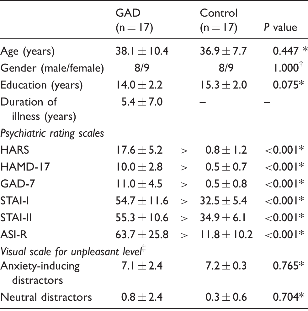
Mann–Whitney U test.
Chi-square test.
Visual scale for unpleasant level: 11-point visual scale ranging from 0 (least unpleasant) to 10 (most unpleasant).
ASI-R, Anxiety Sensitivity Index-Revised; GAD-7, Generalized Anxiety Disorder Scale 7; HAMD-17, Hamilton Rating Scale for Depression 17; HARS, Hamilton Anxiety Rating Scale; STAI-I, State-Trait Anxiety Inventory I; STAI-II, State-Trait Anxiety Inventory II.
The participants underwent structured clinical interviews for their DSM-IV diagnoses. Their anxiety levels were then assessed using various psychiatric rating scales: Hamilton Anxiety Rating Scale (HARS); Hamilton Rating Scale for Depression 17 (HAMD-17); Generalized Anxiety Disorder Scale 7 (GAD-7); State-Trait Anxiety Inventory I (STAI-I); State-Trait Anxiety Inventory II (STAI-II); and Anxiety Sensitivity Index-Revised (ASI-R) (Table 1).
Paradigm for brain activation
To induce emotional responses of participants, both of the neutral and anxiety-inducing images were presented. Neutral images consist of scenic pictures such as natural landscapes, natural parks, mountains, and forests, which is capable of inducing a comfortable feeling. Anxiety-inducing images included the photographs of life-threatening behaviors, traffic accidents, disasters, and other misgivings or anxious events.
After the fMRI experiments, the participants rated the unpleasant level from the neutral and anxiety-inducing images using an 11-point visual scale ranging from 0 (least unpleasant) to 10 (most unpleasant) (Table 1).
The activation paradigm (7) consisted of a string of “cue and encoding (6 sec)-delay (4 sec)-distractor (6 sec)-button ready (2 sec)-retrieval (2 sec)-intertrial rest (12 sec)” (Fig. 1) and the string repeated 20 times as a total of 20 trials using 10 neutral distractor and 10 anxiety-inducing distractor trials (7). In the “cue” period, each trial started with the presentation of the “face” word. In the “encoding” period, three different kinds of the expressionless human faces masked with an oval shape hood were randomly presented once. During the “delay” period, the participants were instructed to maintain working memory of the encoded faces. In the “distractor” period, two neutral images or two anxiety-inducing images were presented as the distractors to disturb the working memory maintenance of targets. During the “retrieval” period, either a new face or the face presented in the “encoding” period was presented. At this time, participants were instructed to perform the button response while viewing the probe face for 2 s. The participants pressed the right-hand button if the probe face was recognized as one of the three previously viewed faces or the left hand button if the face was new. The recognition accuracy (%) of participants was measured as the number of correct response during the 10-face trials. Also, reaction times (ms) were measured from the onset of presenting the probe face to the button press.
Activation paradigm for working memory recognition tasks with the neutral distractor (a) and anxiety-inducing distractor (b). ITR, intertrial rest.
The final activation paradigm for this fMRI experiment was presented to the participants through a monitor interfaced with a Superlab program (Cedure Corporation, San Pedro, CA, USA) in a MRI room.
MRI and fMRI
Participants were examined using a 3 Tesla Magnetom Verio MR Scanner (Siemens Medical Solutions, Erlangen, Germany) with a birdcage head coil. The high-resolution T1-weighted (T1W) images (repetition time [TR]/echo time [TE], 1900/2.35 ms) were obtained with the following parameters: field of view (FOV), 22 × 22 cm; matrix size, 256 × 256; number of excitations (NEX), 1; and slice thickness, 5 mm. Functional images were acquired from a total of 25 axial slices parallel to an AC-PC (anterior commissure to posterior commissure) line using a gradient-echo echo planar imaging (GRE-EPI) with the following parameters: TR/TE, 2000/30 ms; flip angle, 90°; FOV, 22 × 22 cm; matrix size, 64 × 64; NEX, 1; slice thickness, 5 mm without a slice gap.
Post-processing and statistical analysis of MR images
The fMR images were post-processed using the SPM8 package (Wellcome Department of Cognitive Neurology, London, UK). In the course of SPM8 analysis (7), the images were realigned to correct the head movement and were spatially normalized to the Montreal Neurological Institute (MNI) EPI template images. Normalized images were smoothed with 8 mm full-width at a half-maximum (FWHM) Gaussian filter to increase signal-to-noise. To analyze the individual blood oxygenation level-dependent (BOLD) signal, a two-sample t-test was performed in the rest (inter-trial rest) and activation conditions during the neutral and anxiety-inducing distractors.
The group difference of the whole-brain activation between patients with GAD and healthy controls during the neutral and anxiety-inducing distractors was assessed by a two-sample t-test (uncorrected, P < 0.001). The correlations between BOLD signal changes in activation conditions and volume changes, and its correlation with behavioral and cognitive characteristics in both groups were analyzed by Pearson’s correlation test with a 95% confidence interval. The difference of anxiety levels, facial recognition accuracy (%), and reaction time (ms) in both groups were analyzed by Mann–Whitney U test using SPSS 19.0 (SPSS Inc., Chicago, IL, USA).
The MRI data were post-processed using the SPM8 program with diffeomorphic anatomical registration and exponentiated Lie algebra (DARTEL) analysis. The MRIs were reconstructed using an optimized protocol described in our previous study (8,10). Alterations in GM and WM volumes between the patients and the controls were assessed by the independent two-sample t-test (FWE at P < 0.05). In order to quantify the whole-brain volumes, a total intracranial volume (TICV) was calculated by summing the GM, WM, and cerebrospinal fluid (CSF) volumes (mL) in each participant. The MNI coordinates of the maximum t-value in each region were converted to Talairach coordinates.
Results
Assessment of anxiety levels
Table 1 shows the scores of anxiety and depression levels in patients with GAD and healthy controls. Compared to the controls, the patients showed higher scores in all questionnaires (P < 0.001).
Accuracy and reaction time in the facial recognition task
The accuracy (%) and reaction time (ms) for the facial recognition tasks with the neutral distractors were 64.1 ± 0.7% (mean ± standard error of the mean) and 1231.4 ± 9.4 ms, respectively, in patients with GAD, and 65.3 ± 1.0% and 1146.9 ± 7.1 ms in healthy controls; while those with the anxiety-inducing distractors were 52.2 ± 1.3% and 1404.9 ± 10.4 ms in GAD, and 67.6 ± 0.8% and 1220.1 ± 8.4 ms in healthy controls.
Total intracranial volume (TICV)
The TICVs of patients with GAD and healthy controls were 1551.6 ± 167.1 mL and 1584.5 ± 139.4 mL, respectively. There was no significant difference in the TICV between the two groups (P = 0.48). The subtotal volumes of the GM and WM were 668.5 ± 85.9 mL and 502.9 ± 68.9 mL, respectively, in patients with GAD; and 698.2 ± 76.7 mL and 512.7 ± 44.5 mL in healthy controls.
Differential brain activation patterns and regional volume changes
Fig. 2 shows the differential brain activation patterns resulting from neutral and anxiety-inducing distractors in patients with GAD versus healthy controls and their predominant activation areas are summarized in Table 2. In the case of using the neutral distractors, patients showed significantly lower activities in the regions of the fusiform gyrus (FuG), superior parietal gyrus (SPG), precuneus (PCu), superior occipital gyrus (SOG), lingual gyrus (LiG), cuneus (Cun), calcarine cortex (CaC), parahippocampal gyrus (PHG), and cerebellar cortex (Cb) compared with the controls (P < 0.001). In the case of using anxiety-inducing distractors, the patients showed significantly higher activity in the Hip and lower activities in the regions of the DLPFC (Brodmann area [BA] 46), FuG, SPG, PCu, SOG, and Cb (P < 0.001).
Brain areas predominantly activated in healthy controls over patients with GAD (a) and GAD over healthy controls (b) in the working memory tasks with the neutral distractors (upper panel) and anxiety-inducing distractors (lower panel), which resulted from the two-sample t-test (P < 0.001). The color bar represents the t-values. CaC, calcarine cortex; Cb, cerebellar cortex; Cun, cuneus; DLPFC, dorsolateral prefrontal cortex; FuG, fusiform gyrus; Hip, hippocampus; LiG, lingual gyrus; PCu, precuneus; PHG, parahippocampal gyrus; SOG, superior occipital gyrus; SPG, superior parietal gyrus. Brain areas with distinct activation for the working memory tasks with neutral (a) and anxiety-inducing (b) distractors in patients with GAD and healthy controls: two-sample t-test (uncorrected, P < 0.001). L, left; R, right.
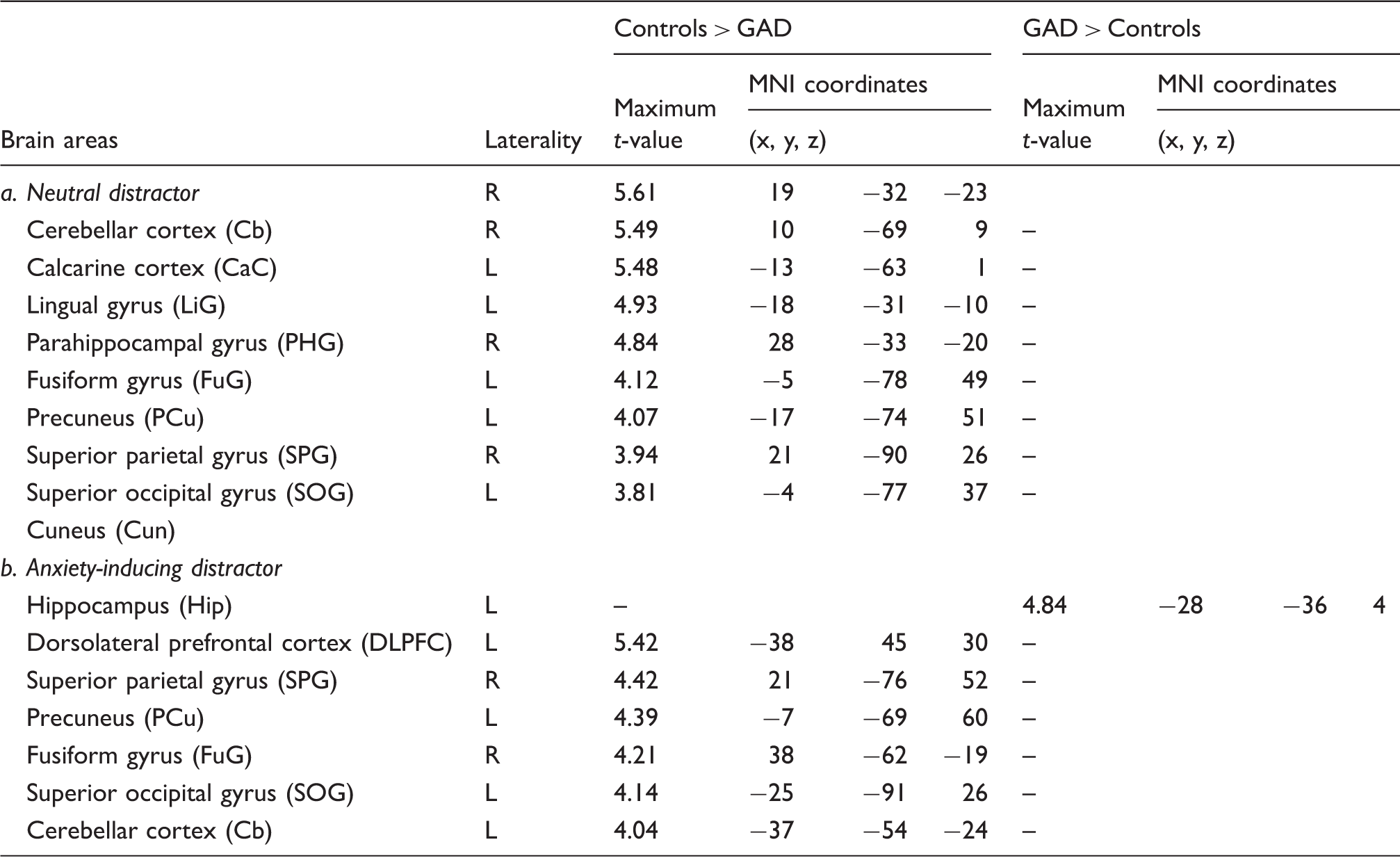
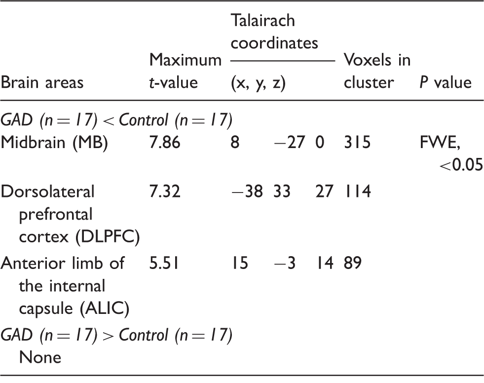
Fig. 3 and Table 3 show the localized WM volume abnormalities in patients with GAD. The patients showed a significant reduction of the WM volumes, particularly in the DLPFC (BA 46), anterior limb of the internal capsule (ALIC), and midbrain (FWE corrected P < 0.05). Contrary to expectations, however, GM volume abnormality in the patients were not observed at a threshold corrected for multiple comparisons (FWE corrected P < 0.05).
Key brain areas demonstrating distinct white matter volume reduction in patients with GAD compared with healthy controls, which resulted from the two-sample t-test (FWE, P < 0.05). The color bar represents the t-values. Regional white matter volume alterations in patients with GAD: two-sample t-test. FWE, family-wise error.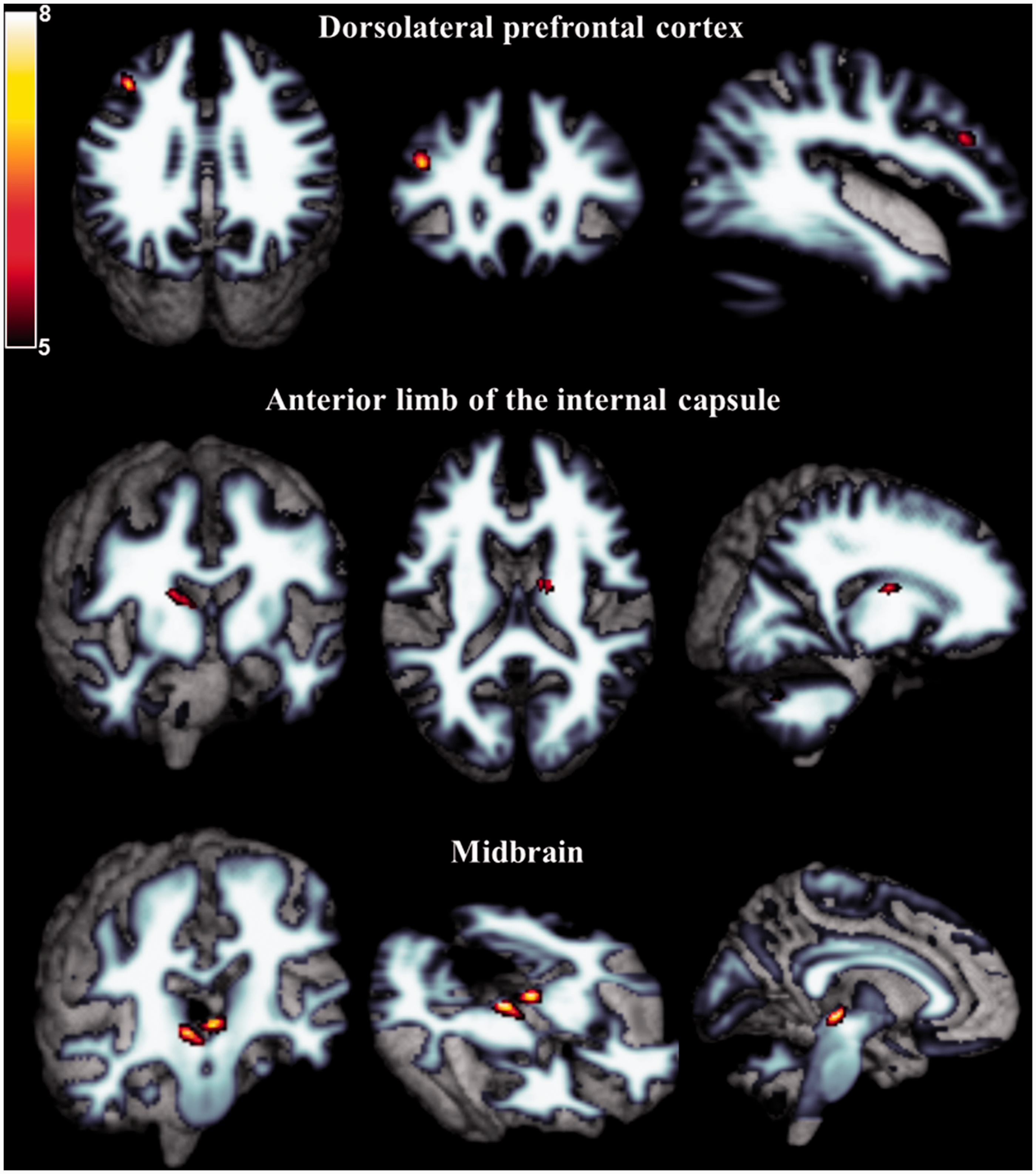
Correlations of the BOLD signal changes with WM volume changes, accuracy, and reaction time
The WM volume change of the DLPFC was positively correlated with BOLD signal change under the anxiety-inducing distractors (Pearson’s correlation coefficient = 0.877, P < 0.0001).
Fig. 4 shows the correlations of BOLD signal changes with accuracy for the recognition task and reaction time under the anxiety-inducing distractors in patients with GAD. The BOLD signal change in the DLPFC (Fig. 4a) is positively correlated with the recognition accuracy (Pearson’s correlation coefficient = 0.758, P < 0.0001), while the BOLD signal change in the Cb (Fig. 4b) is negatively correlated with the reaction time (Pearson’s correlation coefficient = −0.840, P < 0.0001).
The correlations between BOLD signal change in the DLPFC and the accuracy (a) (γ = 0.758, P < 0.0001), and BOLD signal change in the Cb and the reaction time (b) (γ = –0.840, P < 0.001) under the anxiety-inducing distractors in patients with GAD, in which the band with dotted lines shows 95% confidence intervals.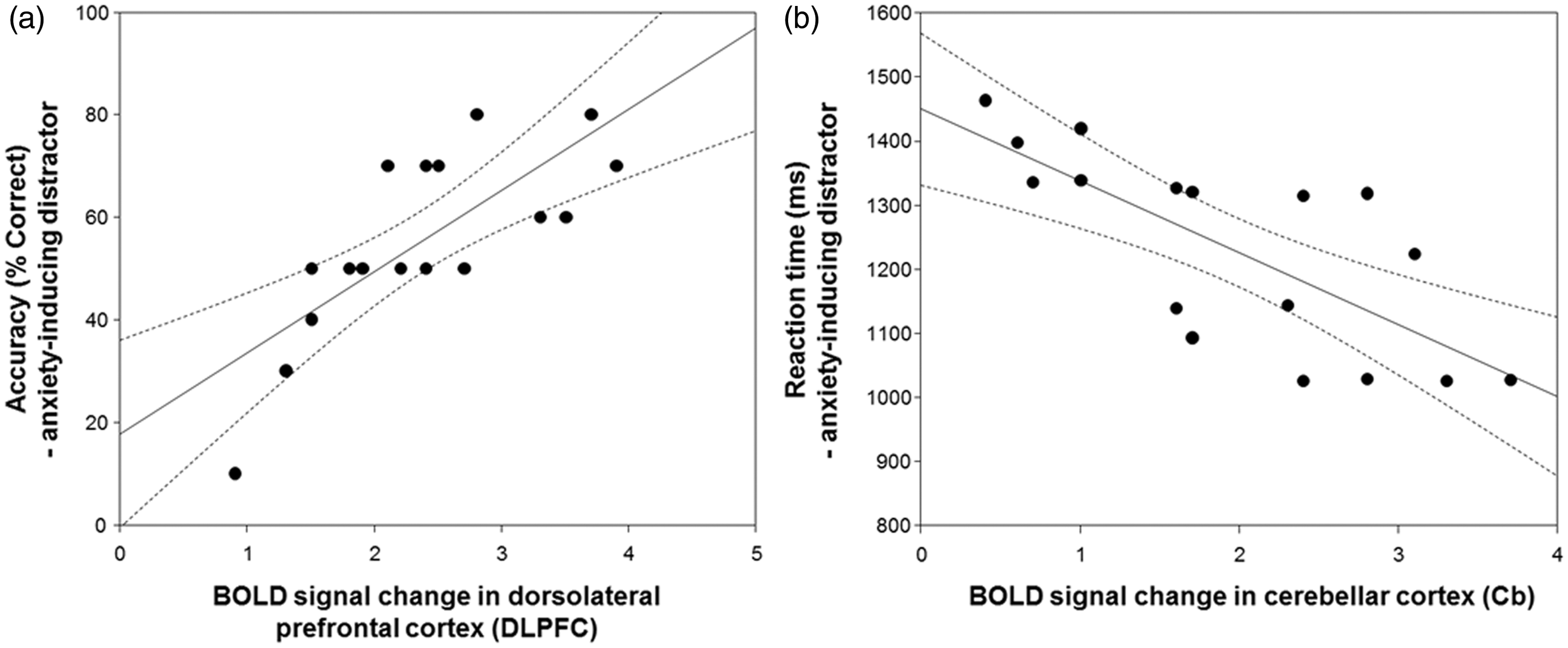
Discussion
The VBM was used to measure the brain anatomical deficits, which may represent more stable and longstanding changes, whereas the fMRI was used to investigate BOLD functional alterations by the indirect physiological changes of regional cerebral blood flow during the activation paradigm (11). Therefore, a combined use of fMRI and VBM is able to provide more valuable information on the neural mechanisms associated with GAD dysfunction.
The most interesting findings in our study are concurrent reductions of WM volume and BOLD signal in the DLPFC in patients with GAD. The DLPFC is a key center involved in cognitive functions including attention and working memory and integration of emotion (6,7). A couple of functional studies (6,12) have suggested that hypo-activation of the DLPFC during viewing negative images reflected attentional deficit and emotional dysregulation in GAD. Moreover, a recent proton MR spectroscopy study (13) demonstrated that GAD is associated with a low level of choline concentration in the DLPFC and the choline plays important roles in the synthesis of various structure and cell signaling molecules, reflecting the symptom severity and cognitive dysfunction in GAD. From these findings, it is assumed that decreased activation of the DLPFC by the anxiety-inducing distractors in patients with GAD is associated with less cognitive function and lower attention in connection with emotional dysregulation compared with healthy controls. Our assumption can be verified by a MRI-based DTI study (14), which demonstrated a significant reduction in fractional anisotropy (FA) in the DLPFC in psychiatric disorders, suggesting the impaired nerve fiber integrity in this region. In the present study, note that WM volume change in the DLPFC was positively correlated with BOLD signal change under the anxiety-inducing distractors; in other words, the DLPFC activation decreases with the WM volume loss. Therefore, it is assumed that activation of the DLPFC in patients with GAD decreased by the nerve fiber degeneration in this area, which is closely connected with the pathological anxiety severity and cognitive dysfunction in working memory tasks with anxiety-inducing distractors (7).
It is also important to note that patients with GAD showed decreased WM volume in the ALIC. Consistent with this finding, a DTI study (15) demonstrated significantly reduced FA in the ALIC of patients with GAD, suggesting that an altered fiber tract in the internal capsule disturbs the connection between the thalamus and neocortex to induce abnormal emotional regulation. The ALIC plays important roles in mediating anxiety, attention, working memory, and language-related brain functions (10,16). WM volume in the midbrain also decreased in patients with GAD, which is consistent with our previous VBM findings showing the decreased GM volume in the midbrain (8). This finding might be associated with reduced glial cell density or glial cell loss in other psychiatric disorders (10). Therefore, alterations in ALIC and midbrain volumes are potentially valuable morphological feature in patients with GAD.
In our fMRI study, another interesting finding is activation of Hip. In patients with GAD, the Hip was predominantly activated in response to anxiety-inducing distractors compared with healthy controls. The Hip is involved in the storage and retrieval of memory, and is also inter-related with the prefrontal cortex associated with various cognitive functions (17). A neuropsychological study (17) demonstrated hyperactivation of the Hip induced by cognitive dysfunction associated with anxiety disorder. Here, we assumed that an increased activation of the Hip in patients with GAD is associated with pathological anxiety response in connection with emotional and cognitive dysregulation. More importantly, the patients showed distinct hyperactivity in the Hip and hypoactivity in the DLPFC simultaneously in the working memory tasks with anxiety-inducing distractors (Fig. 2 and Table 2). Therefore, these findings suggest that the reciprocal brain activation patterns between the Hip and DLPFC are due to dysregulation of the cognitive control and emotional response in GAD and are presumably associated with major symptoms of patients with GAD (7). Together with the Hip, the amygdala (Amg) is involved in emotional regulation in patients with GAD (4), but Amg activation was not found in the current study. A psychopharmacology study (18) reported an increase of Amg activation in response to emotional stimuli in unmedicated patients with GAD, but a decrease of its activation in medicated patients. Here, it is assumed that activation of the Amg would be associated with effects of medication.
Lastly, patients with GAD showed significantly decreased functional activities in the visual center and its association areas (SOG, LiG, CaC, PHG, PCu, Cun, SPG, and FuG) including visual motion area (Cb) in the memory tasks with the neutral and anxiety-inducing distractors. These areas play a critical role in regulation of visual attention and visual information processing (19,20). Particularly, patients with GAD showed longer reaction time in the facial recognition tasks with neutral and anxiety-inducing distractors relative to the healthy controls in our results. Also, the patients showed a negative correlation between BOLD signal change and reaction time in the Cb, which is involved in regulation of eye and cooperative movements (Fig. 4b) (21). From these findings, it is also inferred that the brain functional abnormalities in working memory tasks stems from the hypofunction and/or malfunction of visual attention and information processing in conjunction with GAD symptoms (7).
Our study had several limitations. First, the sample size was small (n = 17) in each group. Future longitudinal studies involving more participants are needed to gain higher statistical power to data. Second, although the GAD patients in this study took medication, the anxiolytic drug effects, which might be linked to the brain activities and volume changes, were not considered. Therefore, a follow-up study is needed to verify the pharmaceutical effects on the variety of type of medications and their dosages and treatment durations (22).
In conclusion, this study demonstrated the functional and morphological alterations in the working memory tasks with neutral and anxiety-inducing distractors in patients with GAD. These findings will be helpful to understand the working memory dysfunction in GAD and to robust the diagnostic accuracy for GAD by additional information on the combined brain functional deficit and cerebral volume change.
Footnotes
Declaration of conflicting interests
The author(s) declared no potential conflicts of interest with respect to the research, authorship, and/or publication of this article.
Funding
The author(s) disclosed receipt of the following financial support for the research, authorship, and/or publication of this article: This work was supported by the funds from the National Research Foundation of Korea (2015R1A2A2A01007827) and Chonnam National University Hospital Biomedical Research Institute (CRI 15011-1).