
Abstract
Background
Renal length, volume, and parenchymal thickness are important clinical parameters, yet data concerning the accuracy and reproducibility of ultrasound (US)-based renal length and volume assessment in patients with chronic kidney disease (CKD) are scarce.
Purpose
To establish whether renal length, volume, and parenchymal thickness can be reliably measured with renal US in patients with CKD.
Material and Methods
All participants underwent renal US, immediately followed by 3-T magnetic resonance imaging (MRI). Renal length, width, transverse diameter, and parenchyma thickness were measured with both methods; renal volume was calculated using the ellipsoid formula. A total of 45 patients with CKD (eGFR [mean ± SD] 57.4 ± 4.4 mL/min/1.73 m2) and 46 participants without CKD (eGFR 97.0 ± 2.4 mL/min/1.73 m2) were included.
Results
US-measured renal length correlated strongly with MRI-measured renal length in no-CKD patients (Spearman’s r = 0.83 and 0.85 for the right and left kidney, respectively; P < 0.005) and CKD patients (r = 0.89 and 0.92 for the right and left kidney, respectively; P < 0.005). There was a significant but weaker correlation between MRI- and US-measured right and left renal volume (r = 0.72, P < 0.005) in no-CKD (r = 0.74 and r = 0.72, respectively; for both: P < 0.005) and CKD patients (r = 0.83 and 0.85, P < 0.005). Weak to moderate correlations were found for parenchyma thickness for the right (CKD group: r = 0.29, no-CKD: r = 0.23; for both: P < 0.05) and left kidney (CKD: r = 0.52, no-CKD group: r = 0.37, P < 0.05). Both intra-observer (Pearson’s correlations of 0.82 for the right and 0.89 for the left kidney) and inter-observer (Lin’s correlation coefficient of 0.90 for the right and 0.82 for the left kidney) reproducibility of US-assessed renal length was high.
Conclusions
US-based assessment of renal length in CKD patients is comparable to MRI measures. Both intra- and inter-observer reproducibility of US-assessed renal length in CKD patients are high. Measurements of US renal volume and parenchymal thickness should, however, be interpreted with caution.
Introduction
Over the last 30 years, renal ultrasound (US) has remained the imaging technique of choice to measure renal length in patients presenting with acute or chronic kidney injury, despite the development of alternative techniques such as computed tomography (CT) and magnetic resonance imaging (MRI) (1). Renal US is widely available, provides reliable anatomic information in experienced hands, and has no side effects. Some studies have reported that renal length measurement is highly reproducible (2–4). However, others have argued that US-based measurements systematically underestimate renal length when compared with MRI-based methods (5). All mentioned studies were performed in healthy volunteers. To the best of our knowledge, no studies have assessed the accuracy and reproducibility of US-based renal length measurement in patients suffering from chronic kidney disease (CKD), although renal length is of the highest importance in clinical decision-making in this patient group. CKD leads generally to a decrease in renal size due to parenchymal atrophy, sclerosis, and fibrosis (6). Renal length is therefore an important clinical parameter, as it provides information on the severity of CKD and its prognosis; besides renal length correlates with estimated glomerular filtration (eGFR) (7,8). Renal length also is associated with histological changes in the kidney, in particular with the prevalence of glomerular sclerosis (9). Finally, renal length can provide information on the underlying disease process. For example, a unilateral small kidney may indicate underlying reflux disease or renal artery stenosis, and progressive shortening of the kidney may be an indication for angioplasty in the latter condition (10,11).
US-determined renal volume was also found to be correlated with glomerular filtration in CKD patients (12). US-based renal volume measurement is considered less reliable and MRI is generally the preferred technique (3,13). Hence, volume cannot be measured directly due to the two-dimensional (2D) nature of US and has to be calculated using the ellipsoid formula or an equivalent, which carries a risk of underestimation of true kidney volume by 21–29% (5). Of note, in clinical practice, the ellipsoid formula is also often used for MR-based volume measurement, instead of other more time-consuming methods such as the disc-summation method. In patients with autosomal dominant polycystic kidney disease (ADPKD), US and MRI measurements of renal length and total renal volume have been shown to be similar (14). However, no studies have compared B-mode US and MRI to assess kidney volume in CKD patients not suffering from ADPKD.
Finally, renal parenchymal thickness is also associated with eGFR and may even be a better indicator of renal function than renal length (15). A longitudinal study in CKD patients suggested that parenchymal thickness measured by ultrasonography has predictive value for renal impairment, at least in an Asian population (16).
The aim of this study was therefore to establish whether renal length, volume, and parenchymal thickness can be reliably measured with renal US in patients with CKD compared with MRI. Furthermore, we aimed to study the intra- and inter-observer reproducibility of US measurements in CKD patients.
Material and Methods
Selection of participants
Participants were selected from an ongoing monocentric research project evaluating the prognostic value of renal tissue oxygenation on the evolution of kidney function (17). All participants underwent a complete physical examination, venous blood sampling, renal US, and a BOLD-MRI exam. BOLD-MRI (blood oxygenation level-dependent MRI) is a non-invasive tool to measure renal tissue oxygenation, based on the paramagnetic properties of deoxy-hemoglobin. The study cohort includes healthy individuals without CKD and patients with CKD (defined as the presence of structural abnormalities detected by imaging or abnormalities detected by histology, estimated glomerular filtration rate [eGFR] < 60 mL/min/1.73 m2 or albuminuria >300 mg/24 h) (18). Patients with CKD were classified into CKD stages according to the Kidney Dialysis Initiative Global Outcomes (KDIGO) work group endorsed guidelines (19).
Inclusion criteria for volunteers without CKD were: eGFR > 90 mL/min/1.73 m2; absence of (micro)albuminuria; and absence of kidney abnormalities at screening renal US.
Exclusion criteria for CKD patients were: ADPKD or eGFR < 15 mL/min/1.73 m2. For the purpose of this study, an additional exclusion criterion for CKD patients was having any renal abnormality hampering measurement of kidney dimensions, such as large cortical cysts or tumors, horseshoe kidneys, or partial nephrectomy. Exclusion criteria for both healthy and CKD individuals were: having a contraindication to MRI (pacemaker, implanted metallic device, claustrophobia); age < 18 years; or incapacity to provide informed consent.
The protocol of the study was approved by the local ethical committee. Written informed consent was obtained from all participants.
Physical examination and laboratory
Body weight was measured with participants in light indoor clothes using a Seca® scale. Height was measured to the nearest 5 mm using a Seca® height gauge. Body surface area (BSA) was calculated using the DuBois formula and body mass index (BMI) was calculated as weight (kg) divided by squared height (m2) (20). eGFR (in mL/min/1.73 m2) was calculated based on the 4D-Modification of Diet in Renal Disease (MDRD) formula (21).
Renal ultrasound
Renal US was performed according to a standardized procedure by two experienced operators using an Aplio XG device (Toshiba Systems, AG/SA Switzerland) equipped with a 2–6 MHz convex transducer. The longitudinal dimensions of each kidney were measured in a section visually estimated to represent the largest bi-polar diameter in inspiration (between the uppermost edge of the upper pole and the lowest edge of the lower pole) (Fig. 1a). Renal length was assessed in the supine position and defined as the maximum length of three consecutive measurements. In case of insufficient image quality, the participant was examined in the left or right decubitus position using a subcostal approach. In order to assess if the respiration phase has an influence on kidney length, measurements were also taken in expiration in 10 participants.
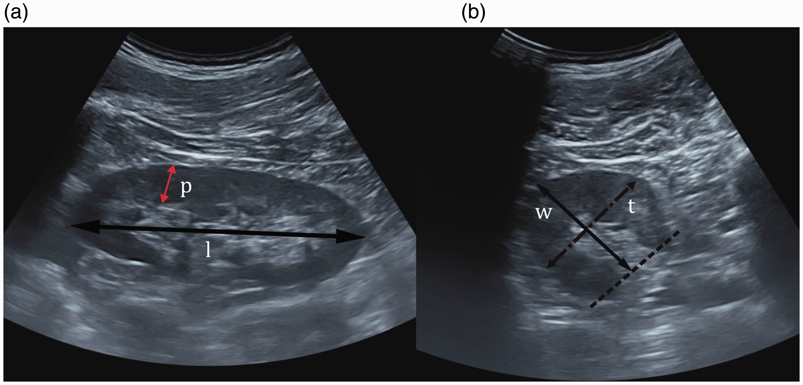
Renal measurements by US B mode. (a) Longitudinal section; (b) transverse section. l, maximum renal pole to pole length; p, parenchymal thickness; w, width; t, thickness. US: ultrasound.
Renal parenchymal thickness was measured as the distance between the sinus fat and the renal capsule, next to the renal pyramids where the thickness had the largest dimension usually at the middle third portion of the kidney (Fig. 1a). The width and thickness were measured in a section perpendicular to the longitudinal axis of the kidney. The level of the transverse section was at the vascular hilum of the kidney. Width and thickness were then measured in two orthogonal directions (Fig. 1b). Renal volume was calculated as length × width × transverse diameter × 0.523 (22). Renal length, parenchymal thickness, and renal volume measurements were performed for each kidney.
As two different physicians performed the US examinations, a concordance study of 20 participants (CKD- and no-CKD patients) was undertaken in which the two operators examined the kidneys on the same day blinded to the other’s results.
In order to assess the reproducibility (intra-observer variability) of US-assessed renal length, the values obtained at the one-year follow-up visit of the cohort were compared with the values of renal length obtained at baseline by the same observer.
Renal MRI
MR measurements were carried out directly after the renal US on a 3-T whole-body MR system (Trio Tim, Siemens Medical Systems, Erlangen, Germany). T1 fat-saturation and T2-weighted (T2W) images were performed in expiration in the sagittal, coronal, and transverse planes for assessment of kidney diameters. Two physicians analyzed the MR images and assessed kidney dimensions. The maximum length of the kidney was measured in a plane which had the largest longitudinal diameter (Fig. 2a). The width and thickness were measured in the transverse plane perpendicular to the longitudinal axis of the kidney at the level of the hilum in two orthogonal directions (Fig. 2b). Renal parenchymal thickness was measured where the thickness had the largest dimension usually at the middle third portion of the kidney. All measurements were made using calipers on a MRI workstation. Renal volume was calculated with the same formula as in US examinations. The two observers performing MRI measurements were not involved in renal US and blinded to the results obtained with US. As for renal US, a concordance study was performed in 20 participants.
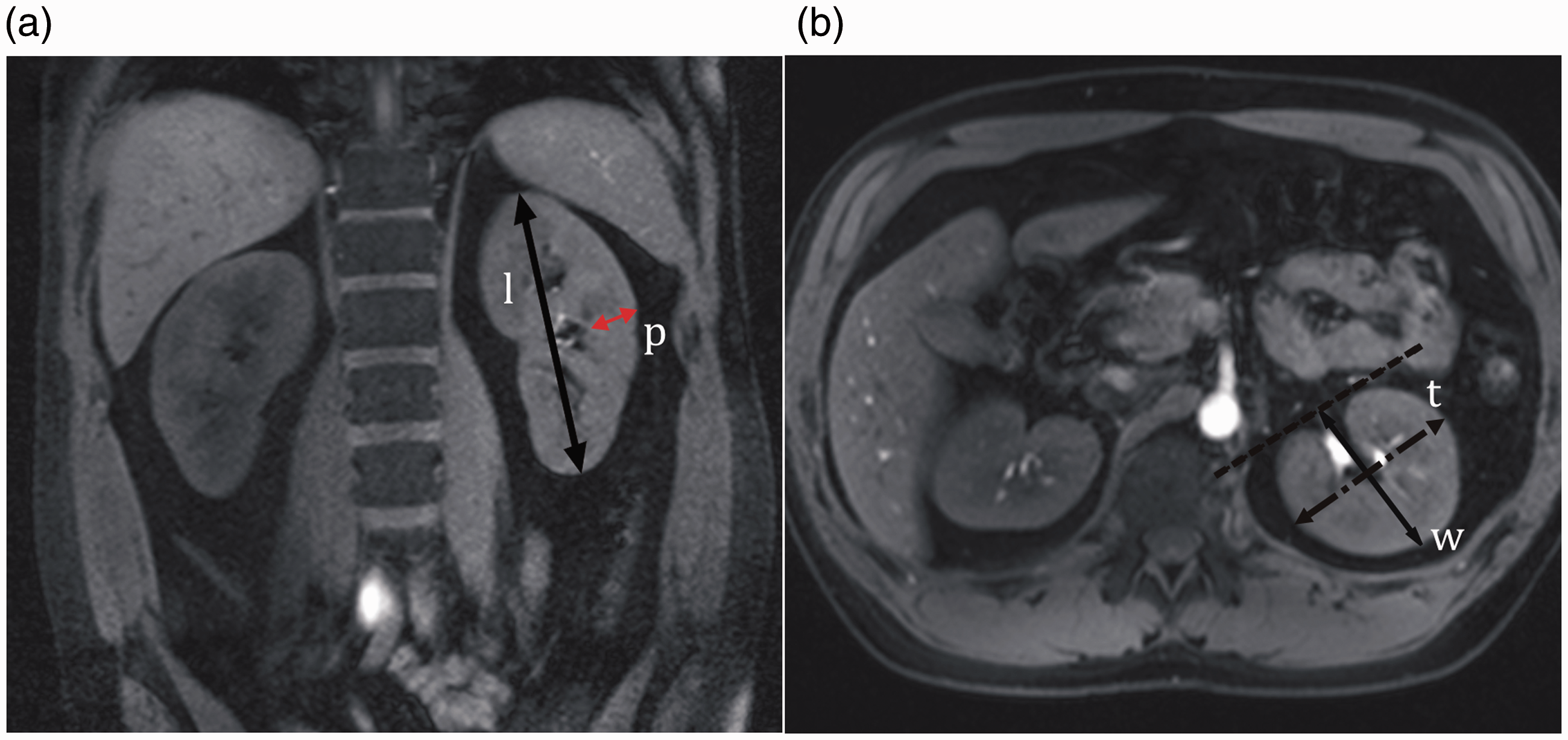
Renal measurements by MRI. (a) Longitudinal section; (b) transverse section. l, maximum renal pole to pole length; p, parenchymal thickness; w, width; t, thickness. MRI: magnetic resonance imaging.
Statistical analyses
Data were expressed as mean and their standard deviation or median (range), as appropriate. In order to compare differences between US- and MRI-assessed dimensions of the right and left kidneys, and differences of US-measured renal length in inspiration or expiration, a paired sample t-test was used.
Accuracy refers to the degree to which a measurement conforms to the correct value. As direct measurement of renal length is not possible in clinical practice, we used MRI-assessed measurement as reference standard. Accuracy of US measures was therefore determined by assessing correlations between US and MRI measurements using Spearman’s rank test. The accuracy of US measurement was further assessed using Bland–Altman plots for agreement between US and MRI measurements for length, volume, and parenchyma thickness of right and left kidneys (23).
Reproducibility refers to the extent to which a test leads to consistent results when repeated (either over time or by another observer). Intra-observer reproducibility of US-assessed renal length was assessed by comparing Pearson’s correlation coefficients and calculating the coefficient of variance (CV), defined as the difference between the baseline and one-year follow-up renal length divided by the mean length of both measurements and expressed in percentage. The inter-observer reproducibility of US and MRI measurements in the concordance study was assessed using Lin’s correlation coefficients. For all tests, P values < 0.05 were considered statistically significant. Statistical analyses were performed with STATA 12.1 software (StataCorp, College Station, TX, USA).
Results
Study population
All 110 participants to the study were screened for eligibility in this analysis. Two people were excluded because they had undergone a partial nephrectomy, one person had horseshoe kidneys, and 16 had other renal abnormalities (mainly cysts) limiting accurate measurement of kidney dimensions, leaving a total of 91 patients for analysis in this study. The group consisted of 45 patients with CKD and 46 participants without CKD. Their baseline characteristics are shown in Table 1. CKD patients were significantly older than healthy controls; other baseline characteristics were comparable between the groups. In the CKD group, eight patients had KDIGO CKD stage 1 (eGFR ≥ 90 mL/min/1.73 m2), eight patients stage 2 (eGFR 60–89 mL/min/1.73 m2), 19 patients stage 3 (eGFR 30–59 mL/min/1.73 m2), and 10 patients stage 4 (eGFR 15–29 mL/min/1.73 m2). The causes of CKD were hypertension (15 patients), glomerulonephritis (six patients), diabetes (six patients), single kidney (six patients), reflux disease (two patients), or not further specified (10 patients).
Baseline characteristics in CKD and no-CKD patients.
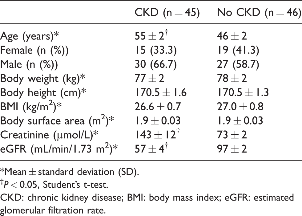
*Mean ± standard deviation (SD).
P < 0.05, Student’s t-test.
CKD: chronic kidney disease; BMI: body mass index; eGFR: estimated glomerular filtration rate.
Accuracy of renal length assessment
No-CKD group
The relationship between US-measured and MRI-measured renal length for participants without CKD is graphically illustrated in Fig. 3. There was a strong correlation between MRI- and US-measured right (Spearman’s rank correlation r = 0.83, P < 0.005) and left renal length (r = 0.85, p < 0.005).
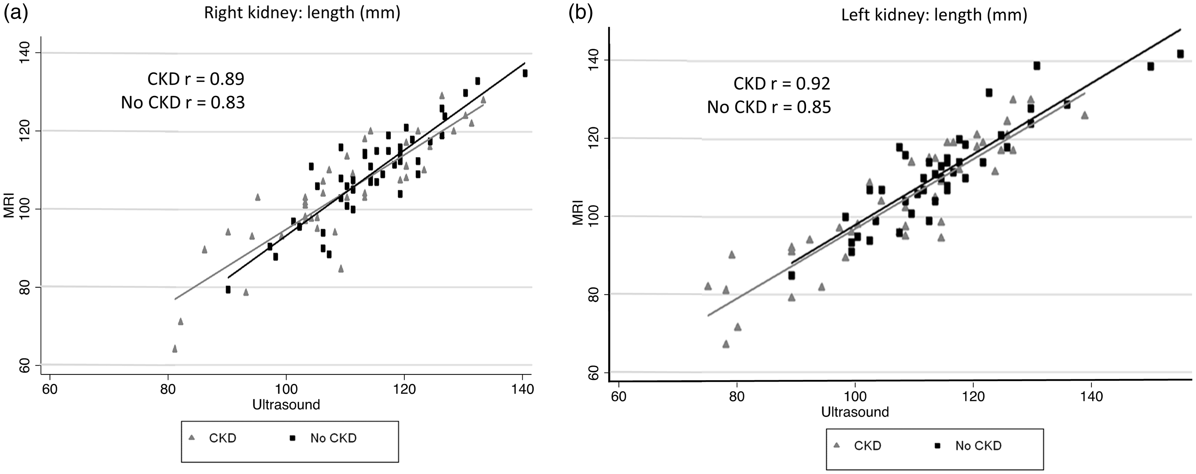
Correlation of MRI and US renal length in no-CKD and CKD patients. (a) Correlation of MRI renal length in mm, US renal length in mm, and Spearman factor correlation (P < 0.01) for right kidney in the no-CDK and CKD groups. (b) Correlation of MRI renal length in mm, US renal length in mm, and Spearman factor correlation (P < 0.01) for left kidney in the no-CDK and CKD groups. CKD: chronic kidney disease; MRI: magnetic resonance imaging; US: ultrasound.
Scatterplots showing the mean of the US- and MRI-measured renal length on the horizontal axis and the difference between the US- and MRI-measured renal length on the vertical axis (Bland–Altman plots) are shown in Fig. 4. The mean difference in renal length between US and MRI was −3.8 mm (95% confidence interval [CI] −5.5–−2.1). Absolute dimensions of US- and MRI-measured renal length in no-CKD participants are shown in Table 2. US-measured renal length was on average 5 mm longer than renal length assessed by MRI: for the right and left kidney, respectively, 5.0 ± 5.4 and 3.9 ± 5.6 mm (P < 0.005).
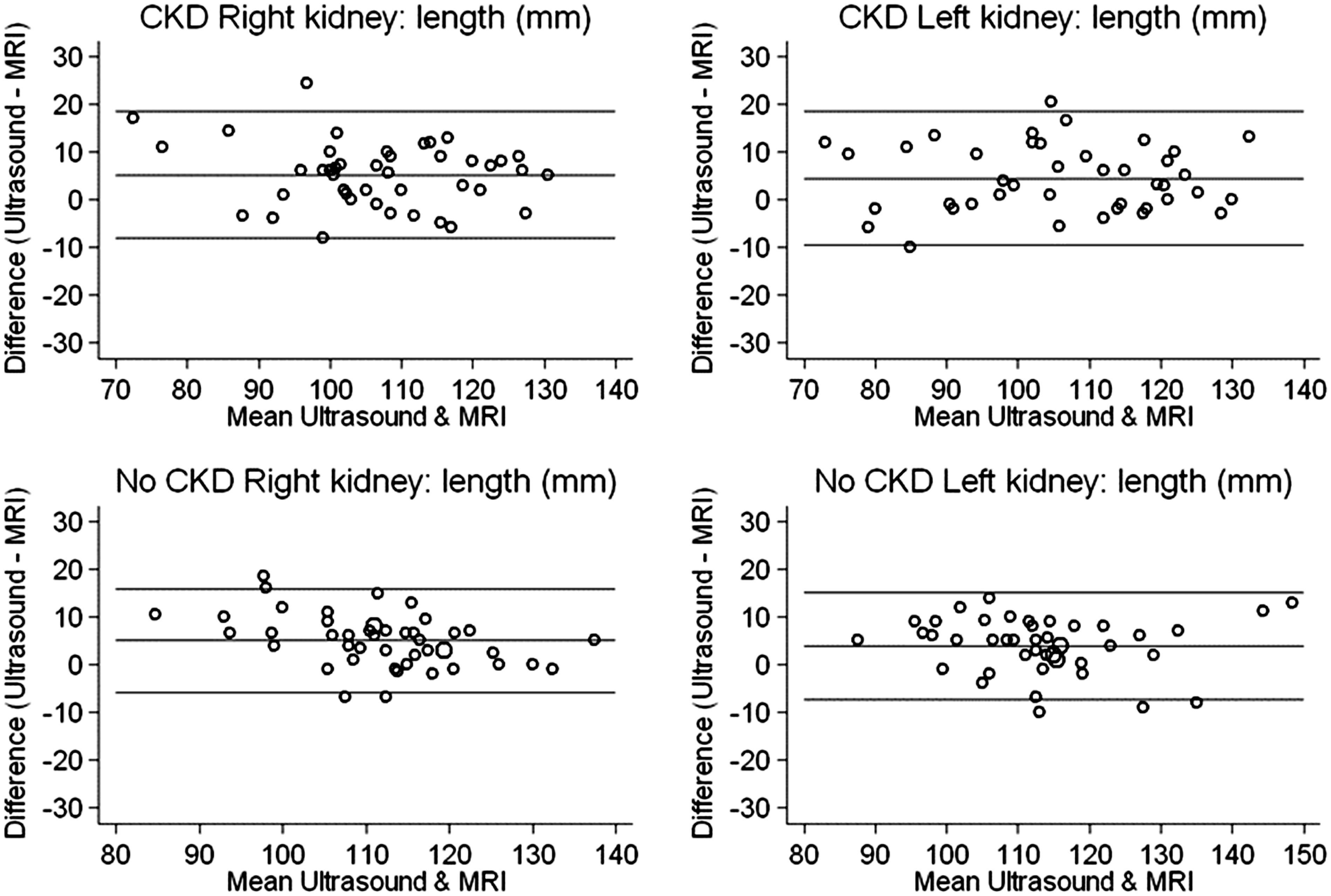
Bland–Altman plot of the difference in renal length (in mm) measured between US and MRI, against the mean measured length in mm. The external lines show the 95% confidence limits on agreement. CKD: chronic kidney disease; US: ultrasound; MRI: magnetic resonance imaging.
Mean renal length, volume, and parenchyma thickness values in CKD and no-CKD participants.
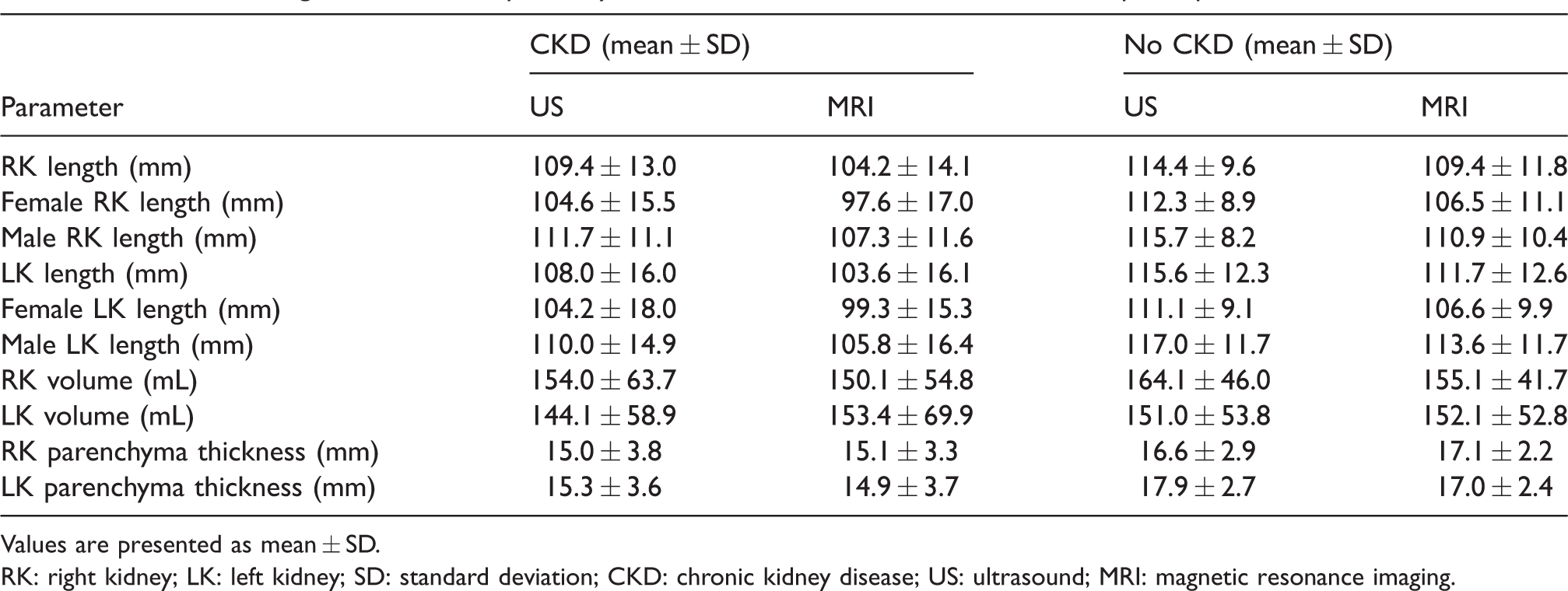
Values are presented as mean ± SD.
RK: right kidney; LK: left kidney; SD: standard deviation; CKD: chronic kidney disease; US: ultrasound; MRI: magnetic resonance imaging.
Left kidneys were longer than right kidneys in the no-CKD group, both with MRI (111.7 ± 12.6 vs. 109.4 ± 11.8 mm, P < 0.05) and US (115.6 ± 12.3 vs. 114.4 ± 9.6 mm).
CKD group
The relationship between US-measured and MRI-measured renal length is graphically illustrated in Figs. 3 and 4. There was a very strong correlation between MRI and US measured right (r = 0.89, P < 0.005) and left renal length (r = 0.92, P < 0.005) in the CKD group. Of note, theses correlations were even stronger than the ones reported for no-CKD patients. The Bland–Altman plots showed a mean difference of −4.4 mm (95% CI = −6.6–−2.1) between US- and MRI-measured renal length.
As for the no-CKD group, US-assessed renal length was ∼5 mm longer than MRI-assessed renal length (right kidney: 5.2 ± 6.7 mm longer; left kidney: 4.4 ± 7.0 mm longer) in absolute dimensions (Table 2). Measurements of renal length according to CKD stages 1–4 are shown in Table 3. As expected, renal length decreased with advancing kidney disease. Through all CKD stages, US-based renal length was about 5 mm longer than MRI measures.
Mean renal length, volume, and parenchyma thickness values according to CKD stages.
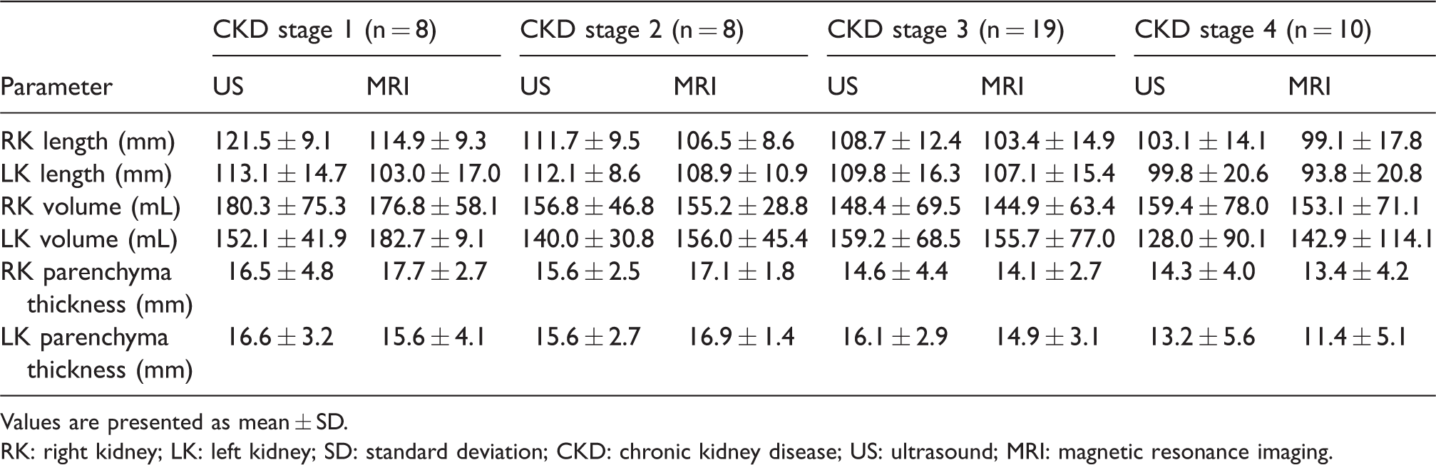
Values are presented as mean ± SD.
RK: right kidney; LK: left kidney; SD: standard deviation; CKD: chronic kidney disease; US: ultrasound; MRI: magnetic resonance imaging.
Accuracy of renal volume and parenchyma thickness assessment
The mean renal volume in the no-CKD group was higher than in the CKD group (Table 2). In the total population, there was no difference in renal volume between the left and right kidney as measured by MRI (152.7 ± 61.0 mL vs. 152.7 ± 48.1 mL; P = 0.71) or US (147.7 ± 56.1 mL vs. 159.3 ± 54.9 mL; P = 0.09). There was a strong correlation between MRI and US measured right (r = 0.83, P < 0.005) and left renal volume (r = 0.85, P < 0.005) in CKD patients (Fig. 5). Significant but weaker correlations were also stated between US and MRI volume measurements for right (r = 0.74) and left kidney (r = 0.72) in the no-CKD group. With increasing CKD stages, renal volume tended to decrease with significant differences between US and MRI measurements, particularly for the left kidney (Table 3).
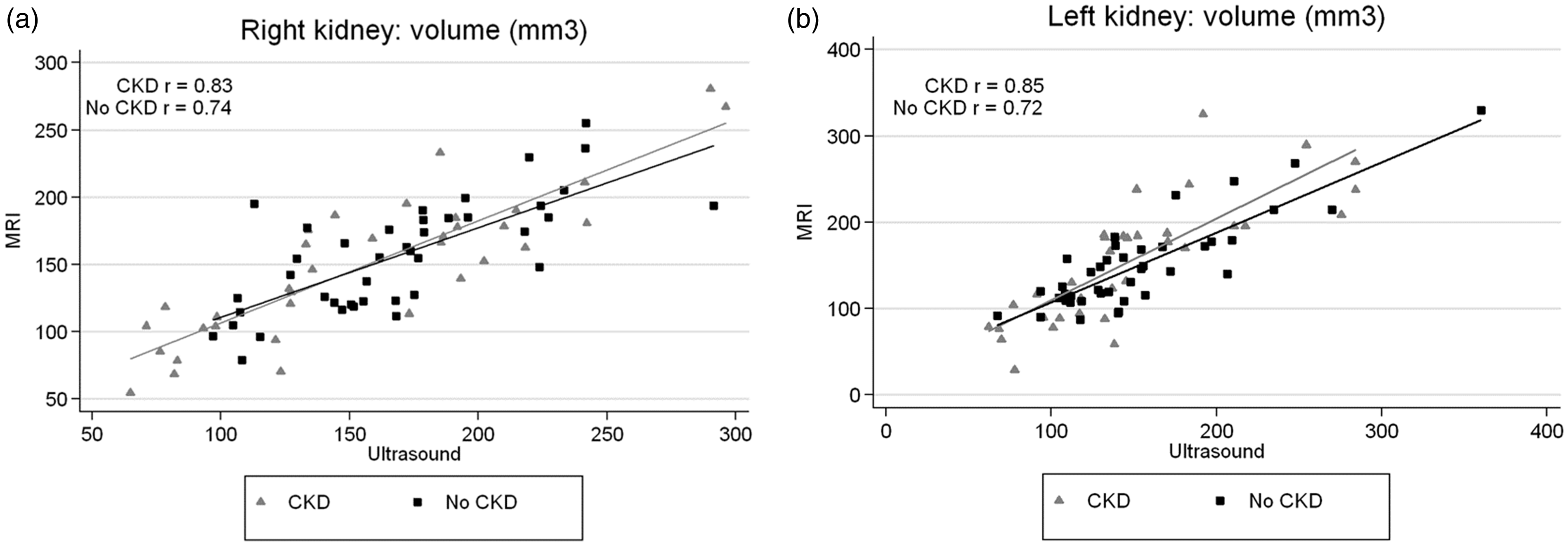
(a) Correlation of MRI renal volume in mm3, US renal volume in mm3, and Spearman factor correlation (P < 0.01) for right kidney in the no-CKD and CKD groups. (b) Correlation of MRI renal volume in mm3, US renal volume in mm3, and Spearman factor correlation (P < 0.01) for left kidney in the no-CKD and CKD groups. CKD: chronic kidney disease; MRI: magnetic resonance imaging; US: ultrasound.
The mean renal parenchyma thickness in the no-CKD group was higher than in the CKD group (Table 2). A weak to moderate correlation between US and MRI parenchyma thickness was found for the right (CKD group: r = 0.29, P < 0.05; no-CKD: r = 0.23, P < 0.05) and left kidney (CKD: r = 0.52, P < 0.05; no-CKD group: r = 0.37, P < 0.05) (Suppl. File; Fig. 1). Renal parenchymal thickness decreased with declining renal function both in US and MRI measures (Table 3).
The Bland–Altman measures of agreement of right and left renal volume and parenchyma thickness in MRI and US for both groups are shown in Fig. 6 and Suppl. Fig. 2. The largest differences were seen for the biggest kidneys.
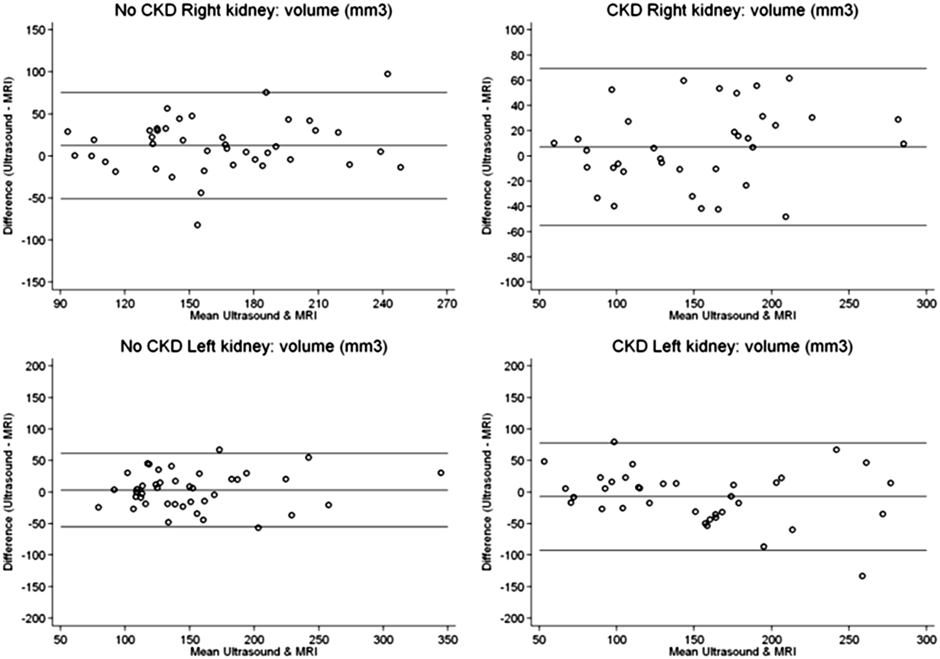
Bland–Altman plot of the difference in renal volume (in mm3) measured between US and MRI, against the mean measured volume (in mm3). The external lines show the 95% confidence limits on agreement. CKD: chronic kidney disease; US: ultrasound; MRI: magnetic resonance imaging.
Intra-observer reproducibility
All included CKD patients underwent a follow-up US one year after the baseline visit. The mean length of the right kidney was 108.3 ± 13.8 mm at baseline versus 106.2 ± 14.0 mm after one year (mean difference = 2.09 mm); for the left kidney, the mean length was 108.1 ± 15.4 mm at baseline versus 107.2 ± 14.4 mm after one year (mean difference = 0.89 mm). The corresponding coefficients of variation were 1.94% for the right kidney and 0.93% for the left kidney; Pearson’s correlations were 0.82 and 0.89, respectively, indicating excellent reproducibility.
Inter-observer reproducibility
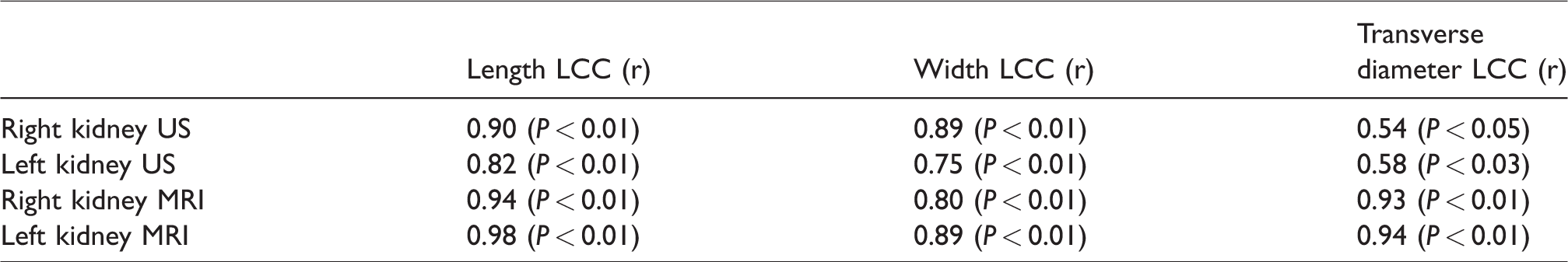
Excellent inter-observer concordance was found for US- and MRI-assessed renal length (Table 4). Concordance was slightly lower, but still good for kidney width. The lowest Lin’s correlation coefficients were found for US-assessed transverse kidney diameter, indicating moderate concordance for that parameter, especially in comparison with MRI result.
Inter-observer Lin’s correlation coefficient (LCC) for US and MRI measurements in a concordance study of 20 randomly selected individuals not included in the analysis of this study.
Influence of the respiration phase on renal length
The mean length of the right kidney was 108.8 ± 4.9 mm in inspiration and 106.3 ± 3.9 mm in expiration (P = 0.141); for the left kidney, the mean length was 110.1 ± 10.3 mm in inspiration and 108.4 ± 9.8 mm in expiration (P = 0.087).
Discussion
The main findings of this study were that: (i) US-measured renal length correlated well with MRI measurements—this was true for the right and left kidney; (ii) the correlation of renal length was even stronger in CKD than no-CKD patients; (iii) US slightly overestimated renal length compared with MRI in both groups and consistently through all CKD stages; (iv) US-based renal volume correlated only moderately with MRI measurements, in particular in no-CKD patients; (v) renal parenchymal thickness correlated only weakly between US and MRI measurements; (vi) intra-observer variability of US-renal length assessment in CKD patients is low indicating a very good reproducibility; (vii) inter-observer variability of renal length was small for renal length, yet large for parenchyma thickness, width, and transverse diameter, underlining that renal US is less appropriate for kidney volume assessment; (viii) a certain degree of inter-observer variability was not only present in US measurements but also in the assessment of kidney dimensions with MRI; and (ix) finally, the respiration phase (inspiration or expiration) does not influence kidney length on US.
Renal US is generally considered as a less reliable technique to assess renal length than CT and MRI, due to its 2D and operator-dependent nature (3,4,24–26). Although renal US has been used for almost 50 years, it is surprising to see that studies assessing the accuracy of US-based measurement of renal length have all been performed during the last decade.
This study shows that renal US is a reliable and reproducible way to assess renal length in patients suffering from CKD. In fact, correlation between US- and MRI-based renal length was even stronger in CKD patients than in no-CKD individuals, although differences were small. This might be partly due to the fact that placement of the longitudinal axis can be challenging with MRI in CKD patients, especially in case of malrotation of the kidneys. Indeed, six cases of malrotation were identified in CKD; the difference between US- and MRI measured length was from 19.5 mm to 26.5 mm in these patients. Contrary to US examination where several longitudinal axes can be placed, the longitudinal axis is usually projected only once in MRI and is dependent of the technician. Hence, in case of patients with malrotation and cross-fused ectopy, the US technique seems to be superior to MRI for renal length measurement.
Despite the fact that the mean differences in measured renal length were small (∼5 mm) and mostly clinically irrelevant, several outliers were found in the CKD and non-CKD groups, with a difference of 1.5–2 cm between both techniques. Most of the outliers had kidneys that were either >125 or <100 mm, suggesting that at the extremes, precision decreases. The four outliers with a renal length in the range of 100–120 mm all had a BMI > 30 kg/m2. This finding underlines the fact that correct renal length measurement with US is more challenging and probably less reliable in obese individuals.
In contrast with previous studies, US-measured renal length was higher than MRI-measured renal length (3,24). This difference was present in both observers, making measurement errors by the observers less likely. Several alternative explanations are possible. First of all, it can be expected that length measurements using MRI and US should differ. While using US the longitudinal renal axis is relatively easy to determine for an experienced operator because the imaging plane in three-dimensional (3D) space can be freely adapted. On MRI, the imaging planes are predetermined unless 3D isotropic sequences are used. Consequently, the greatest renal length on MRI is still probably not measured exactly along the longitudinal renal axis, especially if the imaging plane used for measurements was the true coronal plane. Geometrically, even a small deviation from the exact longitudinal axis would correspond to an underestimation in length measurement and the longitudinal renal axis should rarely lie exactly in the coronal plane. Thus, it seems no surprise that MRI should deliver smaller maximal renal length than US. Second, US measurements were made in inspiration in order to increase visibility of the retro-costal kidneys, whereas MRI measurements were performed in expiration. In theory, the respiration phase might have an influence on renal length due to compression and applanation by the superposed diaphragms, liver, and spleen. However, this hypothesis could not be confirmed as we found no significant difference of kidney length in inspiration and expiration in a sub-group study in 10 participants. Third, US exams were performed by physicians aware of the aim of the study, which might have led to a more focused assessment of renal length. This latter statement remains speculative.
Besides the fact that the measurement of renal length with US is accurate, our results also indicate that it is highly reproducible. High intra-observer reproducibility and inter-observer concordance of measurement techniques are important in everyday clinical practice and these have, to the best of our knowledge, not yet been demonstrated for US-measured renal length in CKD patients.
In the no-CKD group, left kidneys were longer than right kidneys, which is in line with most, but not all, previous studies (2,5,27). This difference has generally been attributed to the fact that the left kidney is less hampered in its growth by the spleen than the right kidney by the liver (2). Besides, the left renal artery is shorter and has a wider diameter than the right one. In contrast, in the CKD group left kidneys were significantly shorter than right kidneys. Why left kidneys were smaller than right kidneys in CKD is unclear. Since left kidney volume was not systematically smaller than right kidney volume, this suggests an alteration in shape with advancing kidney disease that was more pronounced in the left kidney. In a study about the determinants of renal shape in 121 CKD patients, the right and left renal length were similar; however the renal shape index which was defined as renal length/[renal width + renal thickness] was significantly smaller in the left kidney meaning that left kidneys were plumper than right kidneys (28).
The overall correlation between US- and MRI-based renal volume assessment was less strong than for renal length; the inter-observer variability of US-based renal volume measurement was fairly high. For this reason, in our opinion all US analyses concerning renal volume should be interpreted with caution. The most important reason is probably the poor repeatability of the kidney transverse diameter in our study, which has a strong influence on volume measurement (Table 3). Another reason is that measurement errors for the three dimensions are combined for volume calculation, which leads to a greater error and lower correlation of calculated volumes. In clinical practice, the ellipsoid formula is usually used to calculate kidney volume. This method is based on the measurements of the three orthogonal diameters of the kidney and on the assumption that the organ resembles an ellipsoid (22). Of note, MRI- and CT-based methods for fast estimating renal volume also use the ellipsoid formula, although it has been shown that these calculated volumes underestimate real volume by about 25% (3,5,29). Ideally, the disc-summation method, voxel-count method or 3D US should be used for estimation of the volume (3,5,30). Three-dimensional US is a promising technique for renal volume assessment (31). Recent data suggest that morphological kidney parameters provided by 3D US might be more accurate than 2D US (32). However, this technique is not yet established in routine clinical practice.
Finally, US-measured renal parenchyma thickness correlated poorly with MRI measurements. Many problems can be encountered during the measurement of this parameter. The parenchyma thickness varies strongly throughout the kidney and depends on kidney shape. Moreover, cortex measurements are generally taken from the renal capsule to the corticomedullary interface which can be difficult to identify in moderate to advanced CKD patients with poor corticomedullary differentiation and its reproducibility is low (13,15). Renal parenchymal thickness seems to be a more reliable parameter than cortical thickness. Lucisano et al., for example, reported that parenchymal thickness and renal length showed the highest correlation level with eGFR especially after correction of these two parameters by body height (33).
Interestingly, MRI-based renal length measurement was not perfectly concordant between two observers. Since both observers used the same data and length measurement was performed in a pre-defined manner, the only possible explanation is a different appreciation of the longest longitudinal renal axis. This illustrates that any radiologic measurement is subject to inter-individual variability.
This study has several limitations. First, the number of participants was rather small in both groups. Second, patients with renal cysts or ADPKD were excluded from the study; therefore, our findings about accuracy of US-measured length and volume cannot be extended to this patient group. However, strong correlations between US and MRI have previously been reported for ADPKD which suggests that US is also a valid imaging technique in case of renal cysts. Third, renal function evaluation was performed with estimated and not measured GFR. Another limitation is that intra-observer reproducibility was assessed at a one-year interval and renal length might have decreased during this time period. However, no significant difference in length was noticed at one year which makes this hypothesis unlikely. Finally, we compared US with MRI, which, in itself, is not an independent reference standard due to physical and operator dependent measurement errors.
The strengths of this study are the standardization of the radiologic procedures, the fact that US measurements and MRI images were obtained at the same time, and the validation of the measurements in a control group demonstrating low inter-observer variability.
In conclusion, we have shown that renal length measured with US and MRI are similar, both in CKD and no-CKD patients. Surprisingly, the correlation of renal length was even stronger in CKD than no-CKD patients. Besides, renal length assessment with US in CKD patients is also highly reproducible. Renal ultrasound is, however, less appropriate for assessment of renal volume and parenchymal thickness.
Supplemental Material
Supplemental material for How reliable is renal ultrasound to measure renal length and volume in patients with chronic kidney disease compared with magnetic resonance imaging?
Supplemental Material for How reliable is renal ultrasound to measure renal length and volume in patients with chronic kidney disease compared with magnetic resonance imaging? by Philippe Braconnier, Maciej Piskunowicz, Nima Vakilzadeh, Marie-Eve Müller, Emilie Zürcher, Michel Burnier and Menno Pruijm in Acta Radiologica
Footnotes
Declaration of conflicting interests
The author(s) declare no potential conflicts of interest with respect to the research, authorship, and/or publication of this article.
Funding
The author(s) disclosed receipt of the following financial support for the research, authorship, and/or publication of this article: MP is supported by a grant from the Swiss National Science Foundation.
References
Supplementary Material
Please find the following supplemental material available below.
For Open Access articles published under a Creative Commons License, all supplemental material carries the same license as the article it is associated with.
For non-Open Access articles published, all supplemental material carries a non-exclusive license, and permission requests for re-use of supplemental material or any part of supplemental material shall be sent directly to the copyright owner as specified in the copyright notice associated with the article.