






Abstract
Background
Posterior ankle impingement syndrome (PAIS) is a common and debilitating condition, commonly affecting people who participate in activities that involve repetitive ankle plantarflexion. The relationship between clinical and imaging findings in PAIS has not been established.
Purpose
To investigate the relationship between clinical and imaging features in PAIS by reviewing the literature comparing symptomatic patients to asymptomatic controls.
Material and Methods
A systematic literature search was performed to identify all English-language articles that compared imaging features in patients diagnosed with PAIS to imaging in an asymptomatic control group.
Results
A total of 8394 articles were evaluated by title and abstract, and 156 articles were read in full text. No articles compared imaging findings to an asymptomatic control group, thus no articles met the inclusion criteria.
Conclusion
This systematic review found no published research that compared the imaging findings of people diagnosed with PAIS to asymptomatic people. Until this information is available, imaging features in people with posterior ankle impingement should be interpreted with caution.
Introduction
Posterior ankle impingement syndrome (PAIS) is a condition that commonly presents in people who undertake activities requiring repetitive ankle plantar flexion (1–3). It can limit participation and is potentially career-threatening in elite populations (4). It manifests as posterior ankle pain with painful movement restriction and subsequent activity limitation (5–8). Atraumatic PAIS is thought to be caused by cumulative microtrauma secondary to repeated compressive loading in end-range plantar flexion causing impingement of osseous and soft-tissue structures in the posterior ankle region (9).
PAIS is commonly clinically diagnosed in the context of posterior ankle pain associated with ankle plantar flexion (4,10–15). Imaging techniques are frequently employed diagnostically or to guide intervention (13,16–19). Numerous imaging findings have been described in patients with PAIS including anatomical anomalies, bone oedema, soft-tissue inflammation, effusion, and tenosynovitis (Fig. 1) (1,2,5,20,21).
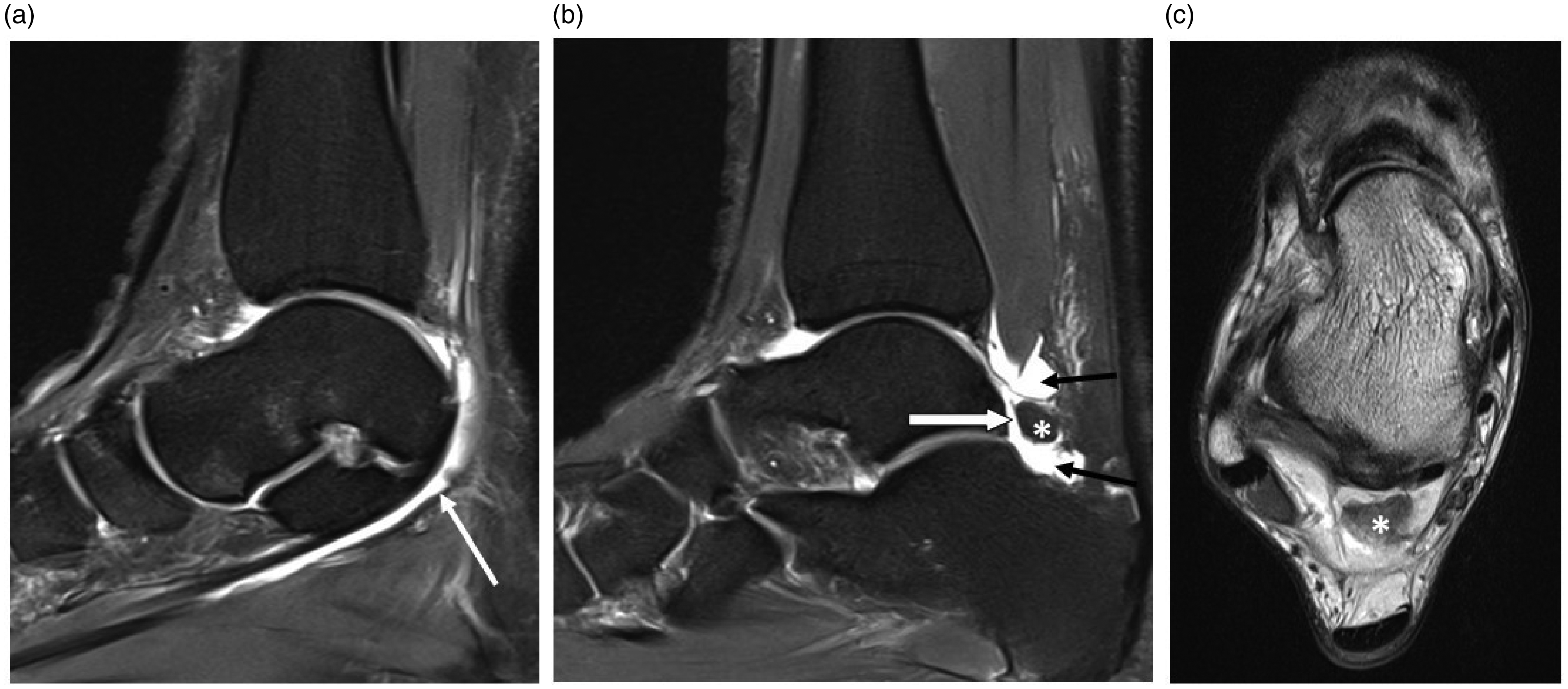
A 29-year-old female soccer player. (a) Sagittal spectral attenuated inversion recovery image through the right ankle reveals flexor hallucis longus tenosynovitis. (b, c) Sagittal spectral attenuated inversion recovery and axial proton-density images through the right ankle demonstrate an os trigonum (*) with increased signal at the synchondrosis (white arrow), and posterior talocrural and subtalar joint synovitis/effusion (black arrows).
There is a lack of consensus regarding the structures and imaging findings that are definitively associated with PAIS (1,2). Osseous and soft-tissue structures occupy space that may compromise the posterior tibio-calcaneal interval. These structures are considered susceptible to compression in plantar flexion and are therefore targets for surgical excision (11,22). The os trigonum, an accessory ossicle in the posterior ankle, has been reported the most common cause of PAIS (Fig. 2) (1,11,23). Early bone scintigraphy studies demonstrated increased uptake in the os trigonum region, labeling the ossicle the source of symptoms (24,25). However, the presence or size of an os trigonum does not necessarily correlate with diagnosis or symptom severity (12,15,26,27).
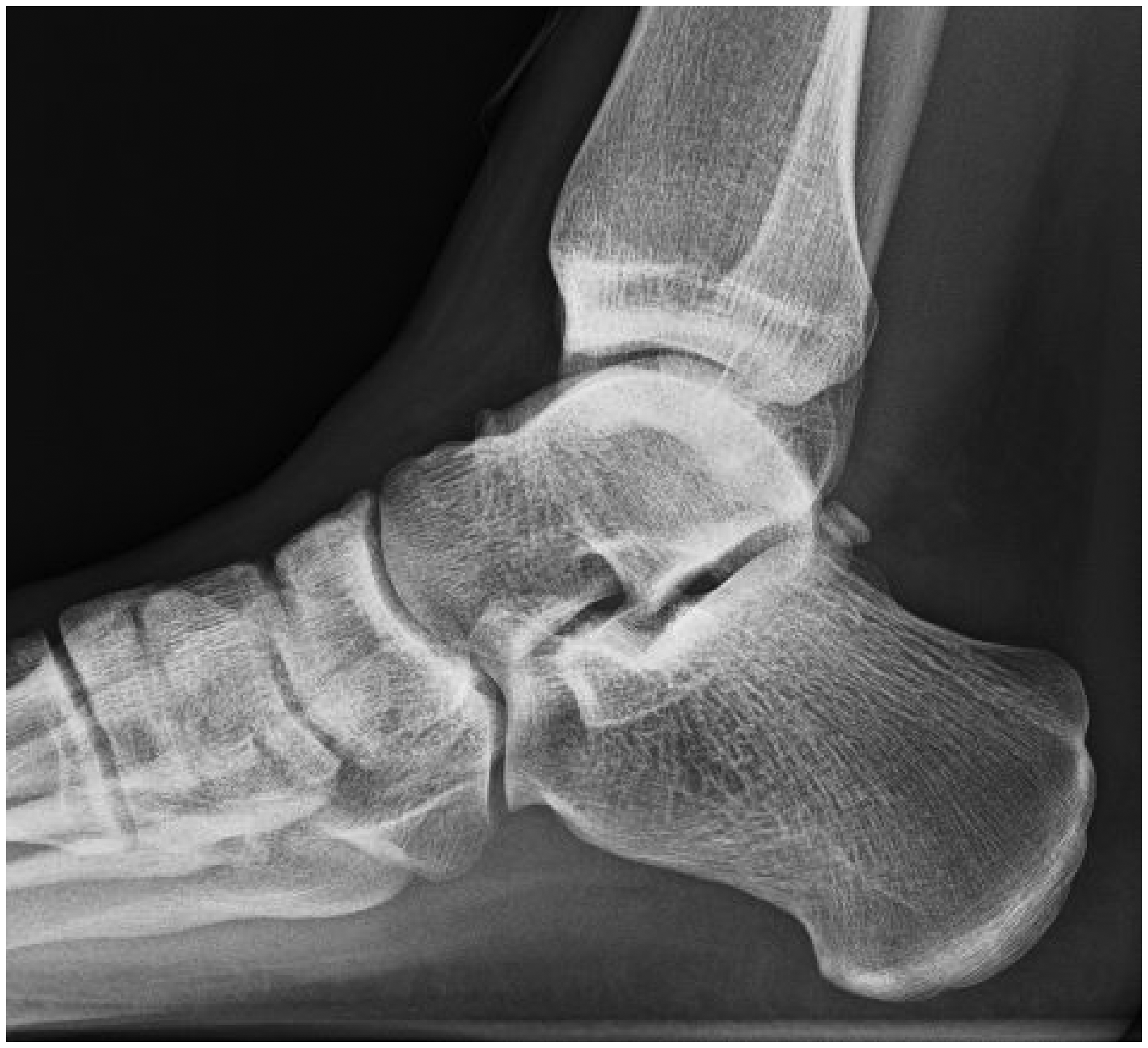
A 25-year-old male ballet dancer. Lateral radiograph reveals an os trigonum.
It has been well established in other body regions that a wide variety of imaging findings in symptomatic patients are also present in asymptomatic individuals (28–35). This has been similarly described in the posterior ankle (36,37). Many foot and ankle imaging studies analyze only symptomatic cases (5,14,20,24), others only asymptomatic (36–38), and some employ either cohort or case-control designs to compare between symptom status (21,39–42). A significant limitation of only analyzing symptomatic cases is the inability to determine the effect of activity exposure on imaging results. As such, rigorous evaluation of the relationship between PAIS-related imaging findings and clinical presentations requires comparison to asymptomatic controls, ideally matched for other confounders such as age and gender.
The aim of the present systematic review was to determine the association between clinical features and imaging findings in PAIS by investigating the differences between symptomatic patients and asymptomatic controls.
Material and Methods
Literature search
A literature search according to PRISMA guidelines (43) was initially conducted in October 2018, and repeated in March 2020, to systematically locate relevant English-language publications reporting imaging features in people diagnosed with PAIS. The following databases were searched: CINAHL, Cochrane, EMBASE, MEDLINE, SPORTDiscus, and Web of Science. Publication year limits were not applied. Search terms were founded on two key concepts: (i) PAIS, and (ii) imaging. Searches used a combination of medical subject headings, keywords, and free text search terms.
Additional methods were employed to identify other relevant publications. The reference lists of all historically key articles in the PAIS literature were manually searched. Abstracts for the 2017–2019 International Association of Dance Medicine and Science conferences were manually searched for contributions referencing PAIS, as substantial PAIS research has been conducted in dancers (1,2). Finally, an electronic search of Google Scholar was conducted using the search terms “posterior ankle impingement” and “imaging” and the first 150 articles by relevance were reviewed.
Study selection
All records returned from database searches were imported into referencing software (Mendeley 1.19.3, Elsevier, Amsterdam, the Netherlands) and duplicates removed. Non-duplicates were imported to a web-based systematic review application (Rayyan QCRI, Doha, Qatar). All studies were assessed for inclusion by two independent reviewers (PB and JL) in two stages (title and abstract, then full text). Disagreements were resolved in consultation with other reviewers (SM and JC).
English-language studies reporting clinical and imaging features in a cohort diagnosed with PAIS compared to an asymptomatic control group were eligible for inclusion. The definition of PAIS must have aligned with symptoms and clinical assessment findings commonly reported in the literature, such as posterior ankle pain with plantar flexion or a positive passive ankle plantar flexion test (10–13,15,16). Common synonyms such as “os trigonum syndrome” or “talar compression syndrome” were included (Table 1). The control group must have had no current or previous clinical ankle symptoms.
Eligibility criteria.
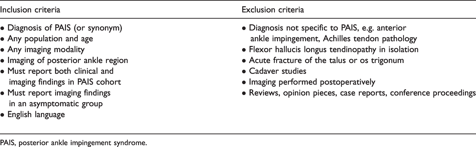
PAIS, posterior ankle impingement syndrome.
Study data extraction, quality assessment, and statistical analyses
Methods for data extraction, study quality assessment and statistical analyses were planned; however, no eligible studies were found.
Results
Literature search
The initial electronic database search (October 2018) returned 7424 unique entries. The initial title and abstract screening phase excluded 7281 articles. Full text screening of the remaining 143 articles returned no eligible studies. The second electronic database search (March 2020) identified 970 articles published since the initial search. Of these, 13 underwent full text screening and no eligible studies were found. No further articles were retrieved via hand searching methods. Ultimately, no studies reported imaging findings in an asymptomatic control group, therefore no studies were eligible for inclusion (Fig. 3).
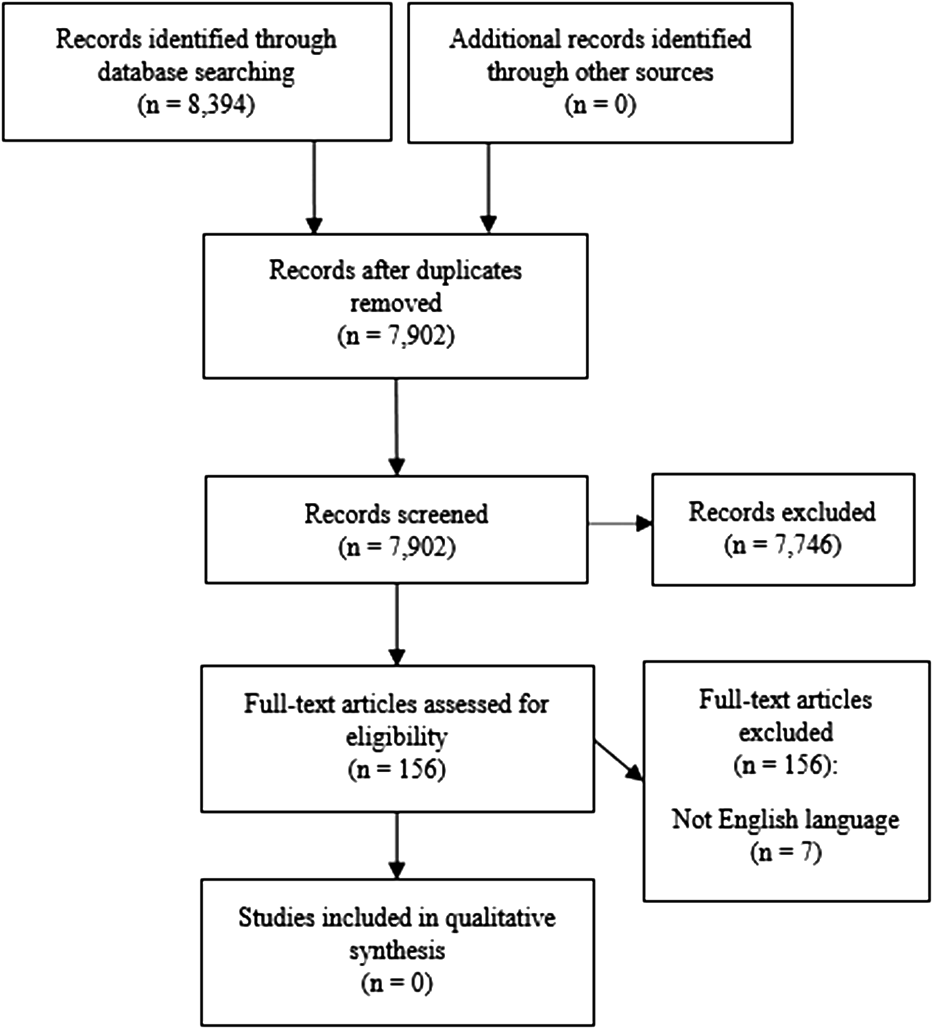
PRISMA flow chart demonstrating study selection process.
Discussion
This review found no studies that compared imaging findings in people with PAIS to asymptomatic people. This result highlights a gap in the understanding of the etiology of PAIS and questions the clinical significance of imaging findings in people with PAIS. Clinical decision-making based on imaging findings may therefore lead to inappropriate management such as surgery, and repercussions in elite populations may be career-ending.
The literature search identified three studies that compared os trigonum prevalence on imaging between patients with clinically diagnosed PAIS and patients with symptomatic, non-PAIS ankles (40–42). Zwiers et al. (40) reported os trigonum prevalence on computed tomography in a group of patients referred to a medical center for various foot and ankle symptoms. Prevalence of os trigonum was 46.4% in patients with posterior impingement complaints, and 30.3% in those without (40). Kushare et al. (42) reported a prevalence of os trigonum of 47% on magnetic resonance imaging (MRI) in a group of pediatric patients diagnosed with PAIS, compared to 5% in an age-matched control group. The control group were symptomatic patients who underwent MRI for acute ankle presentations that were deemed atypical for PAIS. Ozer et al. (41) reported a prevalence of os trigonum of 70.1% in a PAIS “positive” group versus 1.7% in a PAIS “negative” group. Their study population was all clinically diagnosed with either anterior or posterior ankle impingement syndrome, and MRI was used to confirm the clinical diagnosis (41). In this study, the presence of an os trigonum on MRI was a criterion for classifying a patient as PAIS “positive,” which potentially explains why prevalence of os trigonum was so high in their PAIS group compared to the PAIS “negative” group, and greater than that reported in other studies. Prevalence of os trigonum has previously been reported to be in the range of 7%–25% in the general population (25,44). Although these three papers reported a higher os trigonum prevalence in PAIS groups than what is generally considered “normal,” these studies were not included in the systematic review because the comparison groups used were all symptomatic. Symptomatic comparison groups, regardless of diagnosis, may demonstrate concomitant pathology on imaging; therefore, the data are potentially confounded. Furthermore, the comparison groups in these studies were not age-matched (excepting Kushare et al. (42)) nor sex- or activity-matched, potentially further confounding the data.
Research describing musculoskeletal imaging features in areas such as the lumbar spine (28–31), knee (32), hip (33), and patellar tendon (34,35,45) demonstrates a high prevalence of pathological findings and morphologic alterations in the absence of symptoms or dysfunction. The same phenomenon has been observed across several foot and ankle imaging modalities in various high-load populations including asymptomatic marathon runners (38,46), retired ballet dancers (39,47), currently practicing ballet dancers (36,37), and soldiers on active duty (48). In active people, exposure to chronically elevated loads or extreme ranges of motion may contribute to changes on imaging but not necessarily symptoms. As such, the clinical implications of foot and ankle imaging findings in active populations, including those at risk of PAIS, are unclear.
A strength of this review is the application of stringent study inclusion criteria. Investigating the relationship between imaging features and symptoms should not be based on research in only symptomatic groups. A deliberately sweeping search including grey literature was conducted, yet no publications met criteria. A limitation to this review was the inclusion of only studies published in English language, which may have restricted search results.
The imaging features in people with PAIS may not be correlated with the pain and dysfunction experienced clinically. However, this does not mean that these commonly described imaging features (os trigonum, bone marrow oedema, posterior ankle synovitis, etc.) have no connection to clinical presentation. Future research needs to compare imaging findings between people with clear clinically diagnosed PAIS to asymptomatic age-, sex-, and activity-matched groups. Ideally, prospective longitudinal research in at-risk populations would provide greater understanding of the clinical significance of imaging features in PAIS.
In conclusion, there has been no research to date that has compared imaging in people with PAIS to asymptomatic people. There is therefore no evidence of the presence or absence of a causal relationship between imaging findings and clinical presentation in PAIS. Until imaging data comparing people with PAIS to appropriately matched asymptomatic groups are available, the significance of imaging features in people with PAIS should be interpreted with caution.
Footnotes
Declaration of conflicting interests
The author(s) declared no potential conflicts of interest with respect to the research, authorship, and/or publication of this article.
Funding
The author(s) received no financial support for the research, authorship, and/or publication of this article.
