






Abstract
Keywords
Introduction
Breast cancer is one of the most common malignancies among women worldwide and remains the most frequently diagnosed cancer among women in China.1,2 Over the past decade, significant advances in mammography screening, magnetic resonance imaging (MRI), and systemic treatment strategies have improved early detection and clinical management of breast cancer. 3 Despite these improvements, global breast cancer incidence and mortality rates continue to rise, with notable regional disparities. Moreover, current treatment modalities are often limited by systemic toxicity, drug resistance, risk of recurrence, lack of specificity, and collateral damage to surrounding healthy tissues, which collectively contribute to patient suffering and compromised survival.4,5 Therefore, there is an urgent need to develop novel and effective treatment strategies to improve clinical outcomes for patients with cancer.
In recent years, nanotechnology has experienced exponential growth, with nanoparticles demonstrating increasing impact across multiple areas of healthcare. 6 Magnetic nanoparticles (MNPs), in particular, have been extensively explored for biomedical applications such as MRI, 7 targeted drug delivery, 8 gene therapy, 9 magnetic separation of cells or biomolecules, 10 in vivo cell tracking, 11 and cancer hyperthermia.12,13 Under an alternating magnetic field (AMF), MNPs generate localized heat (41°C–46°C) through Brownian and Néel relaxation mechanisms. 14 Tumor cells exhibit greater thermal sensitivity than normal cells, 15 and due to insufficient blood perfusion, cancerous tissues dissipate heat poorly, making them more vulnerable at temperatures above 42°C. 16 Sustained heating above 43°C induces protein denaturation, 17 increases membrane permeability, 18 causes organelle swelling, 19 and results in irreversible DNA damage, 20 with minimal impact on healthy tissues. 21 Compared with conventional radiotherapy and chemotherapy, MNP-mediated magnetic hyperthermia offers a promising alternative with fewer side effects and is increasingly recognized as an important oncotherapeutic approach. 22
Among MNPs, Fe3O4 nanoparticles exhibit superparamagnetic properties (SPIONs) and are widely used in biomedicine because of their excellent magnetism, high stability, excellent biocompatibility, and low toxicity. Magnetic hyperthermia presents several advantages over chemo- or radiotherapy, including efficient uptake of MNPs by cancer cells; non-induction of drug resistance; low systemic toxicity; ease of surface functionalization and size control; high heating efficiency and tissue penetration; and straightforward synthesis, high chemical stability, and long shelf life.23–26 These properties enhance treatment safety and underscore the significant clinical potential of MNPs. Despite promising results, poor targeting efficacy and suboptimal heating performance remain major obstacles to clinical translation. Factors such as particle size, morphology, and surface chemistry critically influence the heating efficiency and targeting specificity of magnetic nanomaterials. 27 Therefore, the development of nanoparticles decorated with specific bioactive molecules capable of recognizing cellular receptors is essential for achieving targeted accumulation and enhanced therapeutic outcomes.
Aptamers, often termed “chemical antibodies,” have emerged as attractive targeting ligands due to their high specificity, low immunogenicity, small size, deep tissue penetration, and ease of synthesis and modification. 28 Compared with conventional antibodies, aptamers offer economic production, facile modification, and minimal immunogenicity, highlighting their promise in detecting breast cancer biomarkers. 29 The S1-4 aptamer is a 48-nucleotide (nt) oligonucleotide that specifically binds to estrogen receptors (ERs) on the surface of MCF-7 breast cancer cells, enabling active targeting. 30 Notably, ER-positive (ER+) breast cancer accounts for approximately 50% of all cases and is primarily treated with endocrine therapy, which can lead to adverse effects such as osteoporosis, endometrial hyperplasia, arthralgia, and even secondary malignancies. 31 Recent studies have demonstrated that aptamer-conjugated MNPs can actively accumulate in cancer cells, thereby improving hyperthermia efficacy. For instance, Zhu et al. 32 developed S2.2 aptamer-conjugated magnetic nanoliposomes for theranostics in breast cancer. Zhang et al. 33 functionalized small extracellular vesicles with the AS1411 aptamer, enhancing tumor targeting and promoting doxorubicin (DOX) accumulation. Jurek et al. 34 reported dextran-coated SPIONs modified with a DNA aptamer for improved hyperthermia in human osteosarcoma. Chen et al. 35 constructed AS1411 aptamer-decorated zinc-doped iron oxide nano-octahedra co-loaded with DOX and HSP90 siRNA, achieving synergistic chemo-hyperthermia under AMF. Collectively, these studies underscore the potential of aptamer-modified Fe3O4 nanoparticles for targeted and biocompatible cancer therapy. However, most previously reported aptamer-functionalized magnetic nanoplatforms have used different aptamers, complex carriers, or combined drug/gene payloads; comparatively few studies have focused on the S1-4 aptamer/membrane ERα axis for magnetic hyperthermia in ER+ MCF-7 cells.
In this study, citric acid (CA)-coated Fe3O4 nanoparticles (FCA) were synthesized via a co-precipitation method. The CA shell not only enhances water solubility and biocompatibility but also provides carboxyl groups for conjugation with amine-modified S1-4 aptamers, resulting in the formation of S1-4-functionalized nanoparticles (FCS). The resulting FCS nanoparticles enable active targeting and accumulation in breast cancer cells, enhancing magnetic hyperthermia efficacy. This work lays a material and theoretical foundation for future in vivo applications of FCS in tumor-specific magnetic hyperthermia. Therefore, the novelty of the present work lies in integrating the ER-targeting S1-4 aptamer with a simple CA-coated Fe3O4 magnetic core to enhance cellular uptake and AMF-triggered tumor cell killing without introducing additional chemotherapeutic or gene payloads.
Materials and methods
Reagents and instruments
S1-4 aptamer was purchased from Qingdao Medicises Biotechnology Co., Ltd (China). FeCl3·6H2O, FeCl2·4H2O, hydrazine hydrate, and CA were obtained from Shanghai Aladdin Biochemical Technology Co., Ltd (China). Fetal bovine serum was purchased from AusgeneX (Australia). MCF-7 cells (Research Resource Identifier (RRID): CVCL_0031) and MEF cells were obtained from the Shanghai Cell Bank, Chinese Academy of Sciences (China). No RRID was available for the MEF cells used in this study. Cells were cultured according to the supplier's instructions. Nanoparticle size and zeta potential were measured using a Zetasizer Nano (Malvern; UK), and transmission electron microscopy (TEM) was performed using a JEM-1400 instrument (JEOL; Japan).
This study was conducted entirely in vitro using established cell lines and did not involve human participants, human tissue, or identifiable personal data. Therefore, ethics approval and written informed consent were not required, and the Declaration of Helsinki was not applicable.
Synthesis of FCA and FCS nanoparticles
Fe3O4 nanoparticles were synthesized via a chemical co-precipitation method. Briefly, 2.7 g of FeCl3·6H2O and 1.0 g of FeCl2·4H2O were dissolved in 20 mL of deionized water. Subsequently, 5 mL of ammonia solution and 2 mL of hydrazine hydrate were added, and the volume was adjusted to 70 mL with deionized water. The mixture was heated to 90°C under mechanical stirring for 30 min. Subsequently, 10 mL of CA (4 g/mL) was added, and stirring was continued for 1.5 h. The resulting black precipitate was collected using a magnet, washed three times with deionized water, and dried at 60°C under vacuum for 12 h to obtain CA-modified FCA.
For aptamer conjugation, 1 mL of FCA suspension (2 mg/mL) was activated with 1-ethyl-3-(3-dimethylaminopropyl) carbodiimide (EDC) and N-hydroxysuccinimide (NHS) under shaking for 30 min at room temperature. The activated nanoparticles were magnetically separated and washed three times. The S1-4 aptamer (5 optical density (OD) units) was denatured at 89°C for 10 min, followed by renaturation on ice for 10 min. The aptamer was then incubated with the activated nanoparticles for 5 h under shaking. The resulting product (FCS) was collected magnetically and washed three times with deionized water.
Characterization of nanoparticles
Crystal structure and phase composition were analyzed by X-ray diffraction (XRD). Morphology and size were examined by TEM. Samples were dispersed in ethanol, dropped onto carbon-coated copper grids, and air-dried before imaging. Hydrodynamic diameter and zeta potential were measured using dynamic light scattering (DLS). Surface functional groups were characterized by Fourier transform infrared spectroscopy (FTIR). Successful aptamer conjugation was confirmed by ultraviolet-visible (UV-Vis) spectroscopy. DLS and zeta potential measurements were obtained from three independent measurements for each sample. Representative TEM, high-resolution transmission electron microscopy (HRTEM), XRD, selected area electron diffraction (SAED), FTIR, and UV-Vis results are shown.
Magnetic hyperthermia setup and heating properties
Magnetic properties were measured using a vibrating sample magnetometer (VSM) with approximately 10 mg of sample. AMF heating experiments were conducted using a commercial magnetic hyperthermia system operating at 474 kHz and 34.7 A. For heating measurements, 1 mL nanoparticle suspensions were placed in nonmetallic tubes and positioned at the center of the induction coil to ensure uniform field exposure. Temperature was recorded in real time using an infrared camera. The induction coil had an inner diameter of 44 mm with 8 turns. All experiments were performed under identical AMF conditions, sample volume, and positioning to ensure comparability. For concentration-dependent heating experiments, FCA and FCS were tested at 0.075, 0.15, 0.3, and 0.6 mg/mL under identical AMF conditions. For cycling stability tests, 0.6 mg of FCA or FCS dispersed in 1 mL deionized water was subjected to five heating–cooling cycles (up to 52°C) under AMF. Deionized water under the same conditions was used as a blank control to account for background heating. Specific absorption rate (SAR) values were calculated from the initial slope of the temperature–time curves obtained at 0.6 mg/mL over 300 s under identical AMF conditions after blank correction. The SAR was determined using Equation (1)
36
and expressed as the energy absorbed per unit mass (W/g):

Biocompatibility assessment
MCF-7 and MEF cells were seeded in 96-well plates and treated with FCA or FCS at concentrations of 0.075, 0.15, 0.3, and 0.6 mg/mL for 24 h. Cell viability was assessed using the 3-(4,5-dimethylthiazol-2-yl)-2,5-diphenyltetrazolium bromide (MTT) assay. Absorbance was measured at 490 nm using a microplate reader. Blank correction was performed using wells containing culture medium and MTT reagent but no cells, and cell viability was calculated as follows:

Three independent biological experiments were performed for each cell line. Within each experiment, each condition was tested in triplicate wells as technical replicates, and the averaged value was used for statistical analysis.
In vitro targeting assay
MCF-7 cells were incubated with FCA or FCS (0.6 mg/mL) for 8 h, fixed with paraformaldehyde, and stained using Prussian blue staining kit according to the manufacturer's instructions. Cells were observed under an optical microscope. The staining experiment was repeated independently three times, and representative images are shown.
Magnetic hyperthermia in tumor cells
MCF-7 cells were treated with FCA or FCS and exposed to AMF (42°C, 10 min). For cell-treatment experiments, samples were exposed using the same commercial AMF system and were placed at the same reproducible central coil position as in the nanoparticle heating studies, so that the AMF exposure conditions were matched as closely as possible across the physical heating and biological assays. Cell viability was evaluated using the MTT assay and live/dead staining (calcein-acetoxymethyl ester (AM)/propidium iodide (PI)). Apoptosis was analyzed by flow cytometry using an Annexin V-fluorescein isothiocyanate (FITC)/PI apoptosis detection kit. For the post-AMF MTT assay, cell viability was calculated using the same blank-corrected formula described above. Three independent biological experiments were performed, with each condition tested in triplicate wells as technical replicates in each experiment. Live/dead staining and flow cytometric apoptosis analyses were each performed in three independent experiments, and representative images/plots are shown where applicable. For half-maximal inhibitory concentration (IC50) determination in the AMF-treated MTT assay, an expanded concentration series was tested to generate complete dose–response curves for FCA and FCS. The resulting viability–concentration data were fitted by nonlinear regression using a four-parameter logistic (4PL) model in GraphPad Prism. IC50 values were determined based on three independent biological experiments, each performed in triplicate.
Statistical analysis
Data are presented as mean ± SD. Unless otherwise stated, in vitro experiments were performed with three independent biological replicates (n = 3), with technical replicates averaged prior to analysis. Nanoparticle characterization measurements were also conducted in triplicate. Normality was assessed using the Shapiro–Wilk test where applicable. Differences between groups were analyzed using one-way analysis of variance (ANOVA) or the Mann–Whitney U test, as appropriate. Statistical analyses were performed using GraphPad Prism, with two-sided tests and p <0.05 considered statistically significant. For AMF-treated dose–response experiments, IC50 values were determined by nonlinear regression using a 4PL model.
Results
Synthesis and characterization of nanoparticles
As shown in Figure 1, TEM images revealed that both FCA (Figure 1(a)) and FCS (Figure 1(b)) nanoparticles exhibited spherical morphology, with no significant change in shape following aptamer modification. HRTEM imaging of FCA (Figure 1(c)) showed a lattice spacing of 0.49 nm. The XRD pattern of FCA (Figure 1(d)) displayed characteristic diffraction peaks at 30.4°, 35.7°, 43.5°, 53.8°, 57.3°, and 63.1°, corresponding to the (220), (311), (400), (422), (511), and (440) crystal planes of Fe3O4, respectively, which aligns well with the standard Fe3O4 reference (PDF#26-1136), indicating high crystallinity. The lattice spacing of the (220) plane observed in XRD corresponded to the 0.49 nm spacing measured using HRTEM. SAED analysis further confirmed the face-centered cubic structure of FCA (Figure 1(e)), consistent with the XRD results.
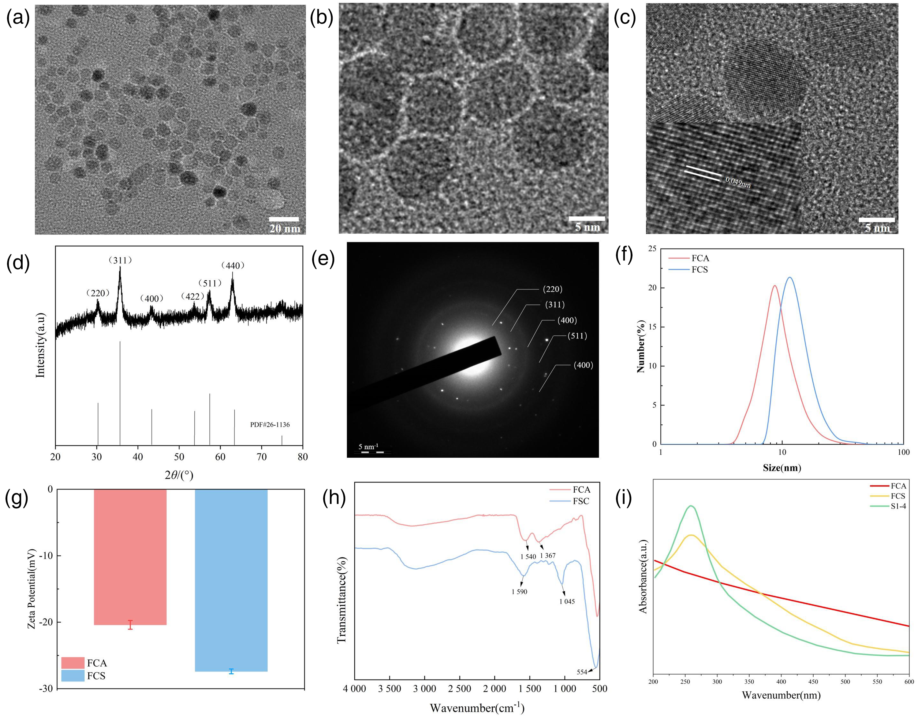
Characterization of the nanoparticles. (a) TEM image of FCA; (b) TEM image of FCS; (c) HRTEM image of FCA; (d) XRD pattern of FCA; (e) SAED pattern of FCA; (f) DLS hydrodynamic size distribution of FCA and FCS; (g) Zeta potential of FCA and FCS (mean ± SD; n = 3 independent measurements); (h) FTIR spectra of FCA and FCS; (i) UV-Vis spectra of FCA, FCS, and S1-4.
DLS measurements indicated a slight increase in hydrodynamic size after aptamer conjugation, with average diameters of 8 ± 3 nm for FCA and 17 ± 4 nm for FCS (Figure 1(f)). Zeta potential values were −19.7 ± 0.5 mV for FCA and −26.7 ± 0.3 mV for FCS (Figure 1(g)), and the polydispersity indices (PDIs) were 0.341 and 0.226, respectively. The increased negative surface charge may help reduce recognition by the reticuloendothelial system (RES), thereby prolonging circulation time and enhancing tumor accumulation, which is a favorable characteristic for subsequent in vivo applications. 37
FTIR spectroscopy (Figure 1(h)) showed a peak at 554 cm−1 in both FCA and FCS, attributed to the Fe–O stretching vibration of Fe3O4. Peaks at 1540 cm−1 and 1367 cm−1 in FCA were assigned to C=O stretching of surface carboxyl groups and C–O stretching of citrate, respectively. After conjugation with the S1-4 aptamer, the C–O peak disappeared, whereas a new peak emerged at 1590 cm−1, corresponding to amide bond formation (C=O and N–H stretching). Additionally, a peak at 1037 cm−1 in FCS was identified as the asymmetric stretching vibration of phosphate groups in the DNA backbone, confirming successful aptamer conjugation. UV-Vis spectroscopy (Figure 1(i)) further supported these findings: FCA showed no significant absorption in the visible range, whereas the S1-4 aptamer exhibited a characteristic nucleic acid peak at approximately 260 nm. The FCS spectrum showed a similar absorption peak at 260 nm, verifying the presence of the aptamer in the nanocomposite. Collectively, these results confirm the successful synthesis of FCA nanoparticles and the effective conjugation of the anionic S1-4 aptamer onto their surface.
Magnetic and AMF-induced heating properties of nanoparticles
Strong magnetic properties and efficient AMF-induced heating performance are critical factors determining the applicability of iron oxide nanoparticles in biomedicine. VSM measurements at 300 K (Figure 2(a)) revealed that both FCA and FCS exhibited hysteresis loops passing through the origin with negligible remanence, confirming their superparamagnetic behavior. The saturation magnetization values were 70.64 emu/g for FCA and 68.32 emu/g for FCS, indicating that the magnetic properties were well preserved after aptamer modification.
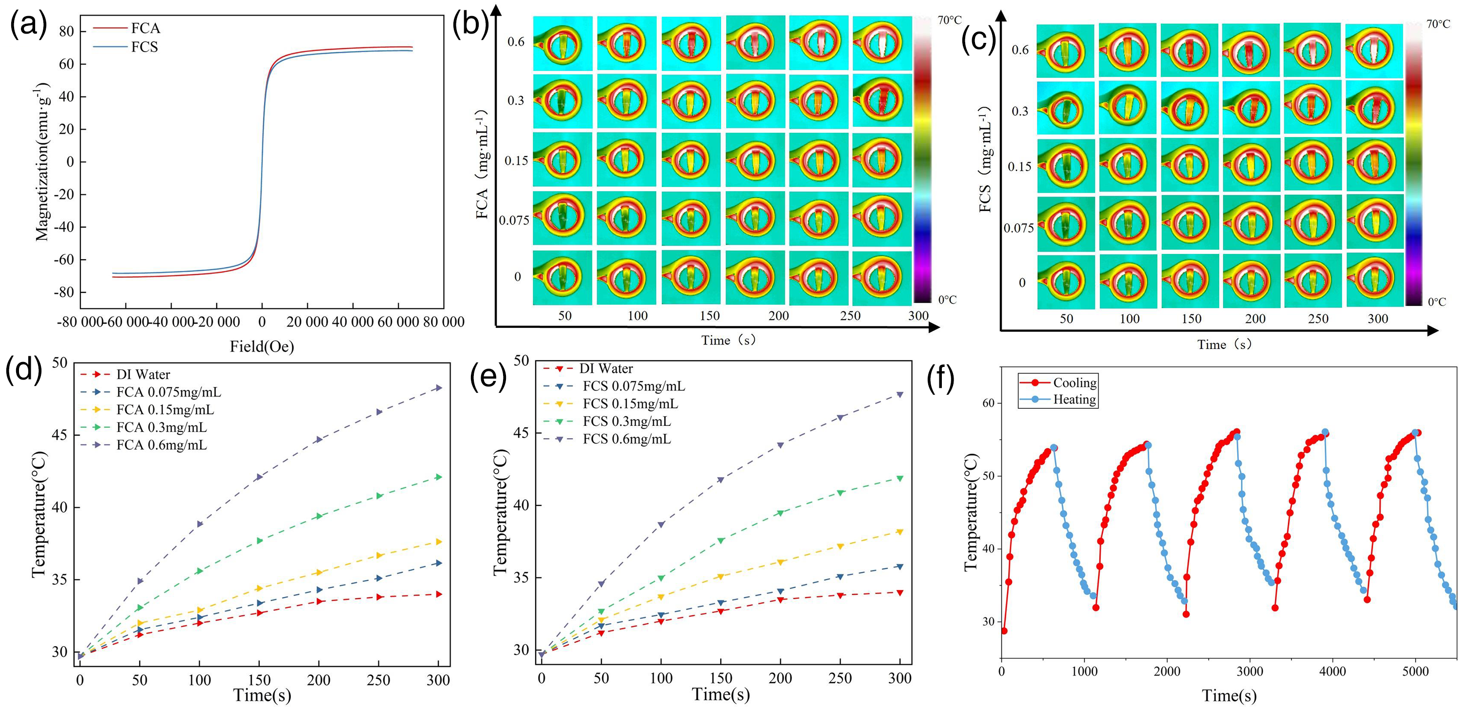
Magnetic hyperthermia performance, SAR analysis, and magnetothermal cycling stability. (a) Hysteresis loops of FCA and FCS, confirming superparamagnetism; (b and c) infrared thermal images of FCA (b) and FCS (c) at different concentrations after AMF exposure; (d and e) time-dependent temperature rise of FCA (d) and FCS (e) under AMF (474 kHz, 34.7 A), indicating AMF-induced magnetic hyperthermia effects. Deionized water exposed to the same AMF served as a blank control for background heating. (f) Cyclic heating–cooling curves demonstrating the excellent magnetothermal (magnetic hyperthermia) cycling stability of FCS during repeated AMF exposure.
The magnetic heating performance of both nanoparticles was further evaluated under an AMF. At a concentration of 0.6 mg/mL, FCA increased from 29.9°C to 48.5°C within 5 min (Figure 2(b) and (d)), whereas FCS reached 47.8°C under the same conditions (Figure 2(c) and (e)). In contrast, deionized water showed only a minor temperature increase to approximately 34°C, which was attributed to nonspecific heating from the AMF setup. This background signal was therefore used as a baseline for data interpretation.
At a lower concentration of 0.3 mg/mL, both FCA and FCS reached approximately 42°C, showing similar heating profiles. Consistently, analysis of the initial heating rates revealed that, after background correction and normalization to the total mass of the composite nanoparticles, FCA and FCS exhibited SAR values of 432 and 409 W/g, respectively, at 0.6 mg/mL, indicating efficient magnetothermal conversion under these conditions.
Magnetothermal cycling stability is an essential parameter for practical hyperthermia applications. As shown in Figure 2(f), FCS maintained highly consistent heating and cooling behavior over five consecutive AMF on/off cycles, with rapid temperature rise upon AMF activation and prompt return to near ambient temperature after deactivation. No significant performance degradation was observed, confirming excellent magnetothermal cycling stability and reusability.
In summary, conjugation of the S1-4 aptamer to form FCS did not compromise the magnetic heating efficiency of the original FCA nanoparticles. Together with their high magnetothermal cycling stability, these properties support the potential of FCS for targeted magnetic hyperthermia applications.
Biocompatibility and in vitro targeting evaluation
The MTT assay was employed to assess cell viability and proliferation after co-culture with nanoparticles at various concentrations. As shown in Figure 3(a) and (b), after 24 h of exposure to FCA or FCS nanoparticles at concentrations up to 0.6 mg/mL, both MEF and MCF-7 cells maintained viability above 80%. No significant difference was observed between the two nanoparticle types, indicating low cytotoxicity and excellent biocompatibility.
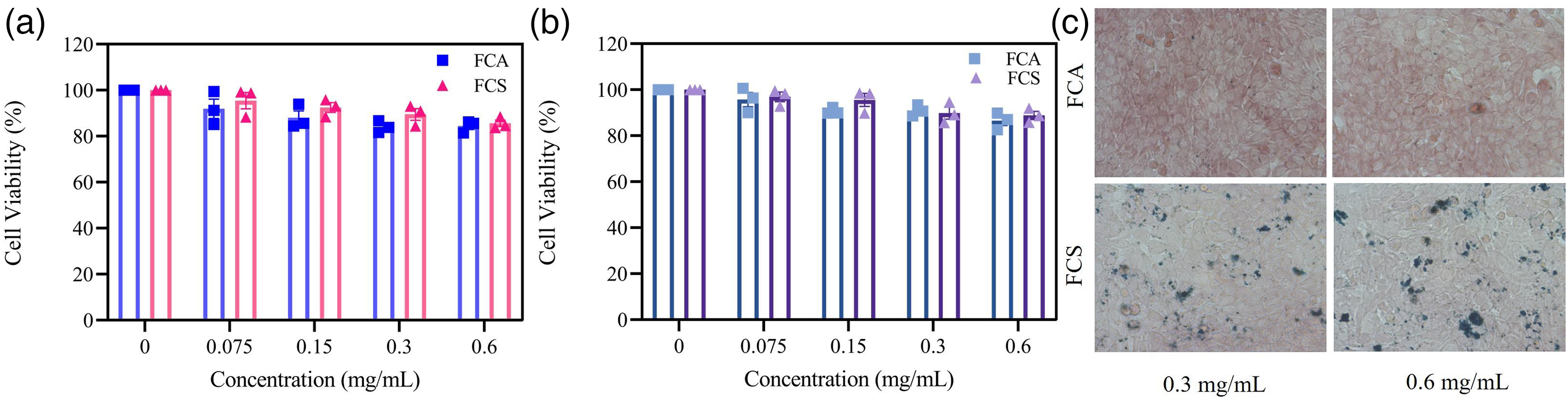
Biocompatibility and targeting of nanoparticles. (a and b) Viability of MEF (a) and MCF-7 (b) cells after 24-h treatment with FCA or FCS (mean ± SD; n = 3 biological replicates). (c) Prussian blue staining shows enhanced cellular uptake of FCS in MCF-7 cells versus FCA.
MCF-7 cells, a classical model for ER+ breast cancer, express high levels of ER alpha (ERα). Although ERα has traditionally been regarded as a nuclear receptor, recent studies confirm its presence on the cell membrane (mERα). The S1-4 aptamer specifically binds to mERα, enabling active targeting of MCF-7 and other ER+ breast cancer cells.
Prussian blue staining was used to evaluate cellular uptake. As illustrated in Figure 3(c), MCF-7 cells treated with FCS exhibited notably stronger staining intensity compared with those treated with FCA, indicating significantly higher uptake of the aptamer-conjugated nanoparticles. This result confirms the targeting capability of FCS toward mERα-expressing breast cancer cells. Because FCA and FCS differed primarily by the presence of S1-4 on the nanoparticle surface, the reproducibly stronger staining in the FCS group supports S1-4-mediated enhancement of nanoparticle association with MCF-7 cells.
Magnetic hyperthermia therapy on tumor cells
To evaluate therapeutic efficacy under AMF, MCF-7 cells were incubated with FCA or FCS at various Fe3O4 concentrations for 24 h, followed by AMF exposure (474 kHz, 34.7 A) for 10 min. MTT assays showed a concentration-dependent decrease in cell viability (Figure 4(a)). At 0.6 mg/mL, viability decreased to 42% for FCA and 18% for FCS. Consistently, IC50 analysis revealed values of 0.442 mg/mL for FCA and 0.259 mg/mL for FCS, indicating a stronger antitumor effect of FCS under AMF conditions.
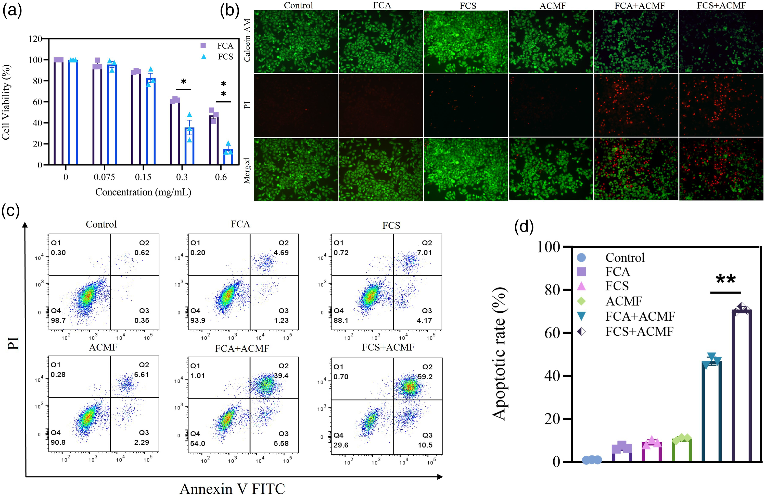
In vitro antitumor efficacy of FCA and FCS with or without AMF exposure.
Subsequently, MCF-7 cells were treated with 0.6 mg/mL Fe3O4 equivalent of FCA or FCS for 24 h and subjected to the same AMF conditions. Live/dead staining using calcein-AM (green, viable cells) and propidium iodide (red, dead cells) was performed immediately after treatment. As shown in Figure 4(b), the FCS group exhibited significantly stronger cancer cell-killing efficacy than the FCA group under AMF exposure.
To further quantify apoptosis, an Annexin V-FITC/PI apoptosis assay was conducted. Cells from each group were collected, digested, centrifuged, and stained according to the kit protocol. Results (Figure 4(c) and (d)) indicated that neither FCS incubation alone nor AMF exposure alone induced significant apoptosis compared with the control group, confirming the excellent biocompatibility of the nanoparticles and the safety of the AMF conditions, consistent with earlier findings. However, in the combined treatment group (nanoparticles + AMF), FCS nanoparticles induced markedly higher apoptosis than FCA. This enhanced effect is attributed to the active targeting capability of the S1-4 aptamer on FCS, which promotes more efficient accumulation within MCF-7 cells, leading to superior hyperthermia-induced apoptosis under AMF.
Discussion
The present study showed that S1-4 aptamer conjugation enabled the construction of a targeted Fe3O4 nanoplatform that retained the superparamagnetic properties and AMF-induced heating capacity of the magnetic core and improved cellular uptake and hyperthermia-mediated antitumor activity in MCF-7 cells. This finding is relevant because the therapeutic performance of magnetic hyperthermia depends not only on heating efficiency in suspension but also on the ability of nanoparticles to accumulate at the tumor cell interface.38,39
Our findings are consistent with previous studies showing that particle size, surface characteristics, and colloidal behavior influence both magnetic performance and biological interactions.40,41 In the present study, aptamer conjugation increased the hydrodynamic diameter and made the nanoparticle surface more negatively charged but did not abolish the heating ability of the Fe3O4 core. These results suggest that rational surface functionalization can improve biological behavior while preserving the magnetic properties required for hyperthermia.39,40
Recent studies have emphasized the importance of surface engineering in magnetic nanoplatform design. Xie et al. reported in 2024 that surface engineering of magnetic iron oxide nanoparticles improved their biomedical performance and targeting potential in breast cancer. 13 Lee et al. and Pawar and Prabhu further highlighted in 2025 the continued development of SPION-based systems for high-performance theranostic and multimodal cancer applications.42,43 Compared with these more complex systems, the present study used a relatively simple Fe3O4-aptamer construct and still achieved enhanced uptake and greater antitumor efficacy under AMF exposure. In contrast to S2.2/DOX magnetic nanoliposomes, AS1411-modified extracellular vesicles, anti-fibroblast growth factor receptor 1 (FGFR1)–tagged SPIONs, and AS1411-dendrimer magnetic nano-octahedra, the present FCS system is ligand-specific for the S1-4/mERα pathway and is designed as a drug-free, structurally simple Fe3O4 platform for ER+ breast cancer hyperthermia. Thus, the main contribution of this study is not the general concept of aptamer-functionalized magnetic nanoparticles but the application of S1-4 aptamer modification to preserve Fe3O4 magnetothermal performance and enhance uptake and AMF-mediated apoptosis in MCF-7 cells.
The targeting results are also in line with previous reports showing that aptamer-based surface ligands can enhance tumor cell recognition and therapeutic efficacy.30,32,33,35 In the present study, FCS showed greater intracellular accumulation, lower post-treatment cell viability, and higher apoptosis than FCA. These findings support the view that active targeting can meaningfully enhance magnetic hyperthermia in ER+ breast cancer cells. Another important finding is that both FCA and FCS showed low cytotoxicity in MCF-7 and MEF cells in the absence of AMF. This suggests that the stronger antitumor effect of FCS was mainly related to AMF-triggered hyperthermia rather than intrinsic nanoparticle toxicity. Regarding receptor specificity, S1-4 has been reported to bind membrane ERα on ER+ breast cancer cells; in the present study, mERα-expressing MCF-7 cells showed stronger Prussian blue staining after FCS treatment than after FCA treatment, together with lower post-AMF viability and higher apoptosis. These comparative findings support receptor-associated targeting, although they remain indirect. Future work should include competitive S1-4 blocking, ERα knockdown or antagonist experiments, and ER-negative control cell lines such as MDA-MB-231 to quantify receptor-specific binding and uptake more rigorously.
Several limitations should be acknowledged. All experiments were performed in vitro, and the present findings cannot predict in vivo behavior, including biodistribution, tumor accumulation, and clearance. In addition, the therapeutic evaluation was mainly conducted in a single ER+ breast cancer cell line, and the uptake mechanism and intracellular fate of FCS were not examined in detail. Further in vivo validation and mechanistic studies are therefore needed. In addition, receptor-blocking experiments, fluorescence-based quantitative uptake assays, and comparisons with ER-negative breast cancer cells were not performed in this study; these analyses will be important for distinguishing S1-4/mERα-specific targeting from nonspecific nanoparticle uptake in future studies.
Overall, the present study indicates that S1-4 aptamer-mediated Fe3O4 nanoparticles provide a feasible strategy for improving the targeting capacity and magnetic hyperthermia effect of iron oxide nanoparticles in breast cancer cells.
Conclusion
FCS nanoparticles showed active targeting to MCF-7 breast cancer cells and improved the antitumor effect of AMF-mediated magnetic hyperthermia with low cytotoxicity under the tested conditions. These findings suggest that FCS nanoparticles may be a feasible platform for targeted magnetic hyperthermia in breast cancer.
Footnotes
Acknowledgments
The authors used assistive AI tools only for language polishing. No AI tools were used in the study design, data analysis, or other research-related methods.

Ethics approval and consent to participate
This study was conducted entirely in vitro using established cell lines and did not involve human participants, human tissue samples, or identifiable personal data. Therefore, ethics approval and informed consent were not required, and the Declaration of Helsinki was not applicable.
Patient consent for publication
Not applicable.
Authors’ contributions
Conceptualization, E.C. and C.N.; methodology, E.C. and Y.Y.; software, E.C.; validation, E.C. and X.W., Y.Y., and X.M.; formal analysis, E.C. and Y.Y.; resources, J.Y. and X.W.; data curation, B.Y. and X.W.; writing—original draft preparation, E.C.; writing—review and editing, E.C. and Y.Y.; supervision, C.N. and X.W.; All authors have read and agreed to the published version of the manuscript.
Funding
The authors disclosed receipt of the following financial support for the research, authorship, and/or publication of this article: This work was supported by the National Natural Science Foundation of China (grant number K112204322).
Declaration of conflicting interests
The authors declared no potential conflicts of interest with respect to the research, authorship, and/or publication of this article.
Availability of data and materials
The data generated in the present study may be requested from the corresponding author.