
Abstract
The development of human uterine cancer is a complex process involving the abnormal expression of tumor suppressors, such as PTEN, ARID1A, and TP53; mismatch repair protein MSH6; and transcription factors, such as PAX2 and PAX8. The functional changes that lead to uterine adenocarcinoma in pet rabbits are not fully understood despite the frequent occurrence of this condition in the species. Thus, an immunohistochemical analysis was performed to visualize the protein expression pattern of carcinogenesis-related molecules in surgical biopsy specimens from 56 uterine adenocarcinomas and 8 uteruses without significant lesions in pet rabbits. Seventy percent of the adenocarcinomas were positive for estrogen receptor (ER), 7% were positive for TP53, and there was a decreased expression in comparison to control uterine epithelium for PAX2 in 54%, for PAX8 in 73%, for ARID1A in 68%, and for MSH6 in 48% of the tumors. TP53 expression was not observed in control uterine tissues. There was a significant negative correlation between nuclear ER and PAX2 immunolabeling in uterine adenocarcinomas. Heat map analysis classified samples into 4 clusters, which revealed that 1 PAX2-positive group had a higher presence of papillary-type uterine adenocarcinomas and a lower prevalence of tubular/solid types compared with the 2 PAX2-negative groups. This study demonstrated that the immunohistochemical phenotype of rabbit uterine adenocarcinoma is comparable to that of human endometrial carcinomas, suggesting the potential for similar oncogenic mechanisms that might prove useful for translational medicine research.
Keywords
Pet rabbits (Oryctolagus cuniculus) are particularly prone to uterine diseases, which account for 13% of all rabbit diseases, and half of all rabbit uterine diseases are neoplastic, with most tumors diagnosed as uterine adenocarcinomas.3,4 About half of uterine adenocarcinomas in rabbits metastasize to the lungs (79%), peritoneum/omentum (33%), and liver (30%).3,4 The high incidence of uterine adenocarcinoma has been suggested to be due to long-term exposure to estrogen. 1 As rabbits are mating oviparous animals with year-round estrus cycles, 16 reproductive tissues are estrogenically stimulated for prolonged periods, leading in some cases to DNA damage; estrogens can be genotoxic in murine models. 5 Rabbit uterine adenocarcinoma is histologically divided into papillary and tubular/solid types, with the tubular/solid type being more invasive and thus carrying increased metastatic risk compared with the papillary type. 1 About 20% of papillary adenocarcinomas are estrogen receptor (ER)-positive, while nearly 90% of tubular/solid adenocarcinomas are ER-positive, suggesting that rabbit uterine adenocarcinoma may arise through multiple carcinogenic mechanisms. 1
In humans, numerous subtypes of the human papillomavirus have been associated with the development of cervical cancer but not with cancers arising from the uterine body. 14 Cancers arising from the uterine body are broadly classified into endometrioid carcinoma, serous carcinoma, and clear cell carcinoma, whereas their carcinogenic mechanisms are unknown. 2 Endometrial cancer is generally classified into estrogen-dependent type I uterine cancers (endometrioid types) and estrogen-independent type II uterine cancers (serous and clear cell types). 7 Risk factors for type I endometrial cancer are related to unopposed exposure of the endometrium to estrogen, including unopposed estrogen therapy, early menarche, late menopause, tamoxifen therapy, nulliparity, infertility or failure to ovulate, and polycystic ovary syndrome. 7 In endometrial carcinomas, oncogenic mechanisms involve mutations and loss of function of paired box 2 (PAX2), paired box 8 (PAX8), tumor protein p53 (TP53), phosphatase and tensin homolog (PTEN), AT-rich interaction domain 1A (ARID1A) genes, and the mismatch repair protein mutS homolog 6 (MSH6) gene.19,34 Serous carcinoma is often positive for TP53 and negative for estrogen receptor (ER), a similar pattern found in many clear cell carcinoma cases. 2
Highly differentiated endometrioid carcinomas are positive for ER, whereas the nuclear immunolabeling for ER in poorly differentiated forms is attenuated and absent in most serous and clear cell carcinomas.22,23 Endometrial stromal cells and smooth muscle cells are also positive for ER, providing a marker to identify uterine origin. 36 PAX2 and PAX8 are transcription factors involved in urogenital differentiation and have been used as highly sensitive and specific markers for tumors of kidney and Müllerian duct origin.20,28,35 PTEN is a tumor suppressor gene that is expressed in a variety of normal cells and is involved in regulating the phosphatidylinositol-3 kinase pathway, cell cycle specification, and cell proliferation. 9 In human uterine cancers, PTEN mutations occur in 52% to 78% of estrogen-dependent type I uterine cancers and 1% to 11% of estrogen-independent type II uterine cancers. 26 ARID1A is a tumor suppressor gene ubiquitously expressed in normal cells, and ARID1A mutations have been detected in various tumors, 6 including in 39% to 44% of human endometrial carcinomas. 33 MSH6 functions as a group of DNA mismatch repair genes, and a deficiency in any of these genes is considered a form of microsatellite instability and may lead to Lynch syndrome. 37 Lynch syndrome is a familial human disease that causes malignant tumors such as colorectal, endometrial, and ovarian cancer, and has not been reported in rabbits. Loss of function of PTEN, ARID1A, and MSH6 acquired by these mutations in human endometrial cancer patients can be confirmed by lack of immunolabeling. 13 However, when the TP53 gene is mutated, as it occurs in human cancers, 15 overexpression of TP53 can be confirmed in the nuclei of tumor cells. 31 However, a completely negative (TP53 null) result may be a truncation mutation that disrupts protein production. In human endometrial cancer, overexpression due to TP53 mutations is found in 20% of type I endometrioid carcinomas and 86% of type II serous carcinomas. 30 With immunohistochemistry (IHC), PAX2, PAX8, ARID1A, MSH6 and PTEN can be evaluated for loss of function by negative nuclear immunolabeling, and for TP53 loss of function by positive nuclear immunolabeling. The changes in expression of these proteins in uterine adenocarcinoma in other animal species, including rabbits, are not known. To address this knowledge gap, this study investigated the protein expression pattern by immunohistochemical analysis of PAX2, PAX8, ARID1A, MSH6, PTEN, and TP53 in rabbit uterine adenocarcinomas.
Material and Methods
Population
The laboratory database of the Department of Veterinary Pathology at Nippon Veterinary and Life Science University was searched for surgical biopsies from pet rabbits submitted between June 1998 and January 2023 using the search terms “rabbit” or “rabbit uterus.” A total of 377 rabbit biopsies were found, of which 166 were uterine biopsies. Uterine adenocarcinoma was diagnosed in 56 cases, and uteruses without significant change in 8 cases. For cases 55 and 56, analysis was performed on metastatic areas of uterine adenocarcinoma only, because primary tumor blocks from the primary site were not available. Breed classification was listed according to the American Rabbit Breeders Association. Details regarding the individual rabbits are summarized in Supplemental Table S1.
Histological Analysis
Tissues were fixed in 10% neutral-buffered formalin immediately after excision and embedded in paraffin within 1 to 3 days to obtain 3-μm-thick sections that were subsequently stained with hematoxylin and eosin. Serial sections were subjected to IHC using the polymer-labeled system Histofine Simple Stain MAX-PO (M) (Nichirei, Tokyo, Japan). Histological diagnosis was made according to the World Health Organization classification of uterine tumors in domestic animals, 21 and uterine adenocarcinoma was subclassified as “papillary” or “tubular/solid” type (Fig. 1a-d) according to the criteria of Asakawa et al 1 Briefly, papillary adenocarcinoma has a papillary to cribriform, expansive growth of glands. In the early stages, myometrial invasion is minimal. The tubular/solid type of adenocarcinoma is characterized by a proliferation of neoplastic cell nests, often forming structures lined by a single layer of epithelial cells, with no papillary proliferation; the neoplastic tissue often infiltrates the deep myometrium.
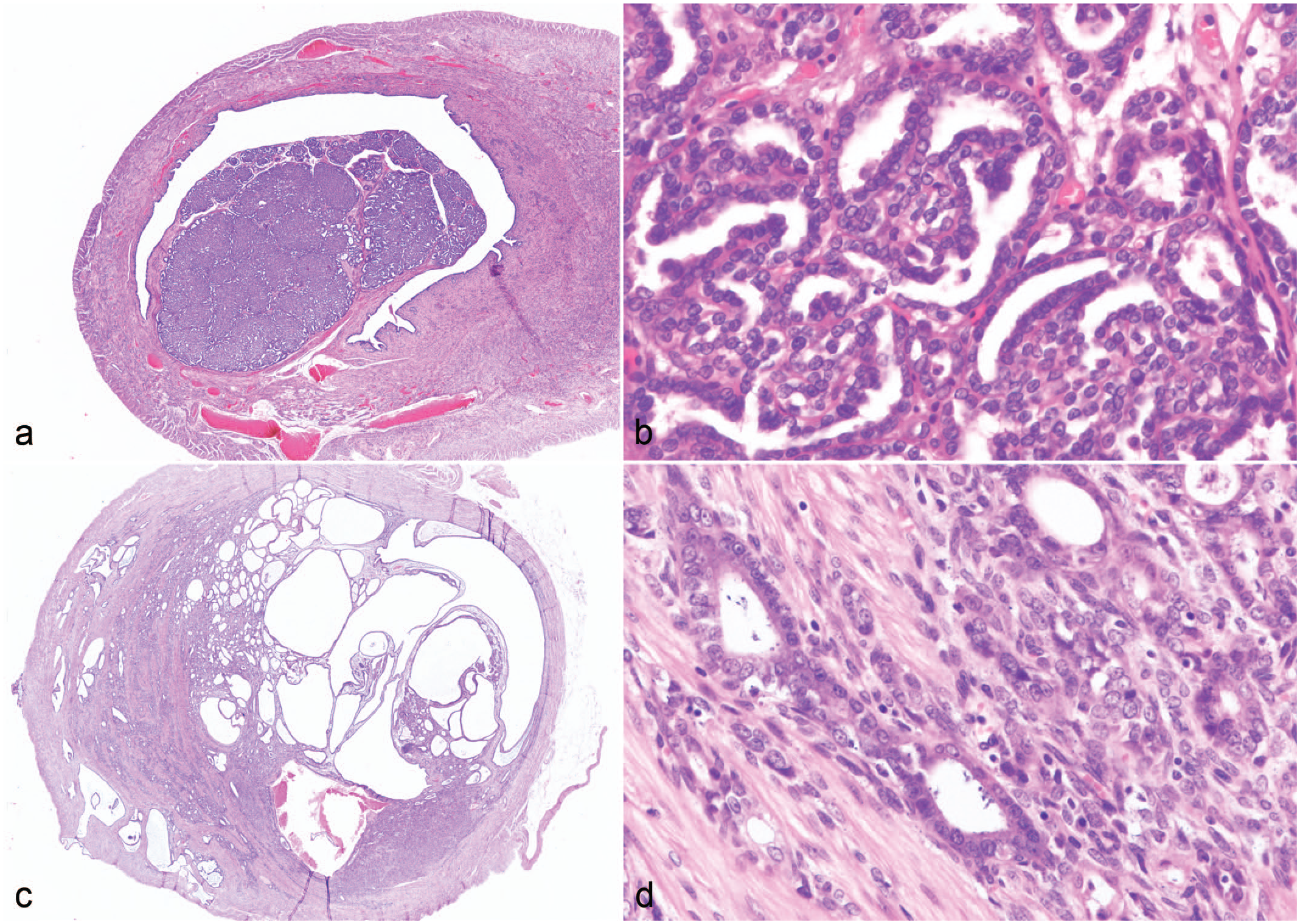
Adenocarcinoma of the uterus in rabbits. Hematoxylin and eosin. (a) Papillary type, low magnification. (b) Papillary type, high magnification. Neoplastic cells grow in a tubular or papillary fashion partially obstructing the uterine lumen. (c) Tubular/solid type, low magnification. (d) Tubular/solid type, high magnification. Neoplastic cells are observed invading the muscle layer.
Immunohistochemical Analysis
IHC of serial sections was performed using the polymeric polymer method with antibodies against cytokeratin (CK), ER, PAX2, PAX8, ARID1A, MSH6, PTEN, and TP53. Details of the primary antibodies and retrieval conditions used in this study are shown in Supplemental Table S2. The immunolabeling results for the control uteruses are shown in Table 1 and Fig. 2. Rabbit uterus was used as positive control for CK, ER, PAX2, PAX8, ARID1A, MSH6, and PTEN. Since TP53 does not show positivity in histologically unaltered tissues, a TP53-positive rabbit uterine adenocarcinoma from the study materials was used as the positive control. For negative controls, the primary antibodies were replaced with normal mouse immunoglobulins.
Expression scores and percentages per immunohistochemical antibody in control rabbit uterus.
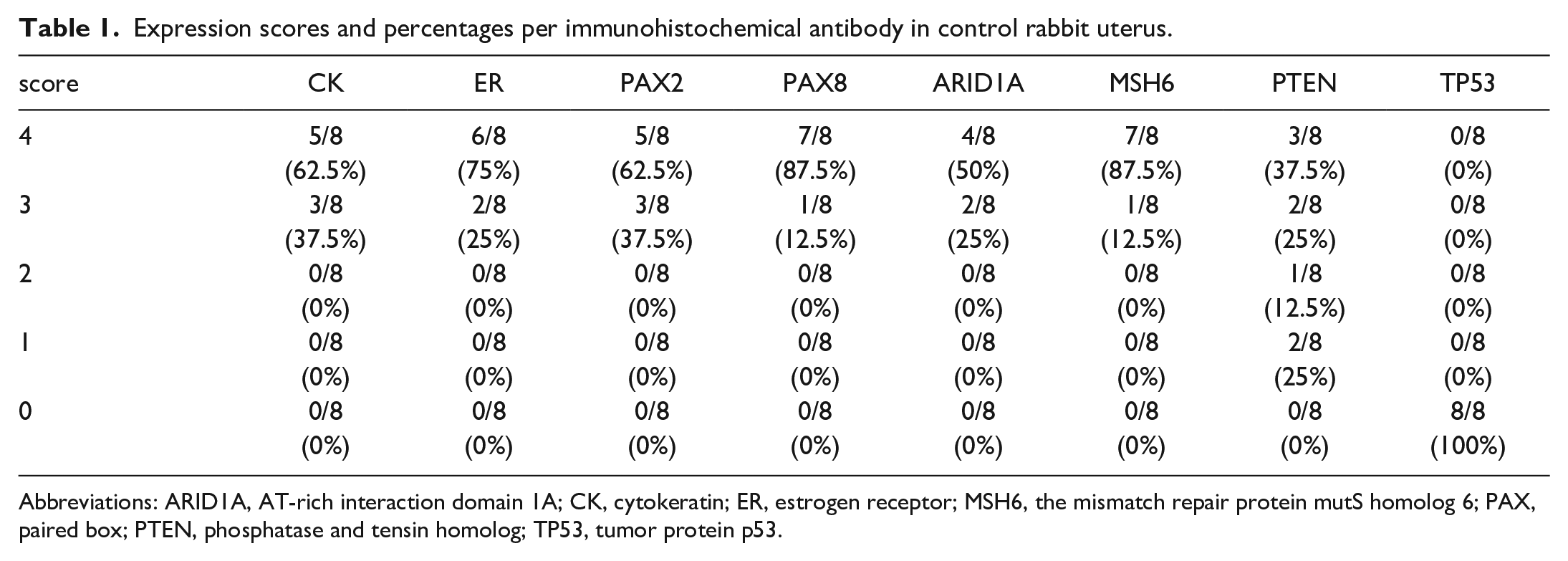
Abbreviations: ARID1A, AT-rich interaction domain 1A; CK, cytokeratin; ER, estrogen receptor; MSH6, the mismatch repair protein mutS homolog 6; PAX, paired box; PTEN, phosphatase and tensin homolog; TP53, tumor protein p53.
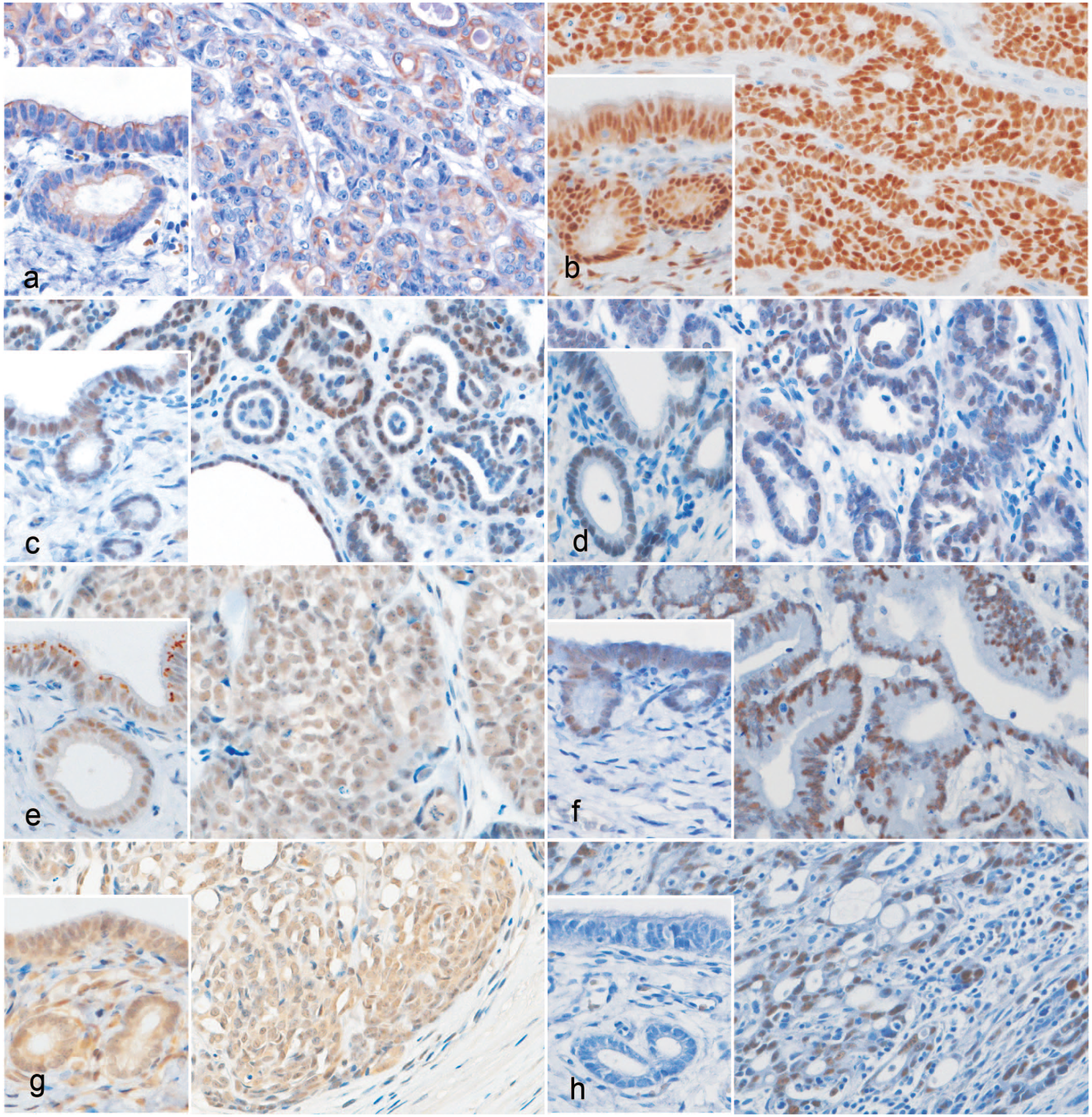
Immunohistochemistry (IHC) of uterine adenocarcinoma and control uterus (insets) in rabbits. (a) The cytoplasm of neoplastic cells has moderate immunolabeling for pan-cytokeratin. (score 4, papillary) Inset: Cytoplasmic immunolabeling of epithelial cells in the control uterus. Cytokeratin AE1/AE3 IHC. (b) Nuclei of neoplastic and stromal cells have estrogen receptor (ER) immunolabeling. (score 4, tubular/solid) Inset: Immunolabeling of nuclei of epithelial and stromal cells in a control uterus. ER IHC. (c) Nuclei of neoplastic cells have PAX2 immunolabeling. (score 4, papillary) Inset: Immunolabeling of nuclei of epithelial cells in a control uterus. PAX2 IHC. (d) Nuclei of neoplastic cells have PAX8 immunolabeling. (score 4, papillary) Inset: Immunolabeling of nuclei of epithelial cells in a control uterus. PAX8 IHC. (e) Nuclei of neoplastic cells have ARID1A immunolabeling. (score 4, tubular/solid) Inset: Immunolabeling of nuclei of epithelial cells in a control uterus. ARID1A IHC. (f) Nuclei of neoplastic cells have MSH6 immunolabeling. (score 4, papillary) Inset: Immunolabeling of nuclei of epithelial cells in a control uterus. MSH6 IHC. (g) Mild to moderate nuclear and cytoplasmic immunolabeling for PTEN is observed in neoplastic cells and stroma cells. (score 4, papillary) Inset: Nuclear and cytoplasmic immunolabeling of epithelial and stromal cells in a control uterus. PTEN IHC. (h) Nuclei of neoplastic cells show moderate to strong immunolabeling for TP53. (score 4, tubular/solid) Inset: immunolabeling for TP53 is absent in a control uterus. TP53 IHC.
Immunolabeling results were classified according to the percentage of positive neoplastic cells and scored on a 5-point scale; 0: 0% to 4%, 1: 5% to 24%, 2: 25% to 49%, 3: 50% to 74%, and 4: 75% to 100%. The immunolabeling for CK was evaluated in the cytoplasm, whereas the immunolabeling for ER, PAX2, PAX8, ARID1A, MSH6, PTEN and TP53 was assessed in the nucleus. Assessments were conducted subjectively by combining immunolabeling percentage estimations performed by 2 pathologists. Discrepancies in percentage estimates were sorted by achieving a consensus.
Statistical Analysis
The evaluation of the association among the antibodies, with scores of 3 or higher for both considered positive (except CK), was performed by Spearman’s rank correlation coefficient. Heat maps were generated in the statistical software R (R Foundation for Statistical Computing, Vienna, Austria) using the contributed packages Biobase 17 and Heatplus. 29 The Ward method was applied for clustering algorithm. 32 The clusters were divided at the third branching point from the top, resulting in four groups. Fisher’s exact test was used to analyze associations between categorial variables, cluster groups, and tissue subtypes, with significant results followed by Bonferroni-corrected post hoc pairwise comparisons. Statistical computations were performed using the statistical software R, and data visualization and additional analyses were conducted in GraphPad Prism version 9 (GraphPad Software, Boston, MA, USA). Significance level was set at 5% for all analysis.
Results
Case Data
The mean age at diagnosis of uterine adenocarcinoma was 5.79 years (range 2 years to 9 years, 5 months; median 6 years). Information on breed was available for 22 of the 56 cases. Thirteen were hybrids, 3 were Holland lops, 2 were Netherland dwarfs, and 1 each was a rex, mini rex, American fuzzy lop, and Jersey wooly. The percentages of papillary and tubular/solid types of uterine adenocarcinoma were 34/56 (61%) and 22/56 (39%), respectively (Fig. 1a-d).
Immunohistochemical Results
The immunohistochemistry scores are presented in Table 2. The cytoplasm of neoplastic cells was positive for CK (Fig. 2a). Nuclei of neoplastic cells were positive for ER, PAX2, PAX8, ARID1A, and MSH6 (Fig. 2b-f). The nuclei and cytoplasm of neoplastic cells were positive for PTEN (Fig. 2g). Nuclei of stromal cells showed ER and PTEN immunolabeling; however, the stromal cells did not display immunolabeling for PAX2, PAX8, ARID1A, or MSH6. Tumors that were positive or partially negative for these markers varied (Table 2). The nuclei of neoplastic cells were generally negative for TP53 but were partially positive in some tumors (Fig. 2h, Table 2).
Expression scores and percentages per immunohistochemical antibody in rabbit uterine adenocarcinoma.
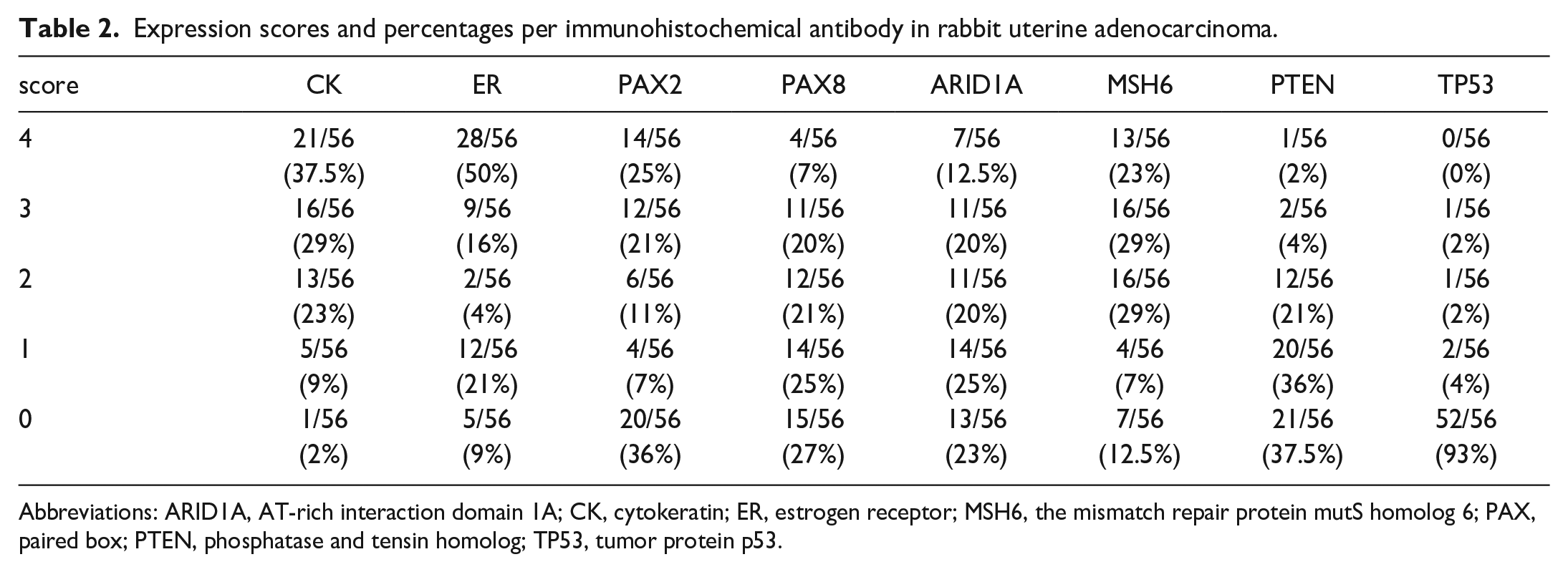
Abbreviations: ARID1A, AT-rich interaction domain 1A; CK, cytokeratin; ER, estrogen receptor; MSH6, the mismatch repair protein mutS homolog 6; PAX, paired box; PTEN, phosphatase and tensin homolog; TP53, tumor protein p53.
Correlation of the immunomarkers
There was a significant negative association between ER and PAX2 immunolabeling (rs = -0.582; P < .001); a significant positive association between PAX2 and PAX8 immunolabeling (rs = 0.509; P < .001); a weak positive association between PAX2 and ARID1A (rs = 0.341; P = .009), MSH6 (rs = 0.340; P = .009) and PTEN (rs = 0.345; P = .008); and a weak positive association between PAX8 and ARID1A (rs = 0.363; P = .005), MSH6 (rs = 0.471; P = .0003), and PTEN (rs = 0.414; P = .001) (Table 3). Heat map analysis divided cases into clusters of ER, PAX2, PAX8, ARID1A, MSH6, PTEN, and TP53 (Fig. 3) immunolabeling results. According to their expression, cases were also classified into 4 clusters: group 1, variable but present nuclear immunolabeling for ER, PAX2, PAX8, ARID1A, and MSH6; group 2, lacks nuclear immunolabeling for ER but has nuclear immunolabeling for PAX2, and more than one of the PAX8, ARID1A, and MSH6; group 3, nuclear immunolabeling for ER and absence of nuclear immunolabeling for either PAX2, PAX8, ARID1A, or MSH6; and group 4, nuclear immunolabeling for ER and absence of nuclear immunolabeling for PAX2, PAX8, ARID1A, and MSH6 (Supplemental Figure S1). The scores for the 4 groups for each antibody are presented in Supplemental Figure S2. In addition, the associations between the above 4 clusters and tissue subtypes were evaluated. In group 1, 13/13 (100%) were papillary and 0/13 (0%) were tubular/solid; in group 2, 12/17 (70.6%) were papillary and 5/17 (29.4%) were tubular/solid; in group 3, 6/16 (37.5%) were papillary and 10/16 (62.5%) were tubular/solid; and in group 4, 3/10 (30%) were papillary and 7/10 (70%) were tubular/solid (Fig. 4a). Fisher’s exact test showed a significant result (P < .001). Post hoc pairwise comparisons showed differences between group 1 and group 3 (P = .002), and group 1 and group 4 (P = .003) with no significant differences among other groups. There were more PAX2-positive cases in the papillary type and significantly more PAX2-negative cases in the tubular/solid type (P < .001) (Fig. 4b). However, ER (Fig. 4c), PAX8, ARID1A, MSH6, PTEN, and TP53 expression did not differ significantly among histologic types.
Spearman’s rank correlation coefficients considering the expression results of each antibody in rabbit uterine adenocarcinoma.
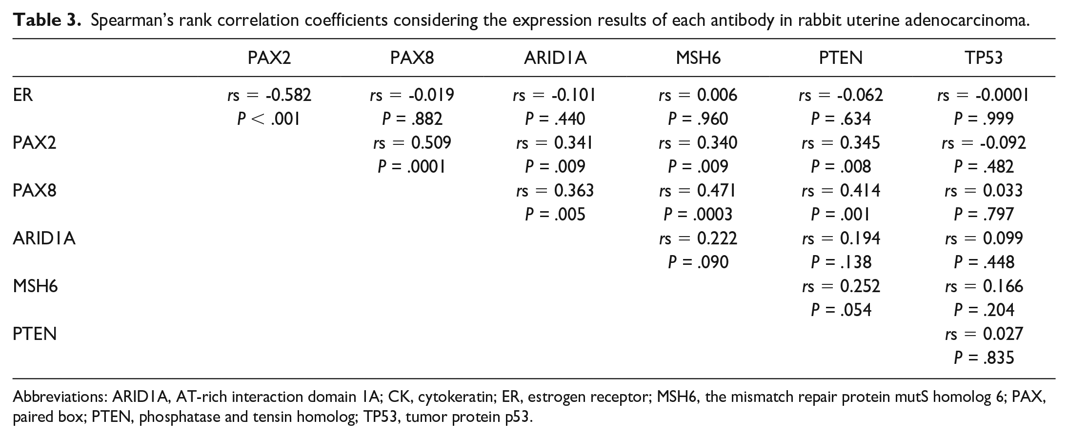
Abbreviations: ARID1A, AT-rich interaction domain 1A; CK, cytokeratin; ER, estrogen receptor; MSH6, the mismatch repair protein mutS homolog 6; PAX, paired box; PTEN, phosphatase and tensin homolog; TP53, tumor protein p53.
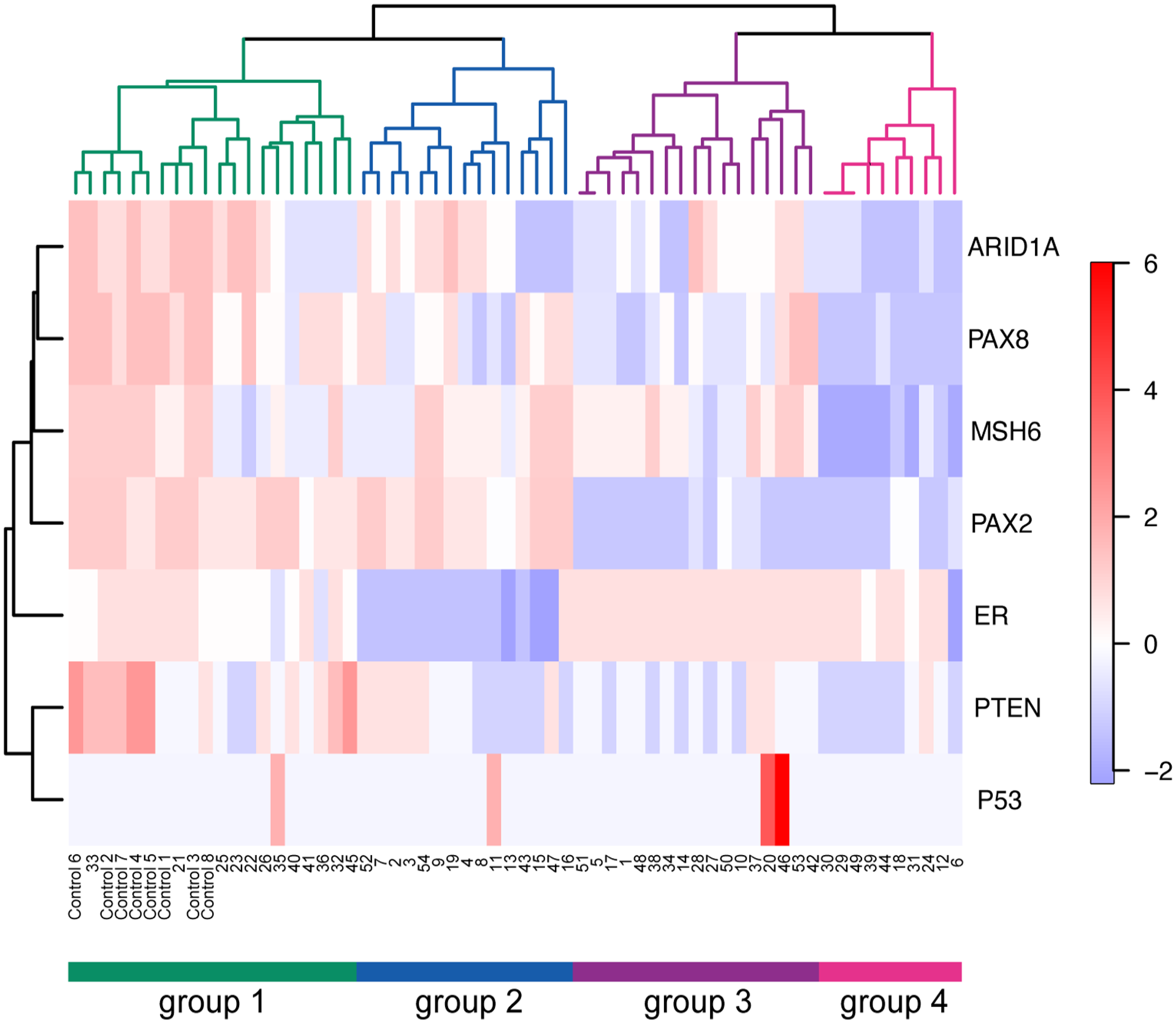
Clustering analysis of immunolabeling results per antibody in rabbit uterine adenocarcinomas. The clusters are divided at the third branching point from the top, resulting in 4 groups.
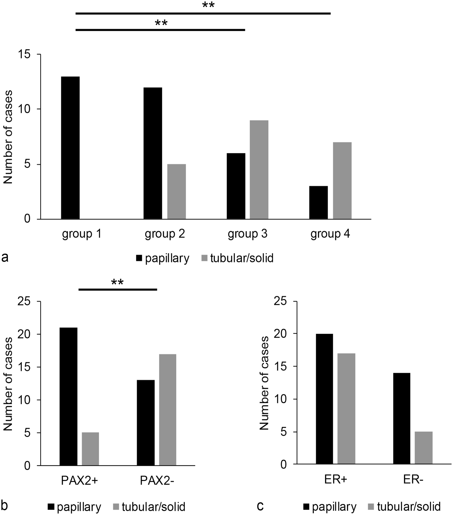
Association with tissue subtypes according to target molecules. (a) Number of cases of papillary and tubular/solid rabbit uterine adenocarcinoma in each of the 4 groups. Bonferroni correction for multiple tests showed P < .01. (b) Relationship between the presence or absence of PAX2 expression and histological type. The Fisher’s extract test showed P < .01. (c) Relationship between the presence or absence of estrogen receptor (ER) expression and histological type. ** P < .01.
Discussion
Uterine cancers in humans have been shown to have an increased loss of PAX2 and PAX8 immunolabeling compared with the control uterus, 24 supporting their roles in tumor suppression and blocking apoptosis, respectively. PAX8 immunolabeling is also known to be reduced in high-grade uterine cancer. 25 In this study, the nuclei of all control uterine mucosal epithelia immunolabeled for PAX2 and PAX8, whereas 24 (43%) uterine adenocarcinomas were PAX2 negative, 29 (52%) were PAX8 negative, and 17 (30%) were negative for PAX2 and PAX8. These results suggest that deletion of nuclear PAX2 and PAX8 may be involved in the carcinogenesis of rabbit uterine adenocarcinoma. A significant negative correlation was also observed between ER and PAX2 immunolabeling scores. This indicates that nuclear PAX2 immunolabeling is decreased in ER-positive uterine adenocarcinomas, further suggesting that the deletion of PAX2 may be involved in tumor development. It has been reported that knockdown of Pax2 increased expression of ER in murine oviduct cells, which is similar to our results. 12 Further, PAX2 expression is suppressed by estrogen prompting its methylation through ER, which suggests a negative correlation between ER and PAX2 immunolabeling. 10 The less invasive papillary type had significantly more nuclear PAX2 immunolabeling than the tubular/solid type, and PAX2 immunolabeling was positively correlated with PAX8, ARID1A, PTEN, and MSH6, suggesting its association with highly differentiated tumor cells. However, PAX2 negativity is not linked to mutation, and the mechanism of loss of expression is not yet understood. 8 Regarding the immunolabeling of ER, Asakawa et al 1 reported that the tubular/solid uterine adenocarcinoma had nuclear immunolabeling for ER more commonly than the papillary type, but no significant difference was found in this study. A possible confounder is the small number of cases analyzed in this study; thus, future studies should aim at increasing sample size to strengthen the confidence and precision of the tumor marker associations.
Mutations in MSH6 cause microsatellite instability, which occur in 28% to 40% of human cases of estrogen-dependent type I uterine cancers and 0% to 2% of estrogen-independent type II uterine cancers. 26 In this study, immunohistochemical analysis of MSH6 showed that 20% (11/56) of the cases had a nuclear immunolabeling score of 0 or 1 in uterine adenocarcinomas, whereas control uterine epithelium had a score of 3-4. This suggests that the observed attenuated expression of MSH6 and microsatellite instability may be involved in the development of rabbit uterine adenocarcinoma, similar to human endometrial tumorigenesis, providing support for a shared carcinogenic mechanism.
In humans, PTEN and ARID1A are mutated in type I endometrial carcinoma rather than type II. 26 Since PTEN and ARID1A were found to be down-regulated in this study as well, it is assumed that their biological functionality is similar to that in human endometrial carcinomas. In this study, a subset of control endometrial epithelial cells had nuclear PTEN immunolabeling; however, immunolabeling for PTEN has been reported to vary in the endometrium during the hormonal cycle in humans, and its stability is still being investigated in rabbits. 27
In this study, elevated nuclear expression of TP53 was observed in 4 cases. In humans, the TP53 gene is mutated in approximately 90% of type II uterine carcinomas. 30 Data of this study indicate nuclear immunolabeling for ER was observed in 2 of the 4 TP53-positive cases. Even estrogen-dependent type I carcinoma can exhibit TP53 positivity when it becomes high-grade. 30 Therefore, the 2 cases in question, which are both ER-positive and TP53-positive, may also be classified under this category.
Positive correlations were found between PAX2 and PAX8; PAX2 and ARID1A; PAX2 and PTEN, PAX2, and MSH6; PAX8 and ARID1A, PAX8, and PTEN; and PAX8 and MSH6. These results suggest that mutations in the PAX family of urogenital transcription factors and mutations in ARID1A, PTEN, and MSH6 might be involved in carcinogenesis in a complex manner. ARID1A binds within an ER-binding enhancer and regulates ER-dependent transcription, and PTEN is down-regulated by estrogen via microRNAs.18,11 Because ARID1A, PTEN, and ER are closely related, they are likely to be involved in carcinogenesis in uterine adenocarcinoma in rabbits.
Heat map analysis classified the cases into 4 clusters, and nuclear PAX2 immunolabeling tended to be higher in the “papillary” compared with “tubular/solid” types, but the detailed mechanism remains unclear and requires further investigation.
Overall, results from the current study suggest that estrogen-induced uterine adenocarcinoma in rabbits may share similar complex carcinogenic mechanisms with its human counterpart, involving dysregulated gene expression. These results suggest that analysis of gene expression patterns could enable the subclassification of uterine adenocarcinoma in rabbits, potentially for more accurate diagnosis. Future investigations directed at correlating expression groups with patient outcomes and determining the molecular drivers behind the development of primary and metastatic rabbit uterine adenocarcinomas may provide new insights. Thus, rabbit uterine adenocarcinomas offer a valuable subject to delve deeper into carcinogenetic mechanisms, clinical staging, histopathology grading, and prognosis, as a potential model for endometrial cancer in humans.
Supplemental Material
sj-pdf-1-vet-10.1177_03009858251332005 – Supplemental material for Immunohistochemical characterization of differentiation-associated transcription factors, tumor suppressor genes, and mismatch repair genes in rabbit uterine adenocarcinoma
Supplemental material, sj-pdf-1-vet-10.1177_03009858251332005 for Immunohistochemical characterization of differentiation-associated transcription factors, tumor suppressor genes, and mismatch repair genes in rabbit uterine adenocarcinoma by Yukino Machida, Sayuri Harashima, Viviana Gonzalez Astudillo and Masaki Michishita in Veterinary Pathology
Footnotes
Author Contributions
YM and SH designed and performed the experiments; YM and MM contributed to the experimental design; YM and SH performed histologic evaluations; YM and SH performed statistical analysis; the manuscript was written by YM, SH, VGA, and MM with contribution from the other authors.
Declaration of Conflicting Interests
The author(s) declared no potential conflicts of interest with respect to the research, authorship, and/or publication of this article.
Funding
The author(s) disclosed receipt of the following financial support for the research, authorship, and/or publication of this article: This work was supported by Grant-in-Aid for Scientific Research for Young Scientists from the Japan Society for the Promotion of Science KAKENHI Grant Number 22K15017, and by the Initiative for Realizing Discovery in the Research Environment from MEXT, Japan.
Supplemental material for this article is available online.
References
Supplementary Material
Please find the following supplemental material available below.
For Open Access articles published under a Creative Commons License, all supplemental material carries the same license as the article it is associated with.
For non-Open Access articles published, all supplemental material carries a non-exclusive license, and permission requests for re-use of supplemental material or any part of supplemental material shall be sent directly to the copyright owner as specified in the copyright notice associated with the article.